A new study from the University of Warwick suggests that the rhythm of human laughter has remained surprisingly consistent for at least 15 million years. By comparing the laughter of humans and other great apes, researchers uncovered evidence that this ancient vocal pattern may offer valuable clues about how human speech gradually evolved.
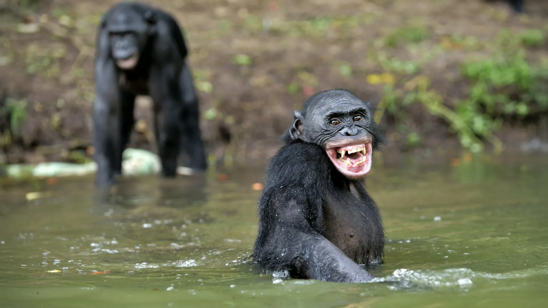
Humans are not the only primates that laugh. Chimpanzees, bonobos, gorillas, and orangutans all produce laughter, but scientists have long wondered how those vocalizations changed over millions of years and whether they could reveal anything about the origins of human language.
To investigate, researchers analyzed laughter recordings from four orangutans, two gorillas, three bonobos, four chimpanzees, and four humans. Their study, published in Communications Biology, examined 140 separate laughter sequences.
Despite the differences between species, the team found a striking similarity. Every species produced laughter with evenly spaced rhythmic intervals between successive sounds.
The researchers believe this shared rhythmic pattern originated in a common ancestor that lived around 15 million years ago. They propose that the basic structure has remained remarkably stable throughout the evolution of all living great apes.
Dr. Chiara De Gregorio, Honorary Research Associate, Department of Psychology, University of Warwick said: “How did humans evolve the remarkable ability to speak? Speech leaves no fossils, and complex language exists only in our own species. But we’ve found a 15-million-year-old clue in an unexpected place: our laughter. Unlike speech, laughter is shared by all living great apes. By comparing how different species laugh, we can see that a basic rhythmic structure has remained unchanged since our last common ancestor. That’s extraordinary.”
Human Laughter Became More Flexible
Although the underlying rhythm appears to have stayed the same, human laughter has become faster, more varied, and far more adaptable than that of other great apes.
People can consciously adjust when and how they laugh depending on the situation. A spontaneous laugh triggered by tickling differs from a polite laugh during a meeting, a nervous laugh after making a mistake, or contagious laughter shared among friends. While each serves a different social purpose, they all retain the same basic rhythmic foundation.
According to the researchers, this growing ability to control vocal timing likely developed gradually over the course of great ape evolution. That increasing level of vocal control, including over laughter, may have provided one of the essential building blocks that eventually made human speech possible.
A Window Into the Evolution of Speech
Because spoken language leaves no direct fossil evidence, scientists have few ways to trace its earliest origins. Laughter, however, is evolutionarily much older than speech and remains common to every living great ape, making it a rare opportunity to study how vocal communication evolved.
Dr. Adriano Lameria, Associate Professor, ApeTank, Department of Psychology, University of Warwick said: “It is impossible to assess the precursor forms of language directly from our extinct ancestors. Laughter, being evolutionarily older and having remained shared between all living great apes, provides a rare evolutionary window into the vocal transformations that unfolded across hominid evolution until the first humans appeared on scene. Contrary to the classic notion that the first humans suddenly acquired vocal control capacities remarkably different from their predecessors, laughter evolution tells us that humans lay on a continuum, a prolongation of vocal control capacities that were already being cumulatively honed in for 15 million years.”