Dr Cyriac Abby Philips has built a large online following, but often clashes with India’s traditional medicine specialists.


Dr Cyriac Abby Philips has built a large online following, but often clashes with India’s traditional medicine specialists.
The Princess of Wales did the endurance event to raise awareness about “holistic healthcare” for cancer patients.

Hawaii faces a growing plastic waste challenge. Recycling on the islands is expensive and difficult, and large amounts of marine debris continue to wash ashore or remain in surrounding waters. Now, researchers are exploring an innovative solution by turning discarded fishing nets and household plastic waste into asphalt for roads. Early results suggest the approach could provide a practical new destination for plastics that might otherwise end up in landfills or the ocean.
Jeremy Axworthy, a researcher at the Center for Marine Debris Research (CMDR) at Hawaiʻi Pacific University, presented the findings at the spring meeting of the American Chemical Society (ACS).
“This work investigates whether it’s responsible to use recycled plastics in Hawaii’s roads,” shares Axworthy. “By reusing plastic waste that is already in Hawaii, we can reduce the environmental and economic impacts of transporting waste plastics from the islands, incinerating it or dumping it in Hawaii’s overflowing landfills.”
Why Hawaii Is Testing Recycled Plastic Roads
Since 2020, most roads in Hawaii have been built using polymer-modified asphalt (PMA), which is designed to improve strength and durability. Compared with conventional asphalt, PMA is more flexible and better able to resist cracking, rutting, and water damage, making it well suited to Hawaii’s tropical climate.
To make PMA, pellets of styrene-butadiene-styrene (SBS; a type of copolymer) are melted into a sticky petroleum-based asphalt binder. That binder is then mixed with heated aggregates (rocks and sand), coating the material before it is laid as pavement.
Researchers wondered whether some of the virgin polymer could be replaced with discarded plastics. They also wanted to know whether roads made with recycled plastics would perform well and whether they might release microplastics or other chemicals into the environment. Those questions led the Hawaii Department of Transportation (HDOT) to partner with environmental chemist Jennifer Lynch, director of CMDR and leader of the research team.
Recycling Fishing Nets Into Asphalt
HDOT asked Lynch’s team to tackle two key tasks. The first was to supply abandoned fishing nets collected from Hawaii’s waters for use in experimental recycled plastic asphalt.
“Foreign plastic derelict fishing gear is the largest contributor of Hawaii’s marine debris problem,” shares Lynch. “To date, CMDR’s Bounty Project, which pays a financial reward to licensed commercial fishers for marine debris removal, has removed 84 tons of large, derelict fishing gear from the Pacific Ocean.”
The second goal was to determine whether pavement made with recycled plastic released more microplastics than standard SBS-modified asphalt.
“CMDR’s laboratory is equipped with state-of-the-art chemical instrumentation for quantifying and characterizing microplastics in environmental samples,” explains Lynch. “This capability is incredibly unique and impactful, especially when coupled to our marine debris-removal project and our mission to recycle the debris into long-term, locally necessary infrastructure products.”
After a U.S. company processed the recovered plastics into materials suitable for asphalt production, HDOT moved the project into the real world. A local paving company resurfaced sections of a residential street on Oahu using three different asphalt mixtures: one with standard SBS, one containing recycled polyethylene from Honolulu’s residential recycling program, and one made with polyethylene recovered from discarded fishing nets.
About 11 months later, Lynch’s team returned to collect road dust from each section so they could measure any microplastic released into the surrounding environment.
Measuring Microplastic Shedding
The scientists separated different types of polymers from the road dust, including microplastics, larger plastic fragments, and tire rubber. They then used pyrolysis gas chromatography-mass spectrometry (Py-GC-MS) to determine where the materials came from. The analysis identified styrene and butadiene from standard PMA, polyethylene from recycled plastic and fishing net pavements, and isoprene and butadiene rubber from vehicle tires.
Early findings showed that pavement containing recycled polyethylene did not release more polymers than conventional SBS pavement. The same pattern appeared in laboratory performance testing and in simulated stormwater collected from the experimental road sections.
Although researchers detected microplastic-sized particles, only a very small number were identified as polyethylene, regardless of which pavement type they came from. The researchers believe this is because the plastic becomes blended into the asphalt binder. As the road wears over time, the particles that break away are made up of rock, asphalt binder, and polymer together rather than plastic by itself.
The team is also comparing polymer release from the pavement with the amount of tire material found in road dust.
“In our initial Py-GC-MS data,” continues Lynch, “we saw tire wear swamps the signal of polyethylene by orders of magnitude, like gigantic peaks! We had to search the weeds of the chromatogram to find signs of polyethylene.”
A Possible New Future for Plastic Waste
More testing is still needed to evaluate how well these recycled plastic roads hold up over the long term. Even so, the researchers believe the approach could eventually reduce both landfill waste and marine debris across Hawaii.
“Some people think plastic recycling is a hoax — that it doesn’t work; it’s too challenging,” Lynch shares. “But this work demonstrates that recycling can work when society prioritizes sustainability.”
The research was funded by the Hawaii Department of Transportation.
Meeting
ACS Spring 2026
Title
Harvesting ocean plastics to pave hawaiian roads: Evaluation of microplastic and plastic additive release from asphalt incorporating recycled plastic from various waste streams
Abstract
Polymer modified asphalt (PMA) is used to increase strength and durability of roads. In Hawaii, PMA is typically produced using the virgin co-polymer styrene-butadiene-styrene (SBS). Recycled plastics, such as high-density polyethylene (HDPE), may also be added to asphalt serving to sequester plastic waste. In the state of Hawaii, derelict fishing gear (DFG) is a significant problem, yet it is also a source of HDPE that can be used in recycling. However, asphalt performance and the consequences of adding recycled polymers to asphalt are not well understood. In collaboration with the Hawaii Department of Transportation (HDOT) and the University of Hawaii (UH), the Center for Marine Debris Research (CMDR) are testing the feasibility of using recycled HDPE in asphalt by quantifying microplastics and plastic additives release from roads paved with asphalts made from different combinations of virgin and recycled polymers. The specific asphalt combinations being tested are: SBS (Control-PMA), DFG with and without SBS (DFG-PMA and DFG-neat), Local Waste recycled HDPE with and without SBS (LW-PMA and LW-neat), and Commercially Available, post-industrial recycled HDPE with and without SBS (CA-PMA and CA-neat). Microplastic and plastic additive release under laboratory conditions were performed using a Hamburg Wheel Tracker Test (HWTT) with water sample analyses. Field trials were conducted on a residential road on the island of Oahu, Hawaii. Road dust was swept and analyzed for microplastics by direct analysis and solvent extraction to separate bound plastic from asphalt and plastic additives by water extraction. Microplastic samples utilized pyrolysis gas chromatography mass spectrometry for analysis. Plastic additives are subjected to solid phase extraction with analysis by gas chromatography mass spectrometry. Results produced using these novel analytical methods provide guidance on the use of recycled plastics over virgin plastics in roadways. Moreover, results of this study may provide a viable end of life fate for plastic marine debris, leading to cleaner and healthier oceans.

Many people notice a familiar change as they get older: the waistline gradually expands, even when overall body weight does not change dramatically. This increase in abdominal fat is more than a cosmetic concern. Excess belly fat has been linked to slower metabolism, accelerated aging, type 2 diabetes, heart disease, and other chronic health problems.
Scientists have long known that body composition changes with age, but exactly why fat tends to accumulate around the midsection has remained unclear.
Now, researchers at City of Hope have identified what may be a key biological driver of age-related belly fat. Their findings, published in the journal Science, point to a newly identified type of stem cell that appears during aging and may help fuel the production of new fat cells. The discovery could eventually lead to new strategies for reducing abdominal fat and promoting healthier aging.
“People often lose muscle and gain body fat as they age — even when their body weight remains the same,” said Qiong (Annabel) Wang, Ph.D., the study’s co-corresponding author and an associate professor of molecular and cellular endocrinology at City of Hope’s Arthur Riggs Diabetes & Metabolism Research Institute, a leading center for diabetes research. “We discovered aging triggers the arrival of a new type of adult stem cell and enhances the body’s massive production of new fat cells, especially around the belly.”
Looking Beyond Enlarged Fat Cells
The research team worked with scientists at UCLA and conducted a series of experiments in mice that were later supported by studies of human cells.
Their investigation focused on white adipose tissue (WAT), the body’s primary fat-storage tissue. White adipose tissue is responsible for storing excess energy and is a major contributor to weight gain and belly fat accumulation.
Scientists have long known that existing fat cells can become larger as people age. However, the researchers suspected that another process might also be contributing to expanding waistlines: the creation of entirely new fat cells.
If true, that would mean aging fat tissue could continue growing not just by enlarging existing cells, but by constantly adding new ones.
To test this idea, the team studied adipocyte progenitor cells (APCs), a type of stem cell found within fat tissue. These cells serve as precursors that can mature into fully developed fat cells.
Older Stem Cells Produced Far More Fat
The researchers transplanted APCs from both young and older mice into a separate group of young mice.
The results were striking. APCs taken from older animals generated large numbers of new fat cells.
The opposite experiment produced a very different outcome. When APCs from young mice were transplanted into older mice, they generated relatively few new fat cells.
This suggested that the ability to aggressively produce fat was built into the older APCs themselves and did not depend on the age of the animal receiving them.
To understand what was happening at a molecular level, the researchers used single-cell RNA sequencing, a technique that allows scientists to examine gene activity in individual cells.
The analysis revealed that APCs were relatively quiet in young mice. In middle-aged mice, however, these cells became highly active and began producing large numbers of new fat cells.
“While most adult stem cells’ capacity to grow wanes with age, the opposite holds true with APCs — aging unlocks these cells’ power to evolve and spread,” said Adolfo Garcia-Ocana, Ph.D., the Ruth B. & Robert K. Lanman Endowed Chair in Gene Regulation & Drug Discovery Research and chair of the Department of Molecular & Cellular Endocrinology at City of Hope. “This is the first evidence that our bellies expand with age due to the APCs’ high output of new fat cells.”
Discovery of a New Age-Related Stem Cell
The scientists found that aging did more than simply activate APCs.
As mice reached middle age, some APCs transformed into a newly identified stem cell population called committed preadipocytes, age-specific (CP-As).
These cells appeared specifically during aging and proved especially effective at producing new fat cells. Their emergence may help explain why older mice gained more fat as they aged.
The researchers then searched for the biological signals controlling this process.
They identified an important signaling pathway known as leukemia inhibitory factor receptor (LIFR). Signaling pathways are communication systems that allow cells to receive instructions and coordinate their behavior. In this case, LIFR appeared to play a major role in helping CP-A cells multiply and develop into fat cells.
“We discovered that the body’s fat-making process is driven by LIFR. While young mice don’t require this signal to make fat, older mice do,” explained Wang. “Our research indicates that LIFR plays a crucial role in triggering CP-As to create new fat cells and expand belly fat in older mice.”
Similar Fat-Producing Cells Found in Humans
To determine whether the findings might apply beyond mice, the team analyzed human tissue samples from people of different ages using the same single-cell RNA sequencing approach.
The researchers identified cells that closely resembled the newly discovered CP-As. These cells were found in greater numbers in tissue from middle-aged individuals.
The human CP-As also showed a strong ability to generate new fat cells, suggesting that a similar biological process may occur in people.
“Our findings highlight the importance of controlling new fat-cell formation to address age-related obesity,” said Wang. “Understanding the role of CP-As in metabolic disorders and how these cells emerge during aging could lead to new medical solutions for reducing belly fat and improving health and longevity.”
A Potential New Target for Age-Related Obesity
Although more research is needed, the discovery provides scientists with a promising new target for future therapies.
Researchers now plan to track CP-A cells in animal studies, investigate how these cells behave in humans, and explore ways to block or eliminate them. If successful, such approaches could potentially help prevent the accumulation of belly fat that commonly accompanies aging.
The study’s first authors were City of Hope researcher Guan Wang, Ph.D., and UCLA researcher Gaoyan Li, Ph.D.
Prof Christopher Balogun-Lynch was “pivotal” in the development of Milton Keynes University Hospital.
Carol Turansky says it was only discovered after she contacted the breast cancer screening unit.
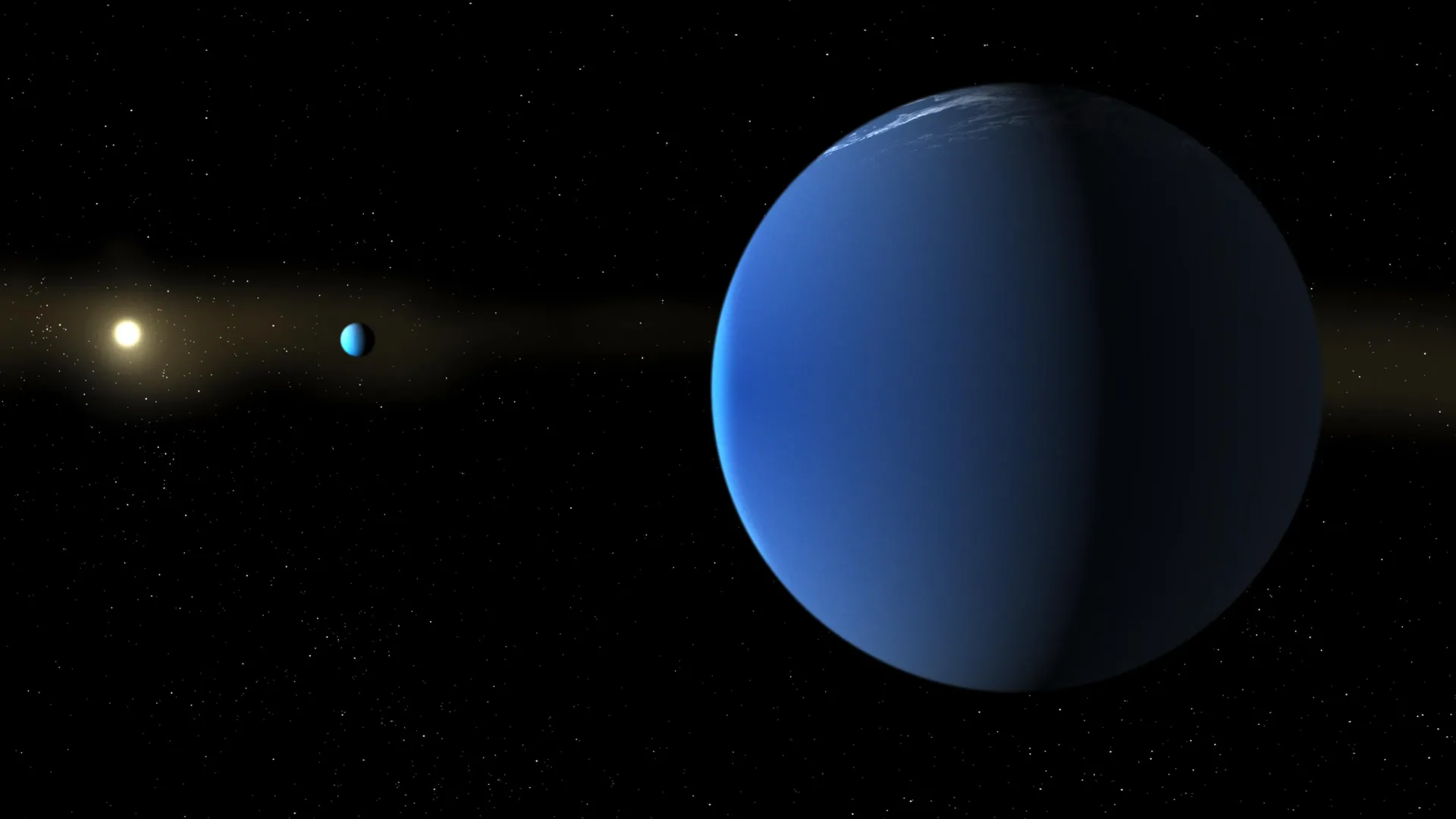
Astronomers have identified two of the fluffiest giant planets ever discovered, with densities so low they are actually less dense than cotton candy. The rare pair of “super-puff” planets was found by an international team led by the University of Oxford, working with Université Côte d’Azur/Observatoire de la Côte d’Azur and the University of Birmingham. The findings were published in Monthly Notices of the Royal Astronomical Society.
The newly confirmed planets, TOI-791 b and TOI-791 c, orbit an F7-type dwarf star about 1,110 light years from Earth in the southern constellation Volans. Although each planet is about the size of Jupiter, both are remarkably lightweight for their size.
TOI-791 b has a density of just 0.038 grams per cubic centimeter, while TOI-791 c measures 0.047 grams per cubic centimeter. Jupiter, by comparison, has an average density of 1.33 grams per cubic centimeter, making it roughly 28 to 35 times denser than these newly discovered worlds.
The comparison becomes even more striking when measured against candy floss, which has a typical density of about 0.05 grams per cubic centimeter. Earth is much denser still, averaging 5.5 grams per cubic centimeter.
Rare Planetary Twins Locked in a Gravitational Dance
Scientists believe the two planets formed together from the same disc of gas and dust surrounding their young star, making them planetary “siblings.”
They are also linked by an unusual orbital arrangement called a 5:3 mean-motion resonance. For every five orbits completed by the inner planet, the outer planet finishes almost exactly three. As they circle their star, their gravity repeatedly pulls on one another, creating small but measurable changes in the timing of each planet’s transit.
Only four other planetary systems are known to contain multiple super-puff planets, making TOI-791 an exceptionally rare opportunity to investigate how these unusual worlds originate and evolve.
Lead author Dr. George Dransfield (she/her) (Department of Physics, University of Oxford and a presenter for BBC Sky at Night) said:
“Only a handful of these super-puffy planets are known, and it is even rarer to find two in the same system. Their extremely low densities make them fascinating targets for understanding how planetary systems form and evolve.”
Citizen Scientists Helped Find the Planets
Volunteers participating in the Planet Hunters TESS citizen-science project first flagged TOI-791 b in 2019 and TOI-791 c in 2023 as possible planets. The project searches observations collected by NASA’s Transiting Exoplanet Survey Satellite (TESS) for signs of previously unknown worlds.
Researchers then combined measurements from telescopes around the world to determine the planets’ sizes and masses, allowing them to calculate their exceptionally low densities.
When a planet crosses in front of its star during a “transit,” it blocks a small amount of starlight. That dip in brightness reveals the planet’s size. In the TOI-791 system, astronomers also detected tiny changes in the timing of the transits caused by the planets’ gravitational interactions. Analyzing those timing variations allowed the team to estimate each planet’s mass.
Antarctica Played a Key Role
The discovery was built on eight years of observations, including data from the ASTEP (Antarctic Search for Transiting ExoPlanets) telescope at Concordia Station in Antarctica. The telescope is jointly operated by researchers from Université Côte d’Azur/Observatoire de la Côte d’Azur and international collaborators.
Antarctica’s long winter nights gave astronomers a major advantage. Months of uninterrupted darkness made it possible to observe the planets’ unusually long transits, each lasting more than 11 hours, without interruption. According to the researchers, these are the longest continuous planetary transits ever fully observed from the ground.
How Do Super-Puff Planets Form?
Scientists are still trying to understand how super-puff planets develop.
One leading explanation is that these worlds possess enormous atmospheres rich in hydrogen and helium that account for a large fraction of their total mass. Researchers think these thick gaseous envelopes may have formed when the planets were much farther from their star, in colder regions of the protoplanetary disc where gas could rapidly accumulate around a solid planetary core.
Future observations are planned to better understand the origins of these unusual planets and test competing theories.
Professor Amaury Triaud (University of Birmingham), the UK Principal Investigator of ASTEP and co-author of the study, said:
“This system offers a unique laboratory for understanding how super-puff planets form and evolve. We propose to carry out space-based observations using the James Webb Space Telescope to assess if the puffy atmosphere contains carbon-, nitrogen-, and oxygen-bearing species, revealing new insight into how these unusual planets formed.”
Professor Tristan Guillot (Université Côte d’Azur), Principal Investigator of ASTEP and co-author of the study, added:
“These multi-planetary systems are complex, with gravitational interactions between the planets that evolve over very long periods, tens of years or more. This discovery highlights the importance of continued international collaboration in astronomy. Bringing together observations from Antarctica, space telescopes and observatories across several continents was essential to revealing the true nature of these extraordinary planets.”
Imagine pouring together two cups of warm water and somehow ending up with a cup of boiling water. That cannot happen in everyday life, but at the quantum level, something similar is possible. Multiple low-energy particles of light can combine their energy to create a single particle with much higher energy.
Researchers at Kyushu University have now created a solid-state molecular material capable of converting visible sunlight into ultraviolet (UV) light under normal outdoor conditions. The new material achieves a photo upconversion efficiency of 1.9%, according to a study published June 23 in Nature Communications.
Why UV Light Matters
Although many people associate UV light with sunburns and skin damage, it plays an important role in numerous technologies. UV light is used for air purification, curing resins in 3D printing, hardening gels in dental fillings, and even applications such as nail treatments.
Despite its usefulness, UV light represents only about 6% of the sunlight that reaches Earth’s surface. Even then, only part of that UV radiation is practical for technological applications.
“What we do here is ‘add together’ the energy from two visible light photons to make one ultraviolet photon. It’s a fascinating process called photo upconversion,” explains Yoichi Sasaki, Associate Professor at Kyushu University’s Faculty of Engineering and the study’s corresponding author.
Turning Visible Light Into UV Light
The process relies on a phenomenon known as triplet-triplet annihilation (TTA). In this approach, a molecule known as a donor absorbs visible light and enters a high-energy triplet state. That energy is then transferred to a nearby acceptor molecule.
When two triplet states encounter one another, they combine and release their energy as a single UV photon.
Scientists have long known that TTA works effectively in liquids because molecules can move freely and interact easily. However, liquid systems often require toxic solvents and may evaporate over time, limiting their practicality. As a result, researchers have spent years searching for a reliable solid-state alternative.
“In solids, molecules are packed tightly, and the π electron clouds — regions of high electron density hovering above and below each molecular plane — can overlap,” says Sasaki. “When that happens, triplets easily fizzle out before they ever meet. Molecules must be close enough for energy to transfer but separated enough to prevent quenching of excitons.”
A New Solid-State Solution
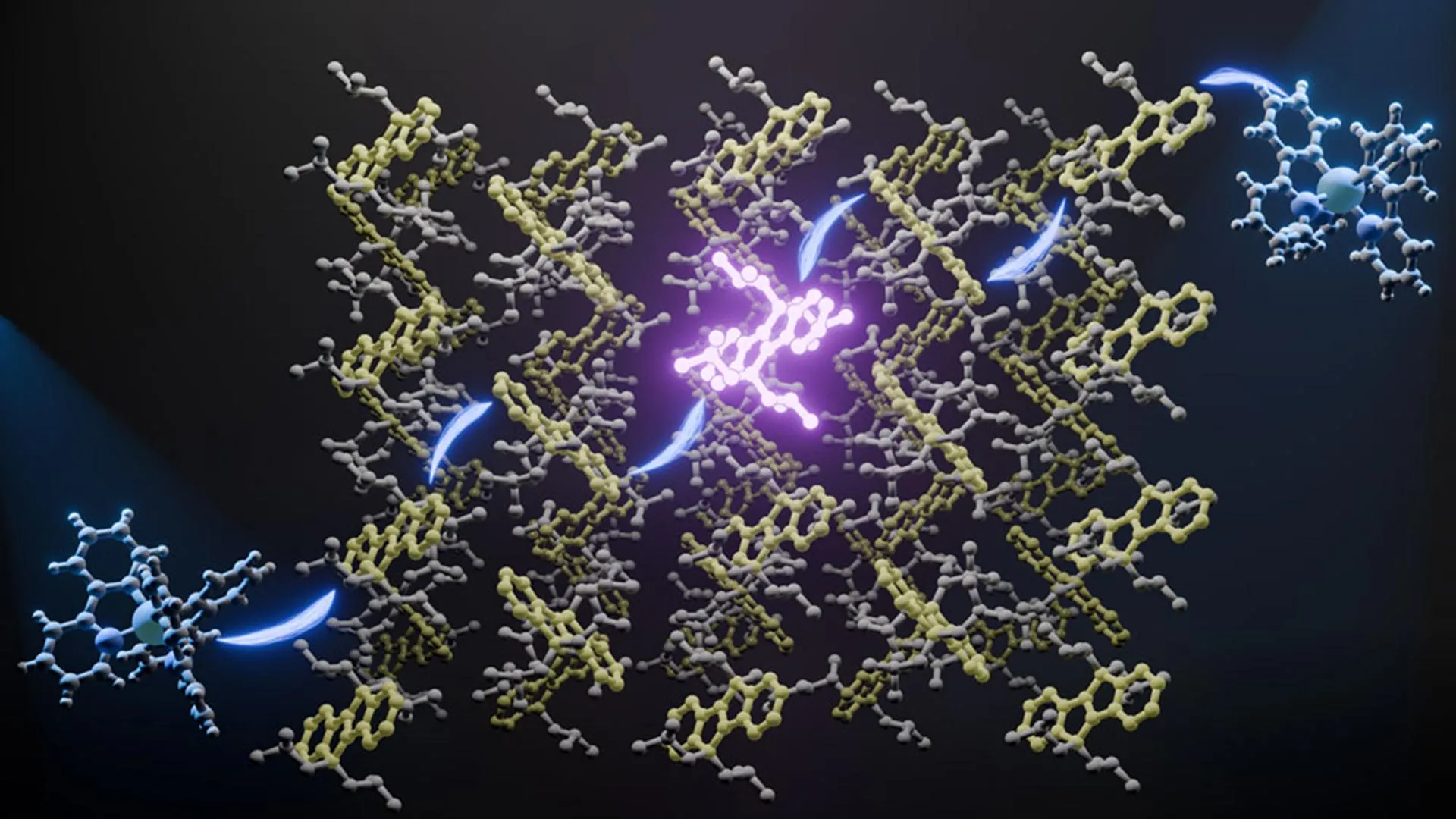
The team’s breakthrough came from an organic semiconductor called dihydroindenoindenedene (DHI).
The researchers modified DHI by attaching alkyl chains to its sp³ carbon atoms — which have four bonds pointing in fixed 3D directions. This design created carefully controlled spacing between neighboring molecules. The molecules remained close enough to transfer energy efficiently while avoiding the strong electronic interactions that can suppress performance.
The resulting material exhibited strong luminescence, long-lived excited states, and highly effective energy transfer. It achieved a solid-state fluorescence quantum yield greater than 60%.
When paired with a donor molecule, the system reached an upconversion efficiency of 1.9%.
“This means roughly two UV photons are produced for every hundred visible-light photons absorbed,” Sasaki adds. “It may sound low, but it runs on natural sunlight alone. Most solid-state materials cannot realize this even at much higher light intensity.”
Potential Applications for Solar-Powered UV Light
The researchers have filed a patent application for the material.
In addition to its performance, the material offers practical advantages. It can be synthesized relatively easily and is made from inexpensive starting materials. The team believes it could eventually be used in solar-powered photocatalysis, indoor air purification systems, and low-intensity 3D printing technologies.
A 14-Year Scientific Journey
For the researchers involved, the achievement represents more than a technical advance.
In 2012, Nobuo Kimizuka, now Professor Emeritus at Kyushu University’s Research Center for Negative Emissions Technologies, began exploring photon upconversion through triplet energy migration in self-assembled molecular systems. His goal was to establish a form of molecular systems chemistry in which self-assembly could perform useful functions.
Over the following years, his group made steady progress using solution-based and gel-based systems. Efficient solid-state upconversion, however, remained difficult to achieve.
A major breakthrough finally arrived in May 2024, less than a year before Kimizuka’s retirement.
The months that followed became an intense push to bring the project to completion. Graduate students Naoyuki Harada, Hayato Shoyama, and Nutnicha Boonmong worked alongside Sasaki and then-Assistant Professor Kiichi Mizukami of Kyushu University’s Faculty of Engineering to consolidate years of research into a final publication.
“We handed the draft to Professor Kimizuka just 11 days before he left the lab, which for us felt like a heartfelt retirement gift,” Sasaki notes.
“This discovery is the culmination of over 14 years of our research and marks a major milestone in photon-upconversion and molecular self-assembly research,” concludes Kimizuka.

Fructose and glucose are two common sugars found in many foods and drinks. Although they contain the same number of calories, new research suggests the brain responds to them in very different ways.
Scientists at the Monell Chemical Senses Center discovered that fructose and glucose communicate with the brain through separate gut-brain pathways. Their findings indicate that these differences may influence food and beverage preferences and could help explain why certain sweetened products are especially appealing.
The study, published June 10 in the journal Neuron, identified a specific signaling route that allows fructose to communicate with the brain. In experiments involving mice, researchers found that this pathway was far less effective than the one used by glucose when it came to reducing activity in neurons associated with hunger.
“This work adds to our growing understanding of how modern diets, especially those high in fructose or high-fructose corn syrup, interact with the neural systems involved in appetite,” said senior author and Monell Member Amber Alhadeff, PhD.
How Fructose and Glucose Affect Hunger Neurons
To investigate how the sugars influence the brain, researchers recorded neural activity in mice after exposure to fructose and glucose.
The team found that fructose increased levels of the gut hormone PYY. That hormone then signaled through the vagus nerve, leading to a modest reduction in the activity of agouti-related protein (AgRP) neurons, which play a major role in driving hunger. When researchers disrupted this pathway, fructose could no longer affect those neurons.
Glucose produced a very different response. According to the researchers, it did not rely on the same PYY-Y2 vagus nerve pathway. Instead, glucose strongly suppressed AgRP neuron activity, resulting in a much larger effect on hunger-related brain signaling.
Sugar Type Influenced Food Preferences
Although fructose and glucose produced similar short-term effects on food intake, the mice eventually developed preferences that corresponded to the degree of AgRP neuron inhibition triggered by each sugar.
The researchers also examined high-fructose corn syrup (HFCS), a widely used sweetener made from a combination of fructose and glucose. The mice showed a preference for HFCS, and the sweetener suppressed AgRP neuron activity more strongly than fructose alone.
According to the researchers, this stronger effect on hunger-related neurons may help explain why foods and beverages containing HFCS can be particularly appealing.
Challenging Assumptions About Calories and Hunger
The results call into question a long-held assumption that AgRP neurons primarily track calorie intake regardless of where those calories come from.
Instead, the findings suggest that these hunger-related neurons can distinguish between different sugars and respond through separate biological pathways. Even though fructose and glucose provide the same amount of energy, the mice’s brains processed them differently.
The study highlights the complexity of nutrient sensing in the body and suggests that even simple sugars can have distinct effects on the gut, the brain, and behavior.
This research was supported by grants R01DK131558, DP2AT011965, R01DK116004, F31DK13558, and S10OD030354 from the National Institutes of Health; the American Heart Association; the New York Stem Cell Foundation; the Klingenstein Fund; the Simons Foundation, the Pew Charitable Trusts, the Penn Institute for Diabetes, Obesity, and Metabolism; the Hearst Fellowship, and the Monell Chemical Senses Center.
Cardiac arrests have gone up during very hot weather, and it’s not just among the elderly and frail, experts are warning.