This CRISPR breakthrough turns genes on without cutting DNA

Scientists at UNSW Sydney have developed a new form of CRISPR technology that could make gene therapy safer while also resolving a decades-long debate about how genes are switched off. The research shows that small chemical markers attached to DNA actively silence genes, rather than simply appearing as harmless byproducts in inactive regions of the genome.

For years, researchers have questioned whether methyl groups, tiny chemical clusters that collect on DNA, merely show up where genes are already turned off or whether they are the direct cause of gene suppression.

In a study published recently in Nature Communications, researchers from UNSW, working with colleagues at the St Jude Children’s Research Hospital (Memphis), demonstrated that removing these chemical tags causes genes to become active again. When the tags were added back, the genes shut down once more. The results confirm that DNA methylation directly controls gene activity.

“We showed very clearly that if you brush the cobwebs off, the gene comes on,” says study lead author Professor Merlin Crossley, UNSW Deputy Vice-Chancellor Academic Quality.

“And when we added the methyl groups back to the genes, they turned off again. So, these compounds aren’t cobwebs — they’re anchors.”

How CRISPR Technology Has Evolved

CRISPR, short for Clustered Regularly Interspaced Short Palindromic Repeats, is the foundation of modern gene-editing technology. It allows scientists to locate specific DNA sequences and make targeted changes, often replacing faulty genetic code with healthy versions.

The system is based on a natural defense mechanism found in bacteria, which use CRISPR to recognize and cut up the DNA of invading viruses.

Early versions of CRISPR tools worked by cutting DNA to disable malfunctioning genes. Later versions became more precise, allowing scientists to correct individual letters in the genetic code. However, both approaches rely on breaking DNA strands, which can lead to unintended changes and increase the risk of serious side effects.

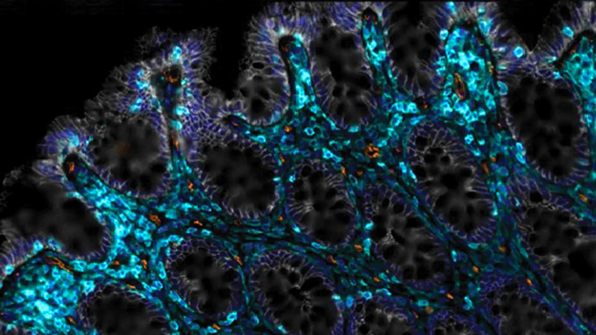
The latest version, known as epigenetic editing, takes a different approach. Instead of cutting DNA, it targets chemical markers attached to genes inside the nucleus of each cell. By removing methyl groups from genes that have been silenced, researchers can restore gene activity without altering the underlying DNA sequence.

New Possibilities for Treating Sickle Cell Disease

The team believes this approach could lead to safer treatments for Sickle Cell-related diseases. These inherited conditions affect the shape and function of red blood cells, often causing severe pain, organ damage, and shortened life expectancy.

“Whenever you cut DNA, there’s a risk of cancer. And if you’re doing a gene therapy for a lifelong disease, that’s a bad kind of risk,” Prof. Crossley says.

“But if we can do gene therapy that doesn’t involve snipping DNA strands, then we avoid these potential pitfalls.”

Rather than cutting DNA, the new technique uses a modified CRISPR system to deliver enzymes that remove methyl groups. This process releases the genetic brakes that keep certain genes switched off. One key target is the fetal globin gene, which helps deliver oxygen before birth. Reactivating this gene after birth could help bypass defects in the adult globin gene that cause Sickle Cell diseases.

“You can think of the fetal globin gene as the training wheels on a kid’s bike,” says Prof. Crossley. “We believe we can get them working again in people who need new wheels.”

What the Research Shows So Far

So far, all experiments have been carried out in laboratory settings using human cells at UNSW and in Memphis.

Study co-author Professor Kate Quinlan says the findings could have far-reaching implications beyond Sickle Cell disease. Many genetic conditions involve genes that are improperly turned on or off, and adjusting methyl groups may provide a way to correct those problems without damaging DNA.

“We are excited about the future of epigenetic editing as our study shows that it allows us to boost gene expression without modifying the DNA sequence. Therapies based on this technology are likely to have a reduced risk of unintended negative effects compared to first or second generation CRISPR,” she says.

Looking ahead, the researchers describe how the therapy might one day work in practice. Doctors would collect a patient’s blood stem cells, which produce red blood cells. In the lab, epigenetic editing would be used to remove methyl tags from the fetal globin gene, reactivating it. The edited cells would then be returned to the patient, where they could settle into the bone marrow and begin producing healthier blood cells.

The Next Steps in Epigenetic Editing

The research teams at UNSW and St Jude plan to test the approach in animal models and continue exploring additional CRISPR-based tools.

“Perhaps the most important thing is that it is now possible to target molecules to individual genes,” Prof. Crossley says.

“Here we removed or added methyl groups but that is just the beginning, there are other changes that one could make that would increase our abilities to alter gene output for therapeutic and agricultural purposes. This is the very beginning of a new age.”


Share Button

Plants can’t absorb as much CO2 as climate models predicted

High levels of carbon dioxide in the atmosphere are a major driver of climate change. At the same time, increased CO2 can encourage plants to grow faster, allowing them to absorb more carbon and potentially slow warming. That benefit, however, depends on whether plants have access to enough nitrogen, a nutrient that is essential for growth. Scientists have only recently taken a closer look at how much nitrogen is actually available in nature. New research involving the University of Graz shows that the so-called CO2 fertilization effect has been significantly overstated.

Plants cannot use nitrogen on their own. The nutrient must first be converted into a usable form through a process called nitrogen fixation, which relies on microorganisms in the soil. This process takes place in natural ecosystems as well as on farmland. “While this process has been significantly overestimated in nature, it has increased by 75 percent over the past 20 years due to agriculture,” says Bettina Weber, a biologist at the University of Graz, summarizing findings from a study published earlier this year.

Building on those results, a new analysis shows that the way nitrogen fixation is calculated in some Earth System models has now been reassessed. These models are widely used to project climate trends and inform major assessments, including the World Climate Report. The updated findings were published in the scientific journal PNAS.

New Findings Prompt Climate Model Revisions

The study was led by Sian Kou-Giesbrecht of Simon Fraser University in Burnaby, Canada. The work was carried out by an international research group focused on biological nitrogen fixation, which includes Bettina Weber. This working group receives support from the U.S. Geological Survey (USGS) John Wesley Powell Centre for Analysis and Synthesis.

“We compared different Earth System models with current nitrogen fixation values and found that they overestimate the nitrogen fixation rate on natural surfaces by about 50 percent,” Weber explains. Because plants depend on this process to access nitrogen, the overestimate has meaningful consequences. According to the study, it results in an overall reduction of about 11 percent in the projected CO2 fertilization effect.

Why Updating Models Is Critical

Weber emphasizes the importance of adjusting climate models to reflect these updated measurements. “This is because gases such as nitrogen oxides and nitrous oxide are produced as part of the nitrogen cycle. These can be released into the atmosphere through conversion processes and alter or disrupt climate processes.” Accurately accounting for nitrogen dynamics, she says, is essential for making reliable predictions about how ecosystems and the climate will respond in the future.

Share Button

Surgeon who ‘stripped naked in cubicle’ struck off

The doctor was working at the Queen Alexandra Hospital in Portsmouth when the incidents happened.

Share Button

Junk food TV and online advert ban comes into force

Soft drinks, chocolate, pizzas and ice creams will be targeted in the UK government’s plan.

Share Button

The myth of willpower – and why some people struggle to lose weight more than others

Thousands of genes that have an influence on weight, say experts – which means weight loss isn’t a level playing field

Share Button

Type 2 diabetes physically changes the human heart, study finds

Researchers at the University of Sydney have uncovered new evidence showing that type 2 diabetes directly changes the heart’s structure and how it produces energy. These findings help explain why people living with diabetes face a much higher risk of developing heart failure.

The study, published in EMBO Molecular Medicine, was led by Dr. Benjamin Hunter and Associate Professor Sean Lal from the School of Medical Sciences. The team examined donated human heart tissue from patients receiving heart transplants in Sydney, comparing it with tissue from healthy donors. Their analysis revealed that diabetes drives specific molecular changes inside heart cells and alters the physical makeup of heart muscle. These effects were most pronounced in patients with ischemia cardiomyopathy, which is the leading cause of heart failure.

“We’ve long seen a correlation between heart disease and type 2 diabetes,” said Dr. Hunter, “but this is the first research to jointly look at diabetes and ischemia heart disease and uncover a unique molecular profile in people with both conditions.

“Our findings show that diabetes alters how the heart produces energy, maintains its structure under stress, and contracts to pump blood. Using advanced microscopy techniques, we were able to see direct changes to the heart muscle as a result of this, in the form of a build-up of fibrous tissue.”

Heart disease remains the leading cause of death in Australia, and more than 1.2 million Australians are living with type 2 diabetes.

Associate Professor Lal said: “Our research links heart disease and diabetes in ways that have never been demonstrated in humans, offering new insights into potential treatment strategies that could one day benefit millions of people in Australia and globally.”

Looking Inside Diseased Human Hearts

To better understand how diabetes affects the heart, the researchers studied heart tissue from both transplant recipients and healthy individuals. This direct examination allowed them to see how diabetes influences heart biology in real human patients rather than relying solely on animal models.

The results showed that diabetes is more than a co-morbidity for heart disease. It actively accelerates heart failure by interfering with essential biological processes and reshaping heart muscle at the microscopic level.

“The metabolic effect of diabetes in the heart is not fully understood in humans,” said Dr. Hunter.

How Diabetes Disrupts the Heart’s Energy Supply

In healthy hearts, energy is mainly generated from fats, with glucose and ketones also contributing. Previous research has shown that glucose use increases during heart failure. However, diabetes interferes with this process by reducing how sensitive heart cells are to insulin.

“Under healthy conditions, the heart primarily uses fats but also glucose and ketones as fuel for energy. It has previously been described that glucose uptake is increased in heart failure, however, diabetes reduces the insulin sensitivity of glucose transporters — proteins that move glucose in and out of cells — in heart muscle cells.

“We observed that diabetes worsens the molecular characteristics of heart failure in patients with advanced heart disease and increases the stress on mitochondria — the powerhouse of the cell which produces energy.”

Structural Damage and Fibrosis in the Heart Muscle

Beyond energy production, the researchers found that diabetes affects the proteins responsible for heart muscle contraction and calcium regulation. In patients with both diabetes and ischemic heart disease, these proteins were produced at lower levels. At the same time, excess fibrous tissue accumulated within the heart, making the muscle stiffer and less able to pump blood efficiently.

“RNA sequencing confirmed that many of these protein changes were also reflected at the gene transcription level, particularly in pathways related to energy metabolism and tissue structure, which reinforces our other observations,” said Dr. Hunter.

“And once we had these clues at the molecular level, we were able to confirm these structural changes using confocal microscopy.”

Implications for Future Treatment and Care

Associate Professor Lal said identifying mitochondrial dysfunction and fibrosis-related pathways opens the door to new treatment approaches.

“Now that we’ve linked diabetes and heart disease at the molecular level and observed how it changes energy production in the heart while also changing its structure, we can begin to explore new treatment avenues,” he said.

“Our findings could also be used to inform diagnosis criteria and disease management strategies across cardiology and endocrinology, improving care for millions of patients.”

Share Button

Scientists found a way to help aging guts heal themselves

Many people notice that certain foods become harder to tolerate with age. One possible reason is damage to the intestinal epithelium, a thin, single layer of cells that lines the intestine. This lining is essential for digestion and overall gut health. In healthy conditions, the intestinal epithelium renews itself every three to five days. Aging or exposure to cancer radiation can disrupt this renewal process, slowing or stopping regeneration altogether. When that happens, inflammation can rise and conditions such as leaky gut syndrome may develop.

Scientists at Cold Spring Harbor Laboratory (CSHL) have now identified a promising way to jump-start intestinal repair. Their strategy relies on CAR T-cell therapy, a powerful form of immunotherapy best known for treating certain cancers. By applying this approach to the gut, the researchers hope to open the door to future clinical trials aimed at improving intestinal health, particularly in people affected by age-related decline.

Targeting Aging Cells That Refuse to Die

This work builds on earlier research led by CSHL Assistant Professor Corina Amor Vegas, whose laboratory studies cellular senescence. As the body ages, it accumulates senescent cells, which no longer divide but also do not die off. These lingering cells have been linked to many age-related conditions, including diabetes and dementia. In earlier studies, Amor Vegas and her team engineered immune cells known as anti-uPAR CAR T cells that selectively remove senescent cells in mice, leading to major improvements in the animals’ metabolism.

The researchers next asked whether removing senescent cells could help restore the intestine’s ability to heal. Amor Vegas partnered with CSHL Assistant Professor Semir Beyaz and graduate student Onur Eskiocak to investigate. They delivered CAR T cells directly to the intestines of both younger and older mice. According to Amor Vegas, the results were striking. “In both cases, we see really significant improvements,” she says. “They’re able to absorb nutrients better. They have much less inflammation. When irritated or injured, their epithelial lining is able to regenerate and heal much faster.”

Protection Against Radiation-Induced Gut Damage

Leaky gut syndrome is particularly common among cancer patients who receive pelvic or abdominal radiation therapy. To model this, the team exposed mice to radiation that damaged their intestinal epithelial cells. Mice treated with CAR T cells recovered far more effectively than those that did not receive the therapy. Notably, a single dose of CAR T-cell treatment continued to support healthier gut function for at least one year.

The researchers also found compelling evidence that anti-uPAR CAR T cells encourage regeneration in human intestinal and colorectal cells, Eskiocak notes. While the precise biological mechanisms behind this effect are still being explored, the findings point to strong therapeutic potential. Beyaz emphasizes the broader significance of the work. “This is one good step toward a long journey in understanding how we can better heal the elderly,” he said.

Share Button

A weak body clock may be an early warning for dementia

  • A large new study suggests the body’s internal clock, known as circadian rhythm, may play an important role in dementia risk.
  • More than 2,000 older adults wore small monitors for about 12 days, allowing researchers to closely track daily patterns of rest and activity.
  • People whose body clocks were weaker or more irregular were significantly more likely to develop dementia over the following years.
  • Those whose activity levels peaked later in the day, rather than earlier, showed a 45% higher risk of dementia.
  • Researchers say future studies exploring circadian rhythm approaches such as light exposure or lifestyle changes could reveal new ways to reduce dementia risk.

Weaker Body Clocks Linked to Dementia Risk

A new study suggests that disruptions in the body’s internal clock may be tied to a higher risk of dementia. Research published on December 29, 2025, in Neurology, the medical journal of the American Academy of Neurology, found that people with weaker and more irregular circadian rhythms were more likely to develop dementia. The study also showed that individuals whose daily activity levels peaked later in the day faced a higher risk than those who peaked earlier. While these findings reveal a strong link, they do not show that circadian rhythm changes directly cause dementia.

What Circadian Rhythms Do in the Body

Circadian rhythm refers to the body’s natural timing system. It controls the 24-hour sleep-wake cycle and helps regulate key functions such as hormone release, digestion, and body temperature. This internal clock is directed by the brain and responds to environmental signals, especially light.

When circadian rhythms are strong, the body stays closely aligned with the daily cycle of light and dark. This leads to consistent patterns of sleep and activity, even when schedules or seasons change. In contrast, weaker rhythms make the body clock more sensitive to disruptions. People with less stable rhythms are more likely to shift their sleep and activity times due to changes in routine or daylight.

Aging, Circadian Changes, and Dementia

“Changes in circadian rhythms happen with aging, and evidence suggests that circadian rhythm disturbances may be a risk factor for neurodegenerative diseases like dementia,” said study author Wendy Wang, MPH, PhD, of the Peter O’Donnell Jr. School of Public Health at UT Southwestern Medical Center in Dallas, Texas. “Our study measured these rest-activity rhythms and found people with weaker and more fragmented rhythms, and people with activity levels that peaked later in the day, had an elevated risk of dementia.”

Who Took Part in the Study

The research followed 2,183 adults with an average age of 79 who did not have dementia when the study began. Among the participants, 24% were Black people and 76% were white people.

Each participant wore a small heart monitor attached to the chest for an average of 12 days. These devices tracked periods of rest and activity, allowing researchers to analyze circadian rhythm patterns. Participants were then monitored for about three years. During that time, 176 people were diagnosed with dementia.

How Researchers Measured Rhythm Strength

Scientists examined the heart monitor data using several indicators of circadian rhythm strength. One key measure was relative amplitude, which reflects the difference between a person’s most active and least active times of day. Higher relative amplitude indicated a stronger and more clearly defined daily rhythm.

Participants were divided into three groups based on rhythm strength. When comparing the strongest and weakest groups, 31 of the 728 people in the high rhythm group developed dementia, while 106 of the 727 people in the low rhythm group did. After accounting for factors such as age, blood pressure, and heart disease, researchers found that those in the weakest rhythm group had nearly two and a half times the risk of dementia. Each standard deviation drop in relative amplitude was linked to a 54% increase in dementia risk.

Later Activity Peaks and Higher Risk

The timing of daily activity also appeared to matter. People whose activity peaked later in the afternoon, at 2:15 p.m. or later, had a higher risk of dementia than those whose activity peaked earlier, between 1:11 p.m.-2:14 p.m. About 7% of participants in the earlier peak group developed dementia, compared with 10% in the later peak group, representing a 45% higher risk.

A later activity peak may reflect a mismatch between the body’s internal clock and environmental signals such as daylight and darkness.

Why Disrupted Rhythms Might Matter

“Disruptions in circadian rhythms may alter body processes like inflammation, and may interfere with sleep, possibly increasing amyloid plaques linked to dementia, or reducing amyloid clearance from the brain,” said Wang. “Future studies should examine the potential role of circadian rhythm interventions, such as light therapy or lifestyle changes, to determine if they may help lower a person’s risk of dementia.”

Study Limitations

One limitation of the research is that it did not include data on sleep disorders, such as sleep apnea, which could have influenced the results.

Share Button

Fossilized bones are revealing secrets from a lost world

For the first time, researchers have successfully examined metabolism-related molecules preserved inside fossilized bones from animals that lived between 1.3 and 3 million years ago. These chemical traces offer rare insight into the animals themselves and the environments they once inhabited.

By analyzing metabolic signals tied to health and diet, the scientists were able to reconstruct details about ancient climates and landscapes, including temperature, soil conditions, rainfall, and vegetation. The results, published in Nature, point to environments that were significantly warmer and wetter than those found in the same regions today.

Studying metabolites — the molecules produced and used in digestion and other chemical processes in the body — can reveal information about disease, nutrition, and environmental exposure. While metabolomics has become a powerful tool in modern medical research, it has rarely been applied to fossils. Instead, most studies of ancient remains rely on DNA, which mainly helps establish genetic relationships rather than day-to-day biology.

“I’ve always had an interest in metabolism, including the metabolic rate of bone, and wanted to know if it would be possible to apply metabolomics to fossils to study early life. It turns out that bone, including fossilized bone, is filled with metabolites,” said Timothy Bromage, professor of molecular pathobiology at NYU College of Dentistry and affiliated professor in NYU’s Department of Anthropology, who led the international research team.

Why Fossil Bones Can Preserve Chemistry

In recent years, scientists discovered that collagen — the protein that provides structure to bones, skin, and connective tissues — can survive in ancient bones, including dinosaur fossils.

“I thought, if collagen is preserved in a fossil bone, then maybe other biomolecules are protected in the bone microenvironment as well,” said Bromage, who directs the Hard Tissue Research Unit at NYU College of Dentistry.

Bone surfaces are porous and filled with tiny blood vessel networks that exchange oxygen and nutrients with the bloodstream. Bromage proposed that during bone growth, metabolites circulating in blood could become trapped inside microscopic spaces within the bone, where they might remain protected for millions of years.

To test this idea, the team used mass spectrometry, a technique that converts molecules into charged particles for identification. Tests on modern mouse bones revealed nearly 2,200 metabolites. The same approach also allowed researchers to detect collagen proteins in some samples.

Testing Fossils From Early Human Landscapes

The researchers then applied this method to fossilized animal bones dating from 1.3 million to 3 million years ago. These samples came from earlier excavations in Tanzania, Malawi, and South Africa, regions known for early human activity.

The fossils belonged to animals with modern relatives still living near those sites today. The team analyzed bones from rodents (mouse, ground squirrel, gerbil) as well as larger animals, including an antelope, a pig, and an elephant. Thousands of metabolites were identified, many of which closely matched those found in living species.

Health Diet and Disease Written in Bone

Many of the detected metabolites reflected normal biological processes, such as the breakdown of amino acids, carbohydrates, vitamins, and minerals. Some chemical markers were linked to estrogen-related genes, indicating that certain fossilized animals were female.

Other molecules revealed signs of illness. In one striking case, a ground squirrel bone from Olduvai Gorge in Tanzania, dated to about 1.8 million years ago, showed evidence of infection by the parasite that causes sleeping sickness in humans. The disease is caused by Trypanosoma brucei and spread by tsetse flies.

“What we discovered in the bone of the squirrel is a metabolite that is unique to the biology of that parasite, which releases the metabolite into the bloodstream of its host. We also saw the squirrel’s metabolomic anti-inflammatory response, presumably due to the parasite,” said Bromage.

Tracing Ancient Diets and Environments

The chemical evidence also revealed what plants the animals consumed. Although plant metabolite databases are far less complete than those for animals, the researchers identified compounds linked to regional plants such as aloe and asparagus.

“What that means is that, in the case of the squirrel, it nibbled on aloe and took those metabolites into its own bloodstream,” explained Bromage. “Because the environmental conditions of aloe are very specific, we now know more about the temperature, rainfall, soil conditions, and tree canopy, essentially reconstructing the squirrel’s environment. We can build a story around each of the animals.”

These reconstructed habitats align with previous geological and ecological research. For example, Olduvai Gorge Bed in Tanzania has been described as freshwater woodland and grassland, while the Upper Bed reflects drier woodlands and marshy areas. Across all studied locations, the fossil evidence consistently points to climates that were wetter and warmer than today.

“Using metabolic analyses to study fossils may enable us to reconstruct the environment of the prehistoric world with a new level of detail, as though we were field ecologists in a natural environment today,” said Bromage.

Research Team and Support

Additional study authors include Bin Hu, Sher Poudel, Sasan Rabieh, and Shoshana Yakar of NYU College of Dentistry; Thomas Neubert, Christopher Lawrence de Jesus, and Hediye Erdjument-Bromage of NYU Grossman School of Medicine; along with collaborators from institutions in France, Germany, Canada, and the United States. The research was supported by The Leakey Foundation, with additional support for the scanning electron microscope provided by the National Institutes of Health (S10 OD023659 and S10 RR027990).

Share Button

Scientists tested intermittent fasting without eating less and found no metabolic benefit

A new study from the German Institute of Human Nutrition Potsdam-Rehbruecke (DIfE) and Charité — Universitätsmedizin Berlin challenges a widely held belief about intermittent fasting. The research shows that time-restricted eating does not lead to measurable improvements in metabolic or cardiovascular health when calorie intake remains unchanged. However, the timing of meals did affect the body’s internal clocks. These findings come from the ChronoFast study led by Prof. Olga Ramich and were published in Science Translational Medicine.

Time-restricted eating (TRE) is a form of intermittent fasting that limits daily food intake to a window of no more than ten hours, followed by a fasting period of at least 14 hours. The approach has become popular as a simple strategy to support weight management and metabolic health. Animal studies show that TRE can protect rodents from diet-related obesity and metabolic problems. In humans, earlier studies have reported benefits such as improved insulin sensitivity, healthier blood sugar and cholesterol levels, and modest reductions in body weight and body fat. As a result, TRE has been widely viewed as a promising tool for preventing insulin resistance and diabetes.

Conflicting Evidence From Earlier Studies

Despite its popularity, past research on TRE has produced mixed results. Many studies have not been able to determine whether observed health improvements came from shorter eating windows, unintentional calorie reduction, or a combination of both. In addition, most earlier trials did not carefully track calorie intake or control for other factors that could influence metabolic outcomes.

To address these gaps, Prof. Olga Ramich, Head of the Department of Molecular Metabolism and Precision Nutrition at the DIfE and Professor at the Charité — Universitätsmedizin Berlin, designed the ChronoFast trial. The goal was to test whether an eight-hour eating window could improve insulin sensitivity and other metabolic markers when calorie intake was kept constant.

How the ChronoFast Study Was Conducted

The study used a randomized crossover design and included 31 women with overweight or obesity. Each participant followed two different eating schedules for two weeks at a time. One schedule involved early time-restricted eating between 8 a.m. and 4 p.m.(eTRE). The other followed a later schedule from 1 p.m. to 9 p.m. (lTRE). Throughout both phases, participants ate nearly identical meals with the same calorie and nutrient content (isocaloric).

Researchers collected blood samples during four clinic visits and performed oral glucose tolerance tests to assess glucose and fat metabolism. Continuous glucose monitoring tracked blood sugar levels over 24 hours while food intake was recorded in detail. Physical activity was monitored using a motion sensor. In collaboration with Prof. Achim Kramer from the Charité — Universitätsmedizin Berlin, the team also examined changes in the body’s internal clock using isolated blood cells.

Measuring the Body’s Internal Clock

Human biology follows internally generated rhythms that roughly align with the length of a day, which is why they are known as circadian clocks (Latin: circa and dia). These rhythms help regulate nearly every physiological process, including sleep and metabolism. Almost all cells in the body contain their own internal clock, which can be influenced by light, physical activity, and food timing.

To measure individual circadian phases, Prof. Dr. Achim Kramer developed the BodyTime assay. This test requires only a single blood sample and provides an objective snapshot of a person’s internal timing. The ChronoFast study used this method and confirmed that eating schedules can shift internal clocks in humans.

No Metabolic Improvements Found

Despite expectations based on earlier research, the ChronoFast study found no clinically meaningful changes in insulin sensitivity, blood sugar, blood fats, or inflammatory markers after the two-week interventions. “Our results suggest that the health benefits observed in earlier studies were likely due to unintended calorie reduction, rather than the shortened eating period itself,” explains Ramich.

While metabolic measures remained largely unchanged, the timing of meals did affect circadian rhythms. Analysis of blood cells showed that the internal clock shifted by an average of 40 minutes during the late eating schedule compared to the early schedule. Participants following the later eating window also went to bed and woke up later. “The timing of food intake acts as a cue for our biological rhythms — similar to light,” says first author Beeke Peters.

Calories and Individual Timing May Matter Most

The findings highlight the importance of calorie balance in achieving health benefits from intermittent fasting. “Those who want to lose weight or improve their metabolism should pay attention not only to the clock, but also to their energy balance,” Ramich concludes.

Future research will need to explore whether combining time-restricted eating with reduced calorie intake produces stronger benefits. Scientists also aim to better understand how individual factors, including chronotype and genetics, may influence how people respond to different eating schedules.

Share Button