An NHS trial over 10 years followed nearly 3,500 men who received focal therapy, a less invasive treatment.


An NHS trial over 10 years followed nearly 3,500 men who received focal therapy, a less invasive treatment.
It will be illegal to sell high-caffeine beverages to under-16s from April next year, but soft drinks with lower caffeine levels will not be affected.
Advisers are asking the government to consider introducing MenB jabs following concerns over an outbreak in Kent earlier this year, in which two people died.
Woman’s Hour discusses fibroids and the need for greater knowledge about the pain they can cause women.
He tells an inquiry he believed a trust was ‘panicking’ and was discouraged from raising rota issues.
A senior Buddhist monk says Herefordshire has “everything” for a “person to be happy”.
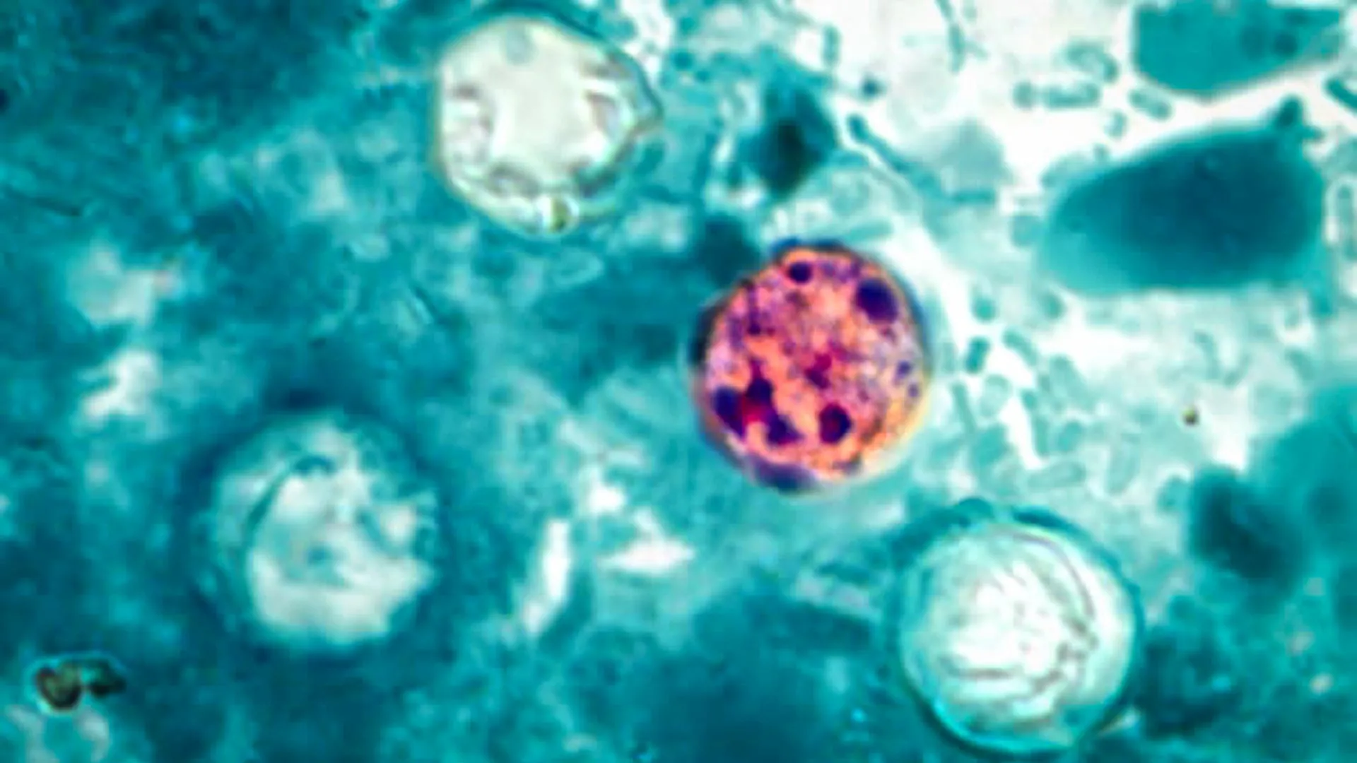
The U.S. Centers for Disease Control and Prevention (CDC) is working with state and federal health agencies to investigate several outbreaks of cyclosporiasis. Efforts to determine and confirm the sources of these outbreaks are still underway.
A large outbreak of cyclosporiasis has been reported in at least four Midwestern states. Public health officials are interviewing people who became sick to learn what foods they ate before their symptoms began.
So far, investigators have not confirmed a specific food as the source. Health agencies are continuing to gather information in an effort to identify what caused the outbreak.
Cases Have Increased Since May
The CDC says it is concerned about the rise in cyclosporiasis cases since the beginning of May. In addition to the large multistate outbreak, federal and state officials are investigating several other clusters of illness across the United States.
Cyclosporiasis is generally not life threatening, but some people can become very sick and may need to be hospitalized. Anyone experiencing possible symptoms should contact a healthcare provider promptly.
CDC and FDA Collecting Outbreak Data
The CDC, public health and regulatory agencies in several states, and the U.S. Food and Drug Administration (FDA) are reviewing multiple types of information as part of the investigation.
As of July 13, more than 400 people infected with Cyclospora had been reported to the CDC in connection with the outbreak. Cases have been identified in Michigan, Ohio, West Virginia, and Kentucky.
The CDC is also aware of additional illnesses that remain under investigation. People linked to the outbreak reported becoming sick on or after June 22, 2026.
Actual Case Count May Be Higher
Health officials believe the true number of illnesses is probably greater than the confirmed total. The outbreak may also extend beyond the four states where cases have already been identified.
Some infected people recover without seeking medical care and are never tested for Cyclospora. Recent illnesses may also be missing from the official count because it can take several weeks to determine whether a case is connected to an outbreak.
To help identify the source, public health officials collect information from patients about their age, race, ethnicity, other demographic details, and the foods they ate before becoming sick. These responses may reveal patterns that help investigators trace the contaminated food.
What To Do If You Have Symptoms
Contact your healthcare provider if you develop symptoms of cyclosporiasis.
Symptoms can vary and usually appear about one week after infection (ranging from 2 days to 2 weeks or more).
Without treatment, symptoms may continue for several days, a month, or even longer.
Help Investigators Find the Contaminated Food
People diagnosed with cyclosporiasis may be contacted by local or state health officials. Investigators may ask what they ate during the two weeks before they became ill.
Providing detailed information can help health agencies identify the food responsible for the outbreak.
How To Reduce Your Risk
Learn which foods are more likely to be associated with cyclosporiasis and what steps can help prevent infection.
Consumers should also stay up to date on food recalls and outbreaks.
Guidance for Healthcare Providers
Healthcare providers should report cyclosporiasis cases to their local health department.
Additional information about symptoms, treatment, and patient management is available through Clinical Care of Cyclosporiasis.

Researchers at the City College of New York are charting a fast-growing area of quantum science centered on materials only a few atoms thick. In these systems, light, electric charge, and magnetism are closely connected rather than behaving independently.
The work comes from physicist Vinod M. Menon’s Laboratory for Nano and Micro Photonics (LaNMP). Researchers believe these unusual interactions could eventually support advanced optoelectronic devices and quantum technologies that manipulate light, charge, and electron spin together.
When Light and Magnetism Interact
In a review published in Nature Materials, titled “Excitons in van der Waals magnetic materials,” the researchers examine recent progress involving layered magnetic semiconductors. These materials allow light-generated excitations called excitons to interact with magnetic order and with magnetic waves known as magnons.
An exciton forms when incoming light energizes an electron and causes it to move, leaving behind a positively charged “hole.” The electron and hole remain linked, forming an electrically neutral particle that can still interact strongly with light. Magnons are different. They are collective waves that travel through the organized magnetic structure of a material.
Scientists have spent years trying to unite the optical properties of exciton-rich semiconductors with magnetism. Earlier strategies included adding magnetic atoms to semiconductors or stacking atomically thin semiconductors on top of magnetic materials.
Van der Waals magnetic semiconductors provide a more direct approach. Within these crystals, excitons and magnetic moments can emerge from the same electronic orbitals. This shared origin allows light and magnetism to influence one another inside the material itself.
“In these materials, light and magnetism no longer operate as separate channels,” said Pratap Chandra Adak, a postdoctoral researcher in Menon’s group and lead author of the Review. “An exciton is not just a passive light-driven excitation sitting on top of the magnetism. It can sense the spin order and magnons, and under the right conditions, even help control the magnetic state itself.”
Reading Magnetic States With Light
The Review examines several important material platforms, including chromium triiodide, nickel phosphorus trisulfide, and chromium sulfur bromide. Research on these two-dimensional magnets has revealed several ways that excitons and magnetic behavior can affect each other.
Excitons can significantly strengthen magneto-optical effects, allowing scientists to identify magnetic states by observing changes in the polarization of light. Magnetic order can also alter the energy of excitons and influence where they are confined within a material.
Interactions between excitons and magnons can connect optical signals with magnetic activity occurring at gigahertz frequencies. The researchers also discuss exciton polaritons, hybrid particles that combine properties of light and matter and can transport optical information through a material.
“Over the past few years, this field has moved from detecting magnetism in atomically thin crystals to actively exploring how magnetic order can control light-matter interactions,” said Menon, professor of physics and senior author of the Review. “The goal of this article is to bring those developments into a coherent framework and identify where the field can go next.”
New Possibilities for Quantum Technology
The researchers identify several potential applications that would depend on precise control of light and magnetism at extremely small scales. These include magneto-photonic memory and data readout, all-optical logic, adjustable light-emitting devices, magneto-optic lasers, and polaritonic technologies.
Another promising application involves quantum transducers. These devices convert signals between microwave and optical frequencies, a capability that could become important for connecting components in future quantum networks.
Major Scientific Challenges Remain
Despite the rapid progress, much of this field remains unexplored. Many possible materials have not yet been studied in detail, and scientists still need better theoretical models that can predict how excitons, electron spins, lattice vibrations, and photons behave when they interact at the same time.
Future research could investigate moiré magnetic excitons, the optical control of spin textures, magneto-photonic devices, magnetic exciton polariton condensation, and the conversion of microwave signals into optical signals for quantum communication.
Other co-authors include Florian Dirnberger of the Technical University of Munich; Swagata Acharya of the National Laboratory of the Rockies; Akashdeep Kamra of Rheinland-Pfälzische Technische Universität Kaiserslautern-Landau; and Xiaodong Xu of the University of Washington.
The work at CCNY was supported by DARPA and the Gordon and Betty Moore Foundation.
Relationship OCD is gaining awareness, but limited data means little in known about how common the condition is.
All newborn babies are set to have a heel prick test for Spinal Muscular Atrophy (SMA) in England.