Organisers say they are seeing more demand for wellness events such as yoga and running clubs.


Organisers say they are seeing more demand for wellness events such as yoga and running clubs.
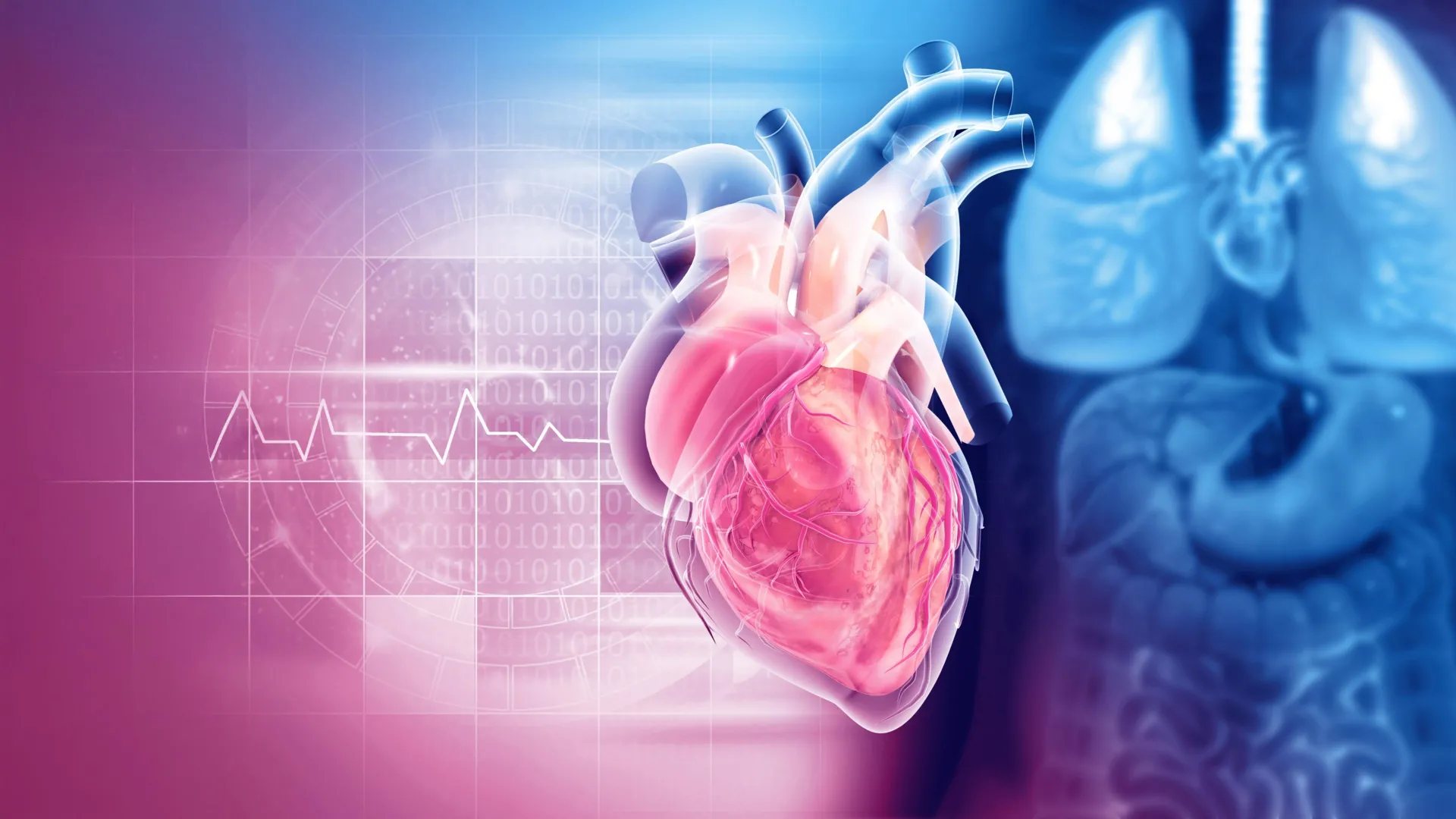
Serotonin is usually associated with mood, sleep, and digestion. But research published in 2023 suggests this chemical messenger may also influence a small structure that performs a vital job during every heartbeat: the mitral valve.
The multicenter investigation found evidence that reduced activity of the serotonin transporter may accelerate damaging changes in valves already affected by degenerative mitral regurgitation (DMR). Researchers at Columbia University’s Department of Surgery led the work in collaboration with the Pediatric Heart Valve Center at Children’s Hospital of Philadelphia (CHOP), the University of Pennsylvania, and the Valley Hospital Heart Institute.
The study was supported by the National Heart, Lung, and Blood Institute and was co led by Columbia’s Giovanni Ferrari, PhD, and CHOP’s Robert J. Levy, MD. The findings were published in Science Translational Medicine in 2023.
A Small Heart Valve With a Critical Role
The mitral valve sits between the heart’s left atrium and left ventricle. The left atrium receives oxygen rich blood returning from the lungs, while the left ventricle pumps that blood throughout the body.
Each time the heart contracts, the mitral valve should close firmly. It functions like a one way gate, keeping blood from flowing backward into the upper chamber.
DMR is one of the most common forms of heart valve disease. As the valve tissue degenerates, the thin flaps that should meet neatly can thicken, stretch, or lose their normal shape. The valve can no longer seal completely, allowing blood to leak backward into the left atrium (regurgitation).
This leak can raise pressure toward the lungs while reducing the amount of oxygen rich blood that moves forward through the heart. Some people have no noticeable symptoms at first. As the condition progresses, however, fatigue and shortness of breath can develop.
The heart must also work harder to maintain circulation. Over time, that added strain can permanently damage the heart and contribute to serious conditions such as atrial fibrillation and heart failure. Atrial fibrillation is an irregular heart rhythm, while heart failure means the heart cannot pump enough blood to meet the body’s needs.
Medicines can ease symptoms and help manage complications, but they cannot reverse the underlying degeneration of the mitral valve.
“Certain medications can ease the symptoms and prevent complications, but they do not treat the mitral valve,” says Ferrari, scientific director of the Cardiothoracic Research Program at Columbia. “If the degeneration of the mitral valve becomes severe, surgery to repair or replace the valve is needed.”
Current medical guidelines evaluate heart valve disease by considering symptoms, valve anatomy, the severity of the leak, imaging results, and how the heart and lung circulation are responding.
Why Serotonin Matters Beyond Mood
Serotonin is a chemical messenger with jobs throughout the body. It contributes to emotional state, digestion, sleep, memory, and blood clotting. In the brain, it acts as a neurotransmitter involved in regulating mood.
Changes in serotonin signaling have long been associated with conditions such as anxiety and depression. That relationship is complex, however, and neither condition can be explained simply as a shortage of one chemical.
Serotonin communicates by attaching to receptors on a cell’s surface. This contact sends a signal that tells the cell how to respond.
A protein called the serotonin transporter (SERT or 5-HTT) helps end that signal. It carries serotonin into the cell so the chemical can be reabsorbed and recycled. This process is known as serotonin reuptake.
Selective serotonin reuptake inhibitors (SSRIs) reduce the transporter’s ability to take serotonin back into cells, leaving more serotonin available for a longer time. This effect can help relieve symptoms of mood disorders.
SSRIs are among the most widely prescribed antidepressants. They include fluoxetine (Prozac) and sertraline (Zoloft).
Because SSRIs reduce SERT activity, the researchers wanted to know whether the same mechanism could unintentionally affect heart valve tissue, particularly in people whose valves were already degenerating.
What Researchers Found in Patients and Mice
The team reviewed clinical information from more than 9,000 patients who had undergone mitral valve repair or replacement for DMR. The researchers also evaluated 100 mitral valve biopsies, which are small tissue samples examined in a laboratory.
“Studying the data of these patients, we found that taking SSRIs was associated with severe mitral regurgitation that needed to be treated with surgery at a younger age than for patients not taking SSRIs,” says Ferrari.
That finding showed an association, not cause and effect. The patient data could not prove that SSRIs caused the disease to progress more quickly. Observational studies cannot rule out other differences between groups that might influence when patients require surgery.
The researchers therefore explored the possible biological mechanism in mice and human valve cells.
They studied transgenic mice that lacked the SERT gene and found that the animals developed thicker mitral valves. Normal mice treated with high doses of SSRIs also developed thickened valves. These experiments supported the possibility that unusually low SERT activity can contribute to structural remodeling of the valve.
A Genetic Clue to Greater Valve Vulnerability
The researchers also examined 5-HTTLPR, a region of the SERT gene that helps control how active the serotonin transporter is.
They identified genetic variants that changed SERT activity in mitral valve cells. A “long” variant was associated with lower activity, particularly in people who inherited two copies (one maternal and one paternal).
Patients with DMR who carried the “long-long” variant underwent mitral valve surgery more often than patients with other variants.
Laboratory experiments offered a possible explanation. Mitral valve cells from patients with the “long-long” variant reacted more strongly to serotonin and produced more collagen. Collagen normally gives tissue structure and strength, but excessive collagen can make a valve thicker and stiffer, changing its shape and movement.
Cells with the “long-long” variant were also more sensitive to fluoxetine than cells carrying other variants. The findings suggest that an already damaged valve may be especially vulnerable when serotonin exposure, reduced transporter activity, and genetic susceptibility occur together.
Could a Simple DNA Test Guide Care?
For patients with DMR and the “long-long” variant, the researchers proposed that taking an SSRI could further reduce SERT activity in the mitral valve.
They suggested testing people with DMR for 5-HTTLPR. The test could be performed using DNA collected from a blood sample or mouth swab. In theory, identifying patients with low SERT activity could help doctors determine who might need closer monitoring or earlier surgery.
“Assessing patients with DMR for low SERT activity may help identify patients who may need mitral valve surgery earlier,” says Ferrari. “Promptly fixing a mitral valve that is very leaky would protect the heart and could prevent congestive heart failure.”
This type of genetic testing has not become a standard part of heart valve care. Major guidelines continue to focus primarily on symptoms, valve structure, leak severity, heart function, and imaging findings. Clinical studies would be needed to establish whether adding genetic testing actually improves treatment decisions and patient outcomes.
What the Findings Do Not Mean
The researchers did not observe harmful effects from normal SSRI doses or the “long-long” variant in cells obtained from healthy human mitral valves.
“A healthy mitral valve can probably stand low SERT activity without deforming,” says Ferrari. “It is unlikely that low SERT can cause degeneration of the mitral valve by itself. SSRIs are generally safe for most patients. Once the mitral valve has started to degenerate, it may be more susceptible to serotonin and low SERT.”
That distinction is important. The findings do not suggest that SSRIs generally damage healthy heart valves. They also do not justify stopping or changing antidepressant treatment without guidance from a prescribing clinician.
The strongest signal appeared in people whose mitral valves had already begun to degenerate. Even in that group, the human findings were observational and could not establish that antidepressants directly caused earlier disease progression.
The original research raised two practical questions for future studies. One is whether patients with DMR who respond well to SSRIs should receive regular monitoring for signs that the valve is worsening. The other is whether patients who do not respond well to an SSRI might benefit from switching to another type of antidepressant rather than increasing the SSRI dose.
Those approaches have not yet been validated in clinical trials.
Later Studies Expand the Serotonin Connection
Research published since 2023 has added support to serotonin’s possible role in heart valve remodeling. Much of that evidence, however, still comes from animals, cells, or relatively small observational studies.
A 2024 study found that mice with deficient SERT activity were more susceptible to fibrotic changes in their cardiac valves and left ventricular heart muscle. Fibrosis is the buildup of stiff, scar like tissue that can interfere with normal movement and function.
That study pointed to HTR2B, one of the cell receptors activated by serotonin, as an important driver of the damaging response. Mitral valve cells appeared particularly responsive to serotonin. The results broadened the concern beyond isolated thickening of the mitral valve, although animal findings cannot determine what happens in people taking standard doses of antidepressants.
Evidence From Another Major Heart Valve
A study published in 2025 investigated serotonin in aortic stenosis, a different type of heart valve disease. The aortic valve controls blood leaving the heart, and aortic stenosis develops when this valve becomes thick, stiff, and narrow.
The researchers compared 38 people with severe aortic stenosis with 38 control participants matched for factors including age, sex, and major medical conditions. Patients with severe aortic stenosis had higher serum levels of serotonin and its primary breakdown product.
The study supported the idea that serotonin signaling may be involved in more than one type of valve disease. However, the research included only 76 people and measured them at a single point in time. It could not determine whether elevated serotonin contributed to the disease, resulted from it, or reflected another biological process.
An Experimental Drug Target Emerges
In February 2026, researchers reported additional evidence connecting low SERT activity with aortic valve disease.
The study examined valve tissue from 66 patients undergoing replacement for severe aortic stenosis and compared it with normal donor valves. Diseased valves showed reduced SERT expression and stronger serotonin receptor signaling.
Researchers then tested the pathway in mice. An experimental compound that blocked HTR2B helped preserve valve structure and improved measurements of blood flow during an early stage of fibrotic remodeling. Human cell experiments also suggested that low SERT activity can make valve cells more sensitive to damaging biological signals.
These results make HTR2B an intriguing possible drug target, but the compound is not an approved treatment for heart valve disease. The mouse model represented early fibrotic changes rather than advanced, heavily calcified aortic stenosis. Additional animal studies and eventual human trials would be necessary before researchers could know whether blocking HTR2B is safe or effective in patients.
A Broader Review Finds an Association
A 2026 systematic review and meta analysis examined clinical studies involving medications that modify SERT activity. The pooled analysis reported a significant association between these drugs and heart valve disease, with an odds ratio of 2.76.
An odds ratio compares the odds of an outcome between groups. It does not directly predict an individual patient’s risk, and it does not prove that the medication caused the outcome.
The review also covered a broader category of SERT modifying drugs, not only commonly prescribed SSRIs. Its authors acknowledged that mechanistic evidence remains limited. More detailed research is needed to separate the effects of different drugs, doses, treatment durations, underlying health conditions, and preexisting valve abnormalities.
A Compelling Clue That Still Needs Clinical Proof
Taken together, the findings published since 2023 make serotonin signaling a more plausible contributor to heart valve remodeling. They suggest that the original mitral valve findings may reflect a broader biological pathway rather than an isolated observation.
The research also raises the possibility that doctors could eventually use genetic information to identify vulnerable patients or that scientists could develop treatments aimed at HTR2B. Such a strategy might block harmful fibrotic signaling without broadly interfering with serotonin’s many essential functions elsewhere in the body.
Important questions remain. Researchers need studies that follow patients over time, compare individual medications and doses, account for other health risks, and determine whether SERT testing changes care in a meaningful way. Human trials would also be required before any treatment targeting HTR2B could enter routine use.
For now, regular cardiology care remains the practical priority for people with degenerative mitral regurgitation. The evidence offers a compelling explanation for why some damaged valves may deteriorate faster, but it does not replace imaging, clinical evaluation, or individualized decisions about antidepressant treatment.
What is driving the UK’s fall in healthy life expectancy?
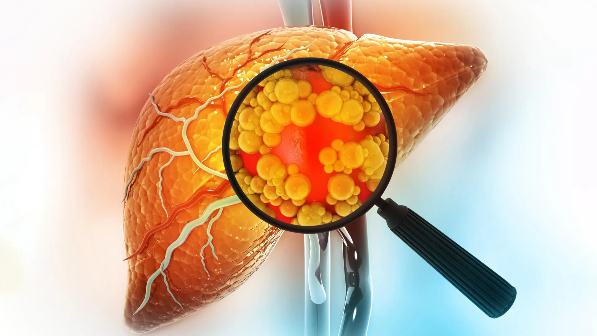
An experimental drug developed at Michigan Medicine has shown the ability to reverse severe fatty liver disease in animal studies by restoring gut health. The findings, published in The Journal of Clinical Investigation, suggest that targeting the connection between the gut and liver could offer a promising new approach for treating metabolic dysfunction-associated steatohepatitis (MASH).
MASH is a serious form of fatty liver disease that affects about 7% of people worldwide. It can progress to cirrhosis, liver cancer, and liver failure, yet effective treatment options remain limited.
The investigational compound, known as DT-109, is a glycine-based tripeptide. Researchers found that it reversed MASH in animal models by interrupting a harmful biological process linking the gut and liver.
“We see clear evidence that DT-109 protects the gut epithelial barrier, reducing the systemic influx of harmful microbial products that are thought to contribute to MASH development and progression,” said Eugene Chen, M.D., Ph.D., senior author of the study and Frederick G. L. Huetwell Professor of Cardiovascular Medicine at the University of Michigan Medical School.
“This compound shows benefits to the gastrointestinal system and has great potential as a treatment for MASH.”
How Gut Bacteria Can Drive Liver Disease
Earlier studies from Chen’s laboratory had already shown that DT-109 could improve MASH in animals. The new research explains how the compound produces those benefits.
The team first identified a major contributor to the disease: an overgrowth of the bacterium Clostridium perfringens, which generates ammonia inside the gut.
High ammonia levels damage the lining of the digestive tract, weakening the intestinal barrier. Once that protective barrier is compromised, harmful microbial products can enter the bloodstream, reach the liver, and trigger inflammatory immune responses, including excessive activation of CD8+ T cells.
Through a series of experiments, the researchers found that DT-109 disrupted this chain of events, helping restore the health of both the gut and the liver.
DT-109 Restores the Gut Barrier
In both mice and nonhuman primates, DT-109 reduced Clostridium perfringens levels and lowered ammonia production in the intestines. As a result, the intestinal barrier became stronger, limiting the movement of harmful substances from the gut into the body.
The results were especially encouraging in nonhuman primates, whose liver biology and gut microbiota more closely resemble those of humans. In these animals, DT-109 reduced liver inflammation and significantly improved the severity of MASH.
“DT-109 connects microbiota modulation with liver protection by restoring gut barrier integrity and limiting the systemic translocation of ammonia and other pro-inflammatory microbial products within the gut-liver axis,” said Jifeng Zhang, Ph.D., co-author and research professor of cardiovascular medicine at U-M Medical School.
“We also found that DT-109 primarily acts in the gastrointestinal tract, but its reach stretches much further.”
Potential Benefits Beyond MASH
The researchers believe DT-109 may have uses beyond treating fatty liver disease.
Previous studies have shown that the compound can reduce the formation of atherosclerosis plaques and prevent vascular calcification in nonhuman primates, suggesting it could also become a treatment for cardiovascular disease.
Because breakdown of the intestinal barrier has also been linked to several digestive disorders, the team believes DT-109 could eventually be explored as a treatment for conditions such as inflammatory bowel disease (IBD).
Future research will focus on additional testing needed to move DT-109 into clinical trials and evaluate its safety and effectiveness in people.
“This study presents novel evidence about the pathogenesis of MASH and provides excitement about a therapeutic avenue to explore for a condition that remains difficult to treat,” said Elliot Tapper, M.D., Academic Director of Hepatology at Michigan Medicine.
“What patients with MASH need is a safe and effective therapy capable of improving their liver and heart health — of course we are excited about these developments.”
Additional authors include Yang Zhao, Ph.D., Ying Zhao M.S., and Yanhong Guo, MD., Ph.D., all of the University of Michigan. Additional co-authors are listed in the published study.
Funding and Disclosures
Ying Zhao, Oren Rom, Jifeng Zhang, and Y. Eugene Chen are inventors on the patent application (Tripeptides and treatment of metabolic, cardiovascular, and inflammatory disorders).
Chen is also an inventor of DT-109. The University of Michigan has patented the compound and licensed it to Diapin Therapeutics. Chen and the university hold an ownership interest in the company. Diapin Therapeutics supplied DT-109 for the study and is continuing to develop the compound.
The study protocol involving humans, all amendments and the informed consent form were reviewed and approved by the Institutional Review Boards at each site, including the First Affiliated Hospital of Xi’an Jiaotong University (approval number: XJTU1AF2023LSK330), and the Institutional Review Board of Jinan University (approval number: 2016-017) and the University of Hong Kong/Hospital Authority Hong Kong West Cluster (approval number: UW 20-700). All experimental protocols involving non-human primates were approved by the Laboratory Animal Care Committee of Xi’an Jiaotong University (approval number: 20191278) and the Institutional Animal Care and Use Committee of Spring Biological Technology Development Co., Ltd. (approval number: 201901). The study was performed in accordance with the National Institutes of Health Guide for the Care and Use of Laboratory Animals.
US officials and diplomats and their families began reporting a mysterious illness a decade ago.

A second pregnancy changes the brain in ways that are both familiar and distinct from a first pregnancy, according to new research from Amsterdam UMC published in Nature Communications. Building on earlier work showing that a first pregnancy reshapes the brain, the researchers found that each pregnancy leaves its own unique imprint on the maternal brain.
In an earlier study, Elseline Hoekzema and colleagues became the first to demonstrate that pregnancy changes the structure of the human brain. They also showed that pregnancy affects how the brain functions. For this latest research, the team followed 110 women over time. Some were expecting their first child, some were pregnant with their second, and others remained childless. By performing repeated brain scans, the researchers tracked how the brain changed throughout the study.
“With this, we have shown for the first time that the brain not only changes during the first pregnancy, but also during a second,” says Hoekzema, head of the Pregnancy Brain Lab at Amsterdam UMC. “During a first and second pregnancy, the brain changes in both similar and unique ways. Each pregnancy leaves a unique mark on the female brain.”
Brain Networks Shift in Different Ways
The researchers found that a first pregnancy produced the largest changes in the structure and activity of the brain’s Default Mode Network, a system involved in self reflection, social thinking, and other important mental functions.
During a second pregnancy, this same network changed again, although to a lesser degree. Instead, the most noticeable changes occurred in brain networks responsible for directing attention and responding to sensory information.
“It appears that during a second pregnancy, the brain is more strongly altered in networks involved in reacting to sensory cues and in controlling your attention,” explains researcher Milou Straathof, who analyzed the data. “These processes may be beneficial when caring for multiple children.”
Brain Changes Linked to Maternal Bonding and Mental Health
The study also uncovered a relationship between pregnancy related brain changes and the emotional bond between mother and child. This connection was stronger after a first pregnancy than after a second.
Researchers also identified links between structural changes in the brain and peripartum depression during both first and second pregnancies. According to the team, this is the first evidence that changes occurring in the brain’s cortex during pregnancy are associated with maternal depression.
The timing of these associations differed depending on pregnancy history. Among first time mothers, they were most apparent after childbirth. For women expecting a second child, they were more noticeable during pregnancy.
“This knowledge can help to better understand and recognize mental health problems in mothers. It is important that we understand how the brain adapts to motherhood.”
Understanding the Maternal Brain
The findings offer new insight into the remarkable adaptability of the maternal brain. Although most women experience one or more pregnancies during their lifetime, scientists are only beginning to understand how pregnancy influences the brain over the long term.
The researchers say these discoveries help fill an important gap in knowledge about women’s biology and could eventually improve care for mothers, including efforts to prevent and treat postpartum depression. The study also highlights the brain’s remarkable ability to continually adapt to major life experiences such as pregnancy and motherhood.

Physicists from the University of Jyväskylä and Aalto University in Finland have successfully created a two dimensional topological crystalline insulator, marking the first experimental realization of a quantum material that scientists had predicted for more than a decade. Until now, attempts to produce it had been held back by difficulties in developing the right materials.
The breakthrough was led by Associate Professor Kezilbeiek Shawulienu in collaboration with Aalto University researchers, including Professor Peter Liljeroth and Professor Jose Lado. The team fabricated the material by growing an atomically thin film consisting of just two layers of tin telluride (SnTe) on top of a niobium diselenide (NbSe2) substrate.
Atomically Thin Crystal Reveals Unique Quantum States
To examine the material’s properties, the researchers used molecular beam epitaxy together with low temperature scanning tunneling microscopy, allowing them to probe its electronic behavior with atomic level precision.
Their measurements revealed pairs of conducting edge states, a defining feature of topological crystalline insulators. These special pathways allow electrons to travel along the edges of the material and are protected by the symmetry of the crystal lattice.
Strain Controls the Material’s Quantum Properties
The conducting edge states appear within a large electronic band gap of more than 0.2 electron volts (eV). The team found that the tin telluride film is compressed by the underlying substrate, creating strain that is essential for stabilizing the material’s topological state.
Even more importantly, the researchers demonstrated that these edge states can be adjusted by changing the strain, offering a practical way to tune the material’s electronic behavior for future technologies.
Potential for Future Quantum Electronics
First principles quantum mechanical calculations confirmed that the observed edge states have a topological origin. The team also examined how neighboring edge states interact, finding that their energy levels shift because of a combination of electrostatic interactions and quantum tunneling.
Because the material has a relatively large band gap, its topological properties are expected to remain stable even at room temperature. That makes it a promising platform for exploring strain tunable two dimensional topological states and could support future advances in spin based electronics and nanoscale devices.
The findings were published in the journal Nature Communications.
Health officials warn little over half of 54-year-olds completed free at-home test kits.
Identical twins Nancy and Margo benefitted from the procedure while in the womb as part of a world-first medical trial.

Smartphones, electric vehicles, and countless portable electronics all depend on batteries. As demand for better energy storage grows, improvements in battery capacity, lifespan, and safety will play a major role in the future of electrification. One of the most promising technologies is the solid-state battery, which could allow smartphones to operate for several days on a single charge and give electric vehicles driving ranges up to three times greater than many current models.
Unlike conventional lithium-ion batteries, which use a liquid electrolyte between two solid electrodes, solid-state batteries replace the liquid with a solid electrolyte. This design offers several potential advantages, including higher energy density, improved safety, and longer battery life. But one stubborn problem has slowed commercial adoption. During charging, tiny tree-like structures called dendrites can grow from the lithium anode, pierce the solid electrolyte, and create internal short circuits.
Now, an interdisciplinary team at the Max Planck Institute for Sustainable Materials (MPI-SusMat) has identified exactly how these dendrites trigger fractures that ultimately lead to battery failure. Their findings were published in the journal Nature.
How Dendrites Crack Solid-State Batteries
Exactly how soft lithium dendrites manage to break through a hard ceramic electrolyte has long puzzled researchers.
“Although the electrodes and the forming dendrites consist of lithium metal, which is soft like a gummy bear, the dendrites are able to penetrate the ceramic electrolyte and lead to a short circuit,” says Dr. Yuwei Zhang, first author of the new publication and head of the group “Chemo-Mechanics of Battery Materials” at MPI-SusMat. “How can soft dendrites fracture the stiff solid ceramic? There are two hypotheses: either internal stress is built up inside the dendrites and induces mechanical fracture of the solid electrolyte. Or, electrons leak along the grain boundaries of the solid electrolyte promoting the formation of lithium nuclei that interconnect later.”
To determine which explanation was correct, the researchers used an advanced combination of sample preparation and materials characterization techniques. Every step was performed under vacuum and at cryogenic temperatures to eliminate interference from oxygen, water, or even the microscopes’ electron beams.
The team examined both the internal stress and the plastic deformation of lithium dendrites trapped inside cracks. Their analysis found no buildup of lithium ahead of the dendrite tip, ruling out one proposed mechanism.
“The soft lithium metal is able to penetrate the stiff ceramic electrolyte, like a continuous waterjet that penetrates a rock. We calculated that hydrostatic stress in the dendrite leads to brittle fracture of the solid electrolyte in the end,” says Zhang.
The researchers also confirmed their conclusions using phase field simulations and electron backscatter diffraction measurements.
New Strategies to Prevent Battery Failure
With a better understanding of how dendrites fracture solid electrolytes, the team is now investigating ways to stop or delay the process.
Potential solutions include making the solid electrolyte tougher so it resists cracking for longer, introducing microscopic voids that redirect dendrite growth and steer cracks away from vulnerable areas, or adding protective coatings to lithium electrodes to reduce dendrite formation in the first place.
The researchers say their work demonstrates the importance of understanding how materials behave at the microscopic level. Those insights could help transform solid-state batteries from a promising concept into a practical technology for future smartphones, electric vehicles, and other electronic devices.
