Popular weight loss drugs linked to a surprising fingernail problem

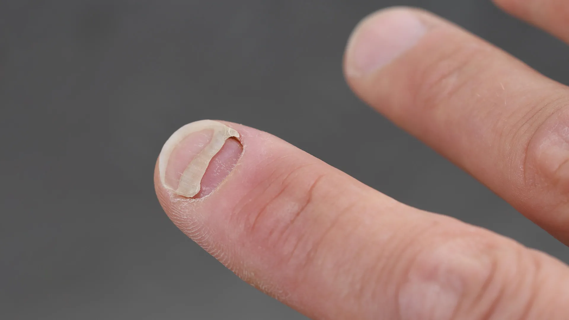
Popular GLP-1 medications used for weight loss and type 2 diabetes (such as Ozempic, Wegovy, Mounjaro, Zepbound, Victoza, and Trulicity) have been linked to an unexpected problem involving fingernails. A multinational study found that people taking these drugs were more than four times as likely to report nail detachment, a condition in which the nail separates from the skin underneath it.

Researchers found that 39% of GLP-1 users experienced nail detachment, compared with just 9% of people who had never taken the medications. Nail discoloration was also considerably more common, and two-thirds (66%) of users reported at least one nail disorder.

The findings, presented at the European Academy of Dermatology and Venereology (EADV) Congress 2026, come from the first controlled study to investigate the possible relationship between GLP-1 medications and nail health.

Unexpected Nail Problems Among GLP-1 Users

GLP-1 receptor agonists have become increasingly popular for managing obesity and type 2 diabetes. These medications act on a hormone signaling system that helps regulate blood sugar and appetite, making them effective tools for improving metabolic health and supporting weight loss.

However, scientists are still investigating their potential effects on other parts of the body. Until recently, information about nail changes during GLP-1 treatment came primarily from individual reports of possible adverse reactions, rather than controlled comparisons.

To investigate the connection, researchers examined 575 people using GLP-1 medications and compared them with 1,329 individuals who had type 2 diabetes and/or obesity but had never used the drugs. Participants were recruited from France, the United States, Brazil, and Mexico.

The results revealed a consistent pattern. Overall, 66% of GLP-1 users reported at least one nail disorder, compared with 53% of the control group. Nail discoloration showed an especially large difference, affecting 46% of users versus 17% of nonusers.

Nail detachment was even more striking, with reports more than four times as common among those taking GLP-1 medications.

Weight Loss Alone May Not Explain the Nail Changes

One important question was whether the nail problems might simply be related to weight loss, rather than to the medications themselves. Rapid changes in body weight can affect nutrition and other physiological processes, potentially influencing nail and hair health.

However, the researchers found that weight loss did not fully account for the differences. Even when they limited their analysis to participants who had lost weight, GLP-1 users continued to report significantly more nail problems than those who had not used the medications.

“Until now, nail changes associated with GLP-1 therapies had essentially been anecdotal,” said study author Dr. Charles Taïeb. “Our findings show a clear association, but they also underline an important point: not everything that happens during GLP-1 treatment is necessarily caused by the treatment itself.”

The researchers emphasized that their findings do not prove that GLP-1 drugs cause nail disorders. Approximately half of the medication users said they had experienced nail problems even before beginning treatment.

When the researchers accounted for existing skin conditions and nutritional deficiencies, some of the differences between the groups became smaller. Nevertheless, nail detachment and discoloration remained approximately two to three times more common among GLP-1 users after these adjustments.

Scientists Also Find Higher Rates of Hair Loss and Scalp Problems

The unexpected findings were not limited to fingernails.

A companion study examining the same participants identified significant differences in hair and scalp health among people taking GLP-1 medications.

Half of GLP-1 users reported unusual hair loss, compared with 34% of nonusers. Reduced hair volume or density was also substantially more common, affecting 42% of users versus 19% of the control group.

Researchers additionally examined hair loss that began after treatment started. This was reported by 26% of GLP-1 users, compared with 12% of nonusers, making it more than twice as common in the medication group.

Scalp redness was approximately three times more frequent among GLP-1 users.

A particularly interesting finding was that hair loss rarely appeared by itself. Most users who reported shedding also experienced other scalp symptoms, including itching, redness or flaking skin.

These accompanying symptoms raised the possibility that inflammation could be contributing to the changes, rather than the hair loss being explained entirely by rapid weight reduction.

“This suggests that what is happening on the scalp may go beyond the classical shedding seen after rapid weight loss and involve an inflammatory component,” explained study author Dr. Bruno Halioua. “Scalp inflammation was the rule rather than the exception, which clinicians do not currently look for and which may be treatable. Taken together, our findings suggest that factors beyond weight loss alone may contribute to the hair, scalp and nail changes observed among GLP-1 users.”

What the Findings Mean for People Taking GLP-1 Drugs

Despite the relatively high frequency of reported symptoms, the researchers stressed that the findings should encourage awareness rather than alarm.

“These changes are frequent, usually benign, and are not a reason to stop an effective treatment on one’s own initiative,” he said. “Anyone who notices hair shedding, an itchy or red scalp or nail changes should mention it to their doctor rather than discontinue therapy.”

There are also important limitations to consider. Both investigations were cross-sectional studies, meaning they examined participants’ experiences without following them over time to establish how symptoms developed. The findings also relied on symptoms reported by participants rather than standardized clinical examinations.

As a result, the studies can reveal patterns and associations, but they cannot determine whether GLP-1 medications directly caused the reported nail, hair, or scalp problems.

To investigate these questions more thoroughly, the research team plans to conduct prospective studies that follow patients from the beginning of treatment. These investigations will include standardized skin, hair and nail examinations along with biological monitoring.

The goal is to establish when the changes first appear, identify the mechanisms behind them, and determine whether they improve or disappear over time.

The research received institutional support from DUCRAY Laboratories (Pierre Fabre Group), a company specializing in dermatological and hair care products developed to help manage skin and scalp conditions and improve quality of life.

Share Button

Scientists find hidden driver of aging and a simple amino acid that fights memory loss in mice

What if some of the changes we associate with aging begin with the loss of a single protein deep inside the brain? Scientists investigating that possibility found that restoring a protein called Menin in older mice improved memory, balance, bone mass, and skin thickness in just 30 days. Even more surprisingly, supplementing the animals with a naturally occurring amino acid improved their learning and memory in only three weeks.

The findings, originally published in March 2023 in PLOS Biology, came from researchers led by Lige Leng of Xiamen University in China. Their experiments identified a previously overlooked connection between inflammation, brain chemistry, and aging throughout the body. Although the results were obtained in mice, they raised an intriguing possibility: Some effects of aging may be influenced by biological processes in the brain that scientists could eventually learn to modify.

Research published since then has added evidence that the brain plays an important role in regulating aging. It has also revealed why the seemingly simple supplement findings deserve careful interpretation.

How a Hidden Brain Protein May Influence Aging

At the center of the discovery is the hypothalamus, a small but powerful region near the base of the brain. It helps regulate essential functions such as body temperature, hunger, metabolism, sleep, and hormone production. Because these systems affect nearly every organ, changes in the hypothalamus can have consequences throughout the body.

Scientists have increasingly recognized the hypothalamus as an important player in aging. One possible reason involves neuroinflammation, the brain’s inflammatory response. Although inflammation helps protect against infections and injuries, persistent inflammation can interfere with normal cellular function and contribute to age-related deterioration.

Earlier work by Leng and colleagues had identified Menin as a protein that helps restrain inflammation in the hypothalamus. Menin also participates in regulating gene activity, allowing it to influence multiple biological pathways.

The researchers wanted to understand what happens when this protective protein begins to disappear with age.

They discovered that Menin levels gradually declined in certain hypothalamic neurons as mice grew older. Interestingly, the same decrease was not observed in astrocytes or microglia, two other important types of brain cells involved in supporting neurons and managing immune responses.

To investigate whether this decline actually contributed to aging, the team developed genetically modified mice in which Menin could be selectively disabled.

The results were striking. Mice with reduced Menin activity developed greater inflammation in the hypothalamus, thinner skin, reduced bone mass, and worsening cognitive performance. Their lifespans were also modestly shortened.

Rather than simply accompanying aging, the loss of Menin appeared capable of helping drive several of its characteristic changes.

Restoring Menin Reversed Several Signs of Aging in 30 Days

The researchers then asked an even more interesting question: If declining Menin contributes to aging, could restoring the protein reverse some of the damage?

To find out, they used a gene delivery technique to increase Menin production in a specific part of the hypothalamus in older (20-month-old) mice. This region, called the ventromedial hypothalamus, helps coordinate metabolism, energy balance, and other bodily functions.

Just 30 days after treatment, the animals showed measurable improvements.

Their skin became thicker, bone mass increased, and performance improved on tests measuring learning, memory, and balance. Researchers also observed reductions in inflammation and improvements in certain metabolic changes associated with aging.

In a separate survival analysis, mice receiving the Menin treatment also lived longer than untreated controls.

These findings suggested that some biological changes associated with old age could be at least partially reversed by restoring Menin activity in a specific group of brain cells.

But the researchers also uncovered an important clue about how Menin produced these effects.

A Simple Amino Acid Offers a Surprising Clue

When Menin levels declined, the researchers noticed that concentrations of D-serine, an amino acid involved in brain signaling, also fell.

Amino acids are best known as the building blocks of proteins, but some perform other important functions. D-serine helps regulate NMDA receptors, specialized proteins on nerve cells that are essential for communication between neurons, learning, and the formation of memories.

D-serine is closely related to L-serine, an amino acid commonly found in foods such as soybeans, eggs, fish, and nuts. The two forms have different biological roles. Although D-serine is also available as a dietary supplement, consuming ordinary serine-rich foods is not equivalent to taking the form used in the experiments.

Further investigation revealed that Menin helps control the production of PHGDH, an enzyme involved in the biochemical pathway that generates serine (which was in turn regulated by Menin). When Menin activity decreased, this pathway became less active, reducing the amount of D-serine available to the brain.

Restoring Menin increased D-serine levels in the hippocampus, a region especially important for learning and memory.

This connection suggested that the cognitive effects of declining Menin might be partly explained by disruptions in D-serine signaling.

To test the idea directly, the scientists gave older mice D-serine in their drinking water for three weeks.

The supplementation improved their performance on learning and memory tests, even without directly restoring Menin.

However, there was an important distinction. While increasing Menin improved both cognitive function and physical signs of aging, D-serine supplementation primarily improved cognition. It did not restore the age-related losses in bone mass and skin thickness.

That difference suggests Menin influences aging through several biological mechanisms, with D-serine accounting for only part of its effects.

Later Research Strengthens the Link Between the Brain and Aging

Since the original 2023 discovery, additional studies have provided evidence that the hypothalamus helps coordinate aging across the body.

In a 2024 study published in Cell Metabolism, researchers at Washington University School of Medicine identified a different group of hypothalamic neurons that communicates with fat tissue. By maintaining or stimulating this communication pathway, they increased physical activity, improved age-related metabolic function, and extended lifespan in mice.

That work involved a different protein and neural circuit from Menin, but supported the broader idea that signals originating in the hypothalamus can influence how the rest of the body ages.

Another 2024 study in the Journal of Physiology and Biochemistry provided additional insight into Menin itself. Researchers found that increasing Menin helped protect cultured hippocampal cells against inflammation and a form of programmed cell death triggered by exposure to the stress hormone corticosterone. Although these laboratory experiments did not establish an anti-aging effect, they supported the idea that Menin has important protective functions in brain cells.

An even larger picture emerged in January 2025, when scientists published a detailed analysis of approximately 1.2 million mouse brain cells in Nature.

The researchers discovered that some of the brain cells most sensitive to aging were concentrated around the third ventricle, a fluid-filled cavity in the hypothalamus. Many aging cells showed reduced activity in genes involved in normal neuronal function and increased activity in genes associated with inflammation and immune responses.

These findings highlighted the hypothalamus as a potential center of vulnerability during brain aging, although they did not directly test the Menin pathway.

New Findings in 2026 Reveal More Aging Pathways

Research published in 2026 has further strengthened the connection between hypothalamic activity and longevity.

In a study published in Cell, scientists found that strengthening daily biological rhythms in a specific hypothalamic region improved several aging-related measurements and extended lifespan in male mice. The intervention also reduced estimates of biological age based on DNA methylation, chemical modifications to DNA that change with age.

Another 2026 study in Cell Metabolism identified a protein called Mimecan that helps skeletal muscles communicate with the hypothalamus and brown fat, a specialized tissue involved in generating body heat. Restoring circulating Mimecan levels in older mice improved this communication and extended lifespan.

Together, these discoveries suggest that aging involves complex communication between the brain and the rest of the body. Rather than depending on one master switch, multiple neural circuits, hormones, and metabolic signals appear to help determine how age-related changes unfold.

Could D-Serine Supplements Protect the Human Brain?

The possibility of improving memory with a relatively simple amino acid is especially appealing. However, evidence that D-serine can reverse age-related cognitive decline in humans remains limited.

A small randomized clinical trial published in 2016 tested D-serine in 50 healthy older adults. Participants performed better on one assessment of spatial learning and problem-solving after receiving the amino acid, but researchers did not find significant improvements across the other cognitive tests or mood measurements.

More importantly, later research suggests that the effects of D-serine depend on the biological circumstances.

In an April 2025 study published in Cellular and Molecular Life Sciences, scientists investigated D-serine in mice genetically engineered to develop features of Alzheimer’s disease.

Unlike the Menin experiments, which associated reduced D-serine with age-related memory impairment, this study found that an early increase in D-serine accompanied disruptions in brain signaling. Removing the enzyme responsible for producing D-serine prevented or reduced several later cognitive impairments in those mice.

These findings do not invalidate the earlier results. Instead, they suggest that D-serine signaling is more complicated than simply having too little or too much. Its effects may depend on the condition being studied, the timing of treatment, and how the brain’s signaling systems are functioning.

There are also unanswered questions about the long-term safety of D-serine supplementation. High doses have raised kidney safety concerns in animal experiments, and research has not established whether prolonged supplementation is a safe or effective way to prevent age-related memory loss in otherwise healthy people.

For now, D-serine should be considered an experimental approach rather than a proven treatment for brain aging.

Can Scientists Eventually Slow Aging by Targeting the Brain?

Despite the encouraging mouse experiments, researchers still need to answer several fundamental questions.

Why does Menin decline in the hypothalamus as animals age? How many of its effects could be reversed by treatment, and how long would those improvements last? Could restoring Menin influence other biological systems in unexpected ways?

It is also unclear whether treatments targeting Menin or D-serine could reproduce similar results in humans.

Nevertheless, the original researchers believe the pathway deserves further investigation.

“We speculate that the decline of Menin expression in the hypothalamus with age may be one of the driving factors of aging, and Menin may be the key protein connecting the genetic, inflammatory, and metabolic factors of aging. D-serine is a potentially promising therapeutic for cognitive decline,” said Leng.

Leng adds, “Ventromedial hypothalamus (VMH) Menin signaling diminished in aged mice, which contributes to systemic aging phenotypes and cognitive deficits. The effects of Menin on aging are mediated by neuroinflammatory changes and metabolic pathway signaling, accompanied by serine deficiency in VMH, while restoration of Menin in VMH reversed aging-related phenotypes.”

The growing body of evidence points toward a potentially important shift in how scientists understand aging. Instead of viewing it only as the gradual deterioration of individual organs, researchers are investigating how specific brain pathways may actively shape the process.

The possibility that restoring those pathways could improve memory, physical function, or longevity remains experimental. But the Menin discovery, together with subsequent findings, offers a compelling reason to explore whether some of the biological changes associated with aging are more reversible than previously thought.

Share Button

Stormont ‘failing to plan’ for ageing population

Politicians are urged to prepare as over-65s are projected to outnumber children in NI by next year.

Share Button

Teen ‘detached from reality’ to cope with return of aggressive cancer

Ethan found out his cancer had returned two weeks after successful treatment.

Share Button

Scientists discover vanilla’s hidden healing potential

The compound responsible for vanilla’s familiar sweet flavor and aroma could have an unexpected future in medicine. Scientists at Flinders University have identified properties in vanillin that could make it a promising ingredient in treatments for chronic wounds, infections, and damaged tissue.

Researchers from the university’s Biomedical Nanoengineering Laboratory found that vanillin has notable antioxidant, anti-inflammatory, and antibacterial properties. Together, these effects could make the compound useful in developing wound care products that address several challenges associated with healing.

Vanillin is the main flavor and fragrance compound found in natural vanilla pods. It can also be manufactured synthetically using materials derived from sources such as clove oil or rice. Although best known for giving foods their distinctive vanilla flavor, the molecule has chemical properties that could make it valuable far beyond the kitchen.

How Vanilla’s Chemistry Could Help Heal Wounds

Vanillin is more than just a flavoring ingredient. Its molecular structure allows it to interact with other substances in ways that could be useful for medicine, particularly in materials designed to protect wounds and deliver treatments.

For example, vanillin can function as a dynamic crosslinker, helping connect different components within a material. This ability could make it useful in creating specialized medical formulations, including materials that carry drugs or support tissue repair.

The Flinders researchers examined vanillin through a framework that connects familiar food ingredients with practical medical applications. Their work explores how the same chemical characteristics that make vanillin useful in everyday products could also contribute to biological effects that benefit human health.

“Vanillin is among the most widely used flavor compounds in the global food system, valued for its sensory attributes, chemical stability and long history of safety – and that can be transferred to other areas, such as medical uses,” says Professor Krasimir Vasilev, Professor of Biomedical Nanotechnology at Flinders University and Director of Flinders’ Biomedical Nanoengineering Laboratory.

A Potential New Treatment for Chronic Wounds and Ulcers

One particularly promising application involves chronic wounds and ulcers, which can be difficult to treat because of persistent inflammation, bacterial contamination, and damage caused by oxidative stress.

Oxidative stress occurs when harmful reactive molecules overwhelm the body’s protective defenses, potentially damaging cells and interfering with tissue repair. A material capable of addressing oxidative stress, inflammation, and bacteria could therefore offer several advantages in wound care.

“Because vanillin’s synthetic form is abundant and cost-effective, we believe vanillin-based formulations may offer safe and multifunctional solutions for ulcer treatment and targeted drug delivery, especially of hydrophobic compounds.

“Owing to its amphiphilic molecular structure, vanillin can interact with reactive oxygen species, cellular membranes, and polymeric matrices, providing a basis for its incorporation into functional formulations.”

Hydrophobic compounds are substances that do not dissolve easily in water, a characteristic that can make certain medications difficult to formulate and deliver effectively. Vanillin’s chemical structure may help researchers design materials that carry these compounds to where they are needed.

Although the findings highlight intriguing possibilities, vanillin-based wound treatments would still need further development and clinical testing before their effectiveness and safety in patients could be established.

From Peppermint Oil to Advanced Medical Coatings

The investigation into vanillin builds on a broader effort at Flinders University to uncover useful medical properties in everyday food ingredients.

In February 2026, the research team published related work in the journal Small, describing a medical coating developed from peppermint oil. The coating, created at the nanoscale, was designed to protect against bacterial infection, inflammation, and oxidative stress while remaining compatible with human tissue.

Because the coating can be applied to medical materials, it could provide a way to introduce several protective functions into products used in healthcare.

Surprisingly, the inspiration for this research came from a simple personal experience.

Professor Vasilev noticed that eating peppermint leaves from his drink significantly eased his sore throat. Curious about the possible biological explanation, he began investigating whether the beneficial properties of peppermint could be incorporated into a durable protective coating.

That idea led him to explore the use of plasma technology, a field he has studied for more than 20 years. Plasma processing can be used to modify material surfaces and create thin functional coatings, potentially allowing biologically active compounds to be incorporated into medical products.

The work with peppermint helped establish an approach that researchers are now extending to vanillin.

Why Vanillin Could Be Especially Useful for Future Medicine

Vanillin offers another important advantage beyond its potential biological activity: it is inexpensive and readily available.

Because synthetic vanillin can be produced in large quantities, researchers believe it could be easier to incorporate into commercially manufactured medical products than some more expensive or difficult-to-source compounds.

Its relatively simple molecular structure and compatibility with established manufacturing processes could also support consistent product quality, reliable supplies, and large-scale production.

These practical considerations are especially important when moving a promising scientific concept from the laboratory toward real medical applications.

The researchers envision vanillin eventually becoming an ingredient in advanced wound dressings, ulcer treatments, targeted drug delivery systems, and therapies designed to support tissue regeneration.

“With coordinated engagement between academia, clinicians, and industry, vanillin has the potential to progress from an underutilized bioactive to a clinically deployable component of next-generation wound care and regenerative therapeutics,” says Professor Vasilev.

The findings suggest that a compound commonly associated with desserts and sweet fragrances could eventually contribute to an entirely different field: helping scientists develop better ways to protect damaged tissue and support healing.

The study, “Vanillin as a bioactive component in biomedical formulations for chronic wound healing and tissue regeneration,” was conducted by Borislav Stoilov, Vi Khanh Truong, Christopher Delaney, and Krasimir Vasilev. It was published in the International Journal of Pharmaceutics.

Share Button

Childhood cancer symptoms can be easily missed – here’s what to look out for

Although rare, cancer is the leading cause of death from disease among children under 15 in the UK.

Share Button

Service increases access to mental health support

The programme covers a range of topics, including managing anxiety and boosting self-esteem.

Share Button

Carys lived with endometriosis pain for decades, then everything changed

Carys Thurlby says “intense pain had become my normal” but surgery changed her life.

Share Button

Scientists build a nuclear clock that could make atomic clocks obsolete

Scientists have reached a major milestone in the quest to build an entirely new kind of clock. After decades of research, a team in Vienna has developed the world’s first self-stabilizing nuclear clock, a device that could eventually measure time with far greater precision than today’s most advanced atomic clocks.

Unlike earlier prototypes, the new nuclear clock can maintain its own stability without depending on a conventional atomic clock. Researchers have demonstrated that it can operate steadily for more than 24 hours without intervention.

The achievement represents an important advance in precision measurement, also known as metrology. By using atomic nuclei as exceptionally stable references, nuclear clocks could eventually allow scientists to measure time and other physical quantities with levels of accuracy that existing technologies cannot reach.

Why Thorium Holds the Key to Nuclear Clocks

The breakthrough relies on an unusual property of thorium atomic nuclei that scientists have investigated for decades.

Atomic nuclei can exist in different energy states, but moving between those states typically requires enormous amounts of energy. Thorium is a rare exception. Two of its nuclear energy states are separated by an unusually small energy gap, making it possible to trigger a transition between them using laser light.

This property is crucial because it allows researchers to control the energy state of a nucleus with a laser, something that is not possible in the same way for most other atomic nuclei. Scientists can then use this extremely precise transition as a reference for measuring time.

A major breakthrough came in April 2024, when researchers led by Prof. Thorsten Schumm at the Institute of Atomic and Subatomic Physics at TU Wien, working with a team led by Prof. Ekkehard Peik at PTB Braunschweig, identified the long-sought nuclear transition experimentally. They demonstrated for the first time that laser beams could excite thorium nuclei.

Further progress followed in the fall of 2024. The researchers connected their thorium excitation apparatus to a conventional optical atomic clock, demonstrating that the nuclei could serve as a highly precise timekeeping reference.

Although this established the basic principle of a nuclear clock, one essential capability was still missing: the ability to maintain its own accuracy independently.

Scientists Build a Nuclear Clock That Stabilizes Itself

“What you really want is a self-stabilizing nuclear clock,” explains Prof. Thorsten Schumm. “The basic idea is simple: you have a laser and you have thorium. The laser changes the energy state of the thorium nuclei, and the thorium nuclei are used to stabilize the frequency of the laser.”

To make this possible, the researchers developed a system built around a special crystal containing thorium atoms, manufactured at TU Wien. A laser shines onto the crystal, interacting with the thorium nuclei inside.

The laser’s light oscillates at a particular frequency, providing a regular rhythm that can be used to measure time. However, even tiny changes in the surrounding environment can interfere with that rhythm.

“The oscillation of this laser light can be used for timekeeping, but the laser frequency can shift slightly from time to time, for example due to temperature fluctuations,” explains Thorsten Schumm. “For high-precision measurements, you therefore need a mechanism to keep the laser frequency exactly stable, so that the clock continues to tick with precisely the same rhythm.”

Traditional atomic clocks solve this problem by using the energy transitions of electrons in atoms as a stable reference. The Vienna team has now demonstrated that the nuclei of thorium atoms can perform the same function.

The mechanism works because thorium nuclei absorb laser light only when the light has precisely the correct frequency. If the laser begins to drift away from that frequency, the amount of light absorbed by the nuclei decreases.

The system detects this change and automatically adjusts the laser to bring it back to the correct frequency. This continuous feedback keeps the clock operating at a consistent rate, without requiring a conventional atomic clock to maintain its stability.

For the first time, scientists have demonstrated a self-regulating nuclear clock that can function independently.

A Clock With an Error of Just One Second in 30 Million Years

The potential advantage of nuclear clocks comes from the extraordinary properties of atomic nuclei.

“The great advantage of the new nuclear clock in Vienna is that, if you use atomic nuclei rather than atoms, much higher precision is possible in principle,” says Thorsten Schumm.

Atomic nuclei are more than 10,000 times smaller than atoms. Because of their extremely small size, they are much less affected by external influences that can disrupt precise measurements.

This resistance to environmental disturbances makes nuclear energy transitions especially promising for ultra-precise timekeeping.

To evaluate their prototype, the researchers measured its performance over the course of a day. The nuclear clock achieved a relative precision of approximately 10 to the power of minus 15, equivalent to an error of roughly one second over 30 million years.

That figure describes the clock’s measured precision rather than an actual test lasting millions of years. Nevertheless, it illustrates the remarkable accuracy the researchers have already achieved with an early prototype.

The new clock is not yet as precise as the world’s leading optical atomic clocks, which remain the benchmark for high-performance timekeeping.

“This is not yet at the level of the world’s best optical atomic clocks, but for a first prototype it is a fantastic result,” says Thorsten Schumm.

The Next Step Toward Even More Precise Timekeeping

The researchers believe their nuclear clock has considerable room for improvement. By using more powerful lasers and developing higher-quality thorium crystals, they hope to substantially increase its precision.

These upgrades could eventually allow nuclear clocks to outperform existing atomic clocks and establish a new standard for measuring time.

The implications extend beyond simply building a better clock. More accurate timekeeping can also improve the measurement of other physical quantities, providing researchers with increasingly sensitive tools for investigating the fundamental properties of nature.

For now, the Vienna team’s achievement demonstrates that a long-pursued scientific concept can operate as a self-sustaining timekeeping system. With the world’s first self-stabilizing nuclear clock now running independently, scientists have taken an important step toward a future in which time can be measured with unprecedented precision.

Share Button

A plastic once thought harmless may be fueling fatty liver disease

Microplastics have spread into nearly every corner of the environment. These tiny plastic particles have been found in ocean water, drinking water, and even inside the human body. Although their presence is well documented, scientists are still working to understand what prolonged exposure could mean for human health.

Researchers at the Texas A&M College of Veterinary Medicine and Biomedical Sciences (VMBS) have now identified a potential health concern involving polyethylene, one of the most widely used plastics in the world. Their findings suggest that polyethylene microplastics may contribute to fatty liver disease, particularly when exposure occurs alongside an unhealthy diet.

Polyethylene accounts for approximately one-third of plastic production worldwide. It is used in numerous everyday products, including food wrappers, storage containers, plastic packaging, and the linings of beverage cups. Despite its widespread use, scientists have devoted relatively little attention to its potential effects on the liver compared with other types of microplastics.

“No studies have really looked into polyethylene’s effect on liver health, and it’s the most widely produced plastic,” said Dr. Adi Joshi, associate professor in VMBS’ Department of Veterinary Physiology and Pharmacology. “What we now know is that these microplastics, especially polyethylene, affect our liver’s natural defense and repair mechanisms.”

Common Microplastics May Contribute to Fatty Liver Disease

To investigate the possible health effects of polyethylene, Joshi and his colleagues examined whether exposure to this common plastic could promote fatty liver disease.

The condition develops when excessive fat accumulates inside liver cells. According to the American Liver Foundation, it affects approximately 25% of people worldwide, making it a major global health concern.

The researchers discovered that polyethylene exposure increased indicators of fatty liver disease even without the added influence of an unhealthy diet. When the plastic was combined with a diet rich in fat, fructose, and cholesterol, the signs of liver disease became more severe.

These results suggest that exposure to environmental pollutants and poor dietary habits may reinforce each other’s harmful effects, potentially worsening liver damage.

“Those who have a more Western-style diet, including foods like burgers and sodas, may have a greater chance of progressing to fatty liver disease if they are also exposed to polyethylene,” Joshi said.

The results were especially unexpected because polyethylene has traditionally been regarded as a relatively biologically inert material. In other words, scientists generally considered it less likely to interact with biological systems than more reactive substances.

However, the findings indicate that polyethylene may influence liver function in ways that were previously overlooked. Its effects appeared both independently and alongside dietary risk factors, raising important questions about how this common plastic interacts with the body’s metabolism and protective mechanisms.

Scientists Pinpoint How Microplastics Affect the Liver

Understanding where and how polyethylene causes biological changes could help explain its potential role in liver disease.

To investigate these effects in greater detail, the Texas A&M team partnered with scientists at the University of Oklahoma and applied a powerful technique called spatial transcriptomics.

This technology allows researchers to study gene activity while preserving information about where individual cells are located within a tissue. Rather than simply measuring which genes are active, scientists can identify precisely where biological changes are occurring.

Using this approach, the team mapped areas of liver damage associated with polyethylene exposure and identified molecular pathways that may help explain the effects.

One important finding involved PPAR-alpha, a protein that helps regulate fat metabolism in the liver. The researchers found that polyethylene activated this protein, suggesting that it plays a significant role in the liver’s response to microplastics.

They also identified ANXA2, a gene associated with tissue repair, as another possible contributor to the disease process.

Together, these discoveries provide a more detailed picture of how polyethylene may interfere with the liver’s normal functions, including the mechanisms involved in protecting and repairing damaged tissue.

Joshi believes that understanding these biological pathways could eventually help researchers identify potential treatments that reduce the harmful effects of microplastic exposure.

Could Microplastics Contribute to More Serious Liver Damage?

Although the findings reveal a previously underexplored connection between polyethylene and fatty liver disease, several important questions remain unanswered.

The researchers plan to investigate whether polyethylene exposure could contribute to more advanced forms of liver disease, including fibrosis. This condition occurs when repeated or ongoing liver damage leads to the accumulation of scar tissue, which can interfere with the organ’s ability to function properly.

The team also intends to examine other molecular processes involved in the liver’s response to polyethylene. One particular focus will be determining whether targeting the PPAR-alpha pathway could help protect the liver against microplastic-related damage.

“This is the pioneering study showing that polyethylene can contribute to fatty liver disease and the use of spatial transcriptomics has determined exactly where the damage has happened within the liver,” Joshi said. “The other microplastics might also be harmful to the liver, and we definitely need to look into other classes of microplastics.”

The findings add to growing concerns about the possible health consequences of microplastics, although further research is needed to establish how these experimental observations translate to everyday human exposure.

For Joshi, the discovery highlights why scientists need to look beyond familiar risk factors such as diet when investigating chronic diseases. As research continues, understanding how common environmental pollutants interact with the body could reveal previously unrecognized contributors to long-term health problems.

Share Button