Scientists discover ancient brain cells that help block distractions

Scientists have identified a group of neurons located in an ancient region of the brain that plays a key role in helping animals focus. These cells appear to improve attention by filtering out distractions and directing the brain toward the most important information.

The discovery, made in mice by researchers at Johns Hopkins University, points to a brain system that is shared by all vertebrates, including humans. The findings could eventually help researchers develop more precise treatments for attention-related disorders.

“A hallmark of ADHD is that even faint distractors draw attention away — and that’s exactly what we see here when these neurons are silenced,” said senior author Shreesh Mysore, a neuroscientist who studies neural circuits tied to behavior. “But the very next day, when the neurons are turned back on, the same animal can ignore distractors again, even very strong ones.”

The federally funded study was recently published in Nature Communications and selected as an editorial highlight.

Ancient Brain Region Linked to Attention

Humans and other animals constantly sort through competing information, focusing on what matters most while ignoring less important signals. This ability, known as selective spatial attention, allows people to follow a conversation in a noisy room or spot a friend in a crowded space. Difficulties with this process are associated with conditions such as autism and Attention-Deficit/Hyperactivity Disorder (ADHD).

For many years, scientists believed that attention was controlled primarily by the prefrontal cortex, a brain region that is especially developed in humans and other primates. However, that explanation leaves an important question unanswered. Many animals can also focus their attention despite lacking a highly developed prefrontal cortex.

“If we really go back in evolution, for hundreds of millions of years, birds have had this ability, fish have had this ability. And they do not typically have a highly developed prefrontal cortex, so how does the brain solve this problem?” said lead author Ninad Kothari, a postdoctoral fellow in the university’s Department of Psychological and Brain Sciences. “We were able to identify an evolutionarily old region in the brainstem which affords this ability.”

Brainstem Neurons Act as a Focus Filter

The researchers found that attention in mice is also regulated by a network of inhibitory neurons located in the brainstem. These neurons are present across vertebrate species, including birds and fish. The decision to investigate these cells in mice grew out of earlier work by Mysore and other researchers studying birds, frogs, and turtles.

To test the neurons’ role, the team designed an attention task similar to those used in human studies. Mice viewed visual cues on a screen and were rewarded when they correctly responded to information displayed directly in front of them while ignoring distracting cues appearing off to the side.

The mice performed the task successfully until researchers temporarily switched off the brainstem neurons.

“When we inactivate these neurons, the mice become hyper distractable,” Kothari said.

Distraction Increases When Neurons Are Disabled

The scientists conducted additional tests to determine whether the mice were failing because of vision problems or movement difficulties. Those possibilities were ruled out.

Instead, the experiments showed that the animals specifically lost the ability to evaluate competing information and focus on the most relevant signal.

“The only thing impaired was their ability to take the competing pieces of information, compare them, and pay attention to the location with the most important information,” Mysore said. “This part of the brain is like an attentional selection engine. It helps solve the question: ‘What is most important information I should pay attention to right now?'”

Potential Implications for ADHD and Autism

The researchers now want to better understand exactly how these neurons influence spatial attention across vertebrate species and whether they serve a similar function in humans.

“All the evidence to date suggests that these neurons exist in humans too,” said Mysore. “But are they responsible for selective spatial attention in humans? An exciting hypothesis is that they play a crucial role.”

Future studies may examine the activity of these neurons in people with ADHD and autism. If researchers find that the cells function differently in those conditions, the discovery could help guide the development of more targeted medications and therapies.

The study’s authors also include Arunima Banerjee, Qingcheng (Jessica) Zhang, and Wen-Kai You of Johns Hopkins University.

Share Button

Many fear the NHS will continue to fail mothers and babies unless there’s a cultural shift

Families will continue to be failed unless the NHS makes meaningful changes, BBC’s Michael Buchanan writes.

Share Button

Why South Africa’s leopards shrank to half their normal size

Animals of the same species don’t always look the same. From birds with different beak shapes to mammals that vary in size or color, populations living in different places can often look very different.

What’s much harder to pin down is why these differences arise. Are they shaped by local environments? Or driven by natural or sexual selection? Or are they simply the result of the random loss of gene variants as populations become isolated and slowly diverge over time?

I’m part of a team of leopard conservationists and researchers who set out to answer some of these questions when we investigated a remarkable population of fewer than 1,000 leopards in South Africa’s Cape Floristic Region, an area that covers the country’s Western Cape, and parts of the Eastern Cape and Northern Cape.

These leopards are much smaller than leopards elsewhere on the continent – in some cases only half the body mass. For decades, researchers and conservationists have debated whether the leopards of this region are truly a separate population in terms of their genes, and if so, what might be driving that difference.

Previous genetic studies offered only limited answers. Most relied on a small number of genetic markers – specific spots in the DNA where mutations tend to happen more. This is useful in finding out large-scale patterns, but misses the finer details needed to understand how populations evolve.

To fill this gap in the research, we turned to whole-genome data. This means that instead of looking for small regions of the DNA where we expect variation, we analyzed the full sequence of paired DNA bases that make up the leopard’s genome (2.57 billion base pairs or roughly 19,000 genes in total). Together with local leopard experts and evolutionary biologists, we collected muscle or skin tissue of the leopards and compared them with genomes of leopards from other parts of Africa.

We found that leopards of the Cape are genetically different from other African leopards. This is because they’ve been isolated from other leopards for a long time and have adapted to one region. This has important implications for conservation.

Leopards in the Cape: smaller, isolated, and genetically unique

Leopards are among the most widespread large carnivores in the world, found across Africa and parts of Asia. Eight subspecies are currently recognized, including the African leopard (Panthera pardus pardus).

The African leopard found across most of sub-Saharan Africa shows extraordinary variation in coat colour, body size and skull shape. In general, leopards living in open habitats tend to be larger and paler, while those in forested areas are often smaller and darker.

The leopards of the Cape Floristic Region (a biodiverse area rich in plants found nowhere else in the world) are an exception to the pattern. They’re relatively small in mass, but until now, no one knew the reason for their distinctive appearance.

Our research found that the leopards of the Cape are not just smaller than other African leopards, they’ve also formed their own genetic group, clearly separated from leopards elsewhere in southern and eastern Africa.

A similar pattern emerged for leopards from Ghana in west Africa. In both cases, there was little evidence of recent genetic mixing with neighboring populations.

Leopards occur and move all along the length of the Cape Fold Belt mountain chain, which serves as a refuge for the cats. Beyond the northern and eastern edge of this mountain chain, it appears that leopard movement stops – the apparent barriers being very dry semi-desert in the north and high human activity in much of the Eastern Cape.

How climate change and human persecution shaped leopards in the Cape over 20,000 years

Looking back in time helped explain why this population is genetically unique. Our analyses suggest that these leopards began diverging from populations further east around 20,000-24,000 years ago, during the Last Glacial Maximum (the coldest phase of the last ice age).

We estimated this by analysing whole-genome DNA to reconstruct when populations split and how much they exchanged genes in the past. (We effectively read their shared evolutionary history, written in the genome.)

During this time, southern Africa became cooler and drier, with fewer grasslands and less food, making it harder for animals to move and survive and causing populations to become separated. More recently, leopard numbers fell sharply in the 1800s and 1900s, likely due to human hunting, habitat loss, and bounty systems that encouraged farmers to kill leopards. In 1968 the leopard bounty ended and the leopard population began to recover as conservation efforts grew.

Because they’d been isolated from other leopards and hunted, we expected our research to show that the leopards of the Cape were genetically depleted (when small populations inbreed and lose genetic diversity). Low genetic diversity makes it harder for populations to adapt to new threats like climate change, disease and human pressure. However, we found they have only slightly lower genetic diversity than other African populations – a really positive finding.

Clues in the genome point to adaptation

We also wanted to find out why the leopards of the Cape are smaller in size.

We found about 90 genes that were more common in these leopards, linked to body size, muscles, bones and energy use. These differences made sense given that the environment they live in has much smaller, more sparsely distributed prey than other leopard habitats. Leopards in the Cape feed mostly on species like rock hyrax (Procavia capensis), klipspringer (Oreotragus oreotragus) and Cape grysbok (Raphicerus melanotis).

Together, these genomic signals suggest that these leopards are small because they’ve adapted that way, and not only because of isolation or genetic drift.

Why this matters for conservation

Populations that are genetically distinct and locally adapted are often described as evolutionarily significant units. This means they represent a unique branch of a species’ evolutionary history and need specific protection so that they can continue to adapt to future change.

Leopards in the Cape Floristic Region occupy a landscape unlike any other in southern Africa, shaped by low prey availability, unique vegetation, and rapidly expanding human populations. Large fenced reserves are rare, and leopards frequently move through agricultural and urban-edge landscapes, where conflict with people is common.

To conserve these leopards, their habitats need to be connected so that they can move around unrestricted and safe from persecution. Poaching and road mortalities are two further threats that need to be addressed to ensure the persistance of leopards in the landscapes. Working in partnership with landowners and communities is essential to protect leopards.

By conserving these leopards, we are not only saving an iconic predator, but also preserving an evolutionary legacy shaped over thousands of years by one of the most distinctive landscapes on the African continent.The Conversation

Share Button

Early humans were bringing fire into caves 1.8 million years ago

Scientists have uncovered new evidence that early human ancestors were using fire in South Africa’s Wonderwerk Cave between 1.07 and 1.79 million years ago. The discovery pushes back one of the earliest known records of fire use linked to hominins and offers fresh clues about how our ancestors first learned to harness fire.

Using a newly developed technique that can detect signs of burning in fossilized bones, researchers identified repeated evidence of fire deep inside the cave. Because these traces were found far beyond the reach of natural wildfires, the findings suggest that early humans were deliberately bringing naturally occurring fire into the cave and keeping it burning.

The research was carried out through an ongoing collaboration led by Dr. Liora Kolska Horwitz of the Hebrew University of Jerusalem’s National Natural History Collections (co-director of the Wonderwerk Cave project with Prof Michael Chazan, University of Toronto) together with an international team of scientists from Spain, Argentina, Canada, USA, South Africa, Portugal and Israel. The project combines archaeology, paleontology, geology, and other scientific approaches to investigate one of the most important developments in human evolution: the use of fire.

Earlier Evidence of Fire Use

The new study builds on earlier work at Wonderwerk Cave, located in South Africa’s Kalahari Desert. In 2012, members of the research team reported evidence of fire dating to about ~1 million years ago (published by members of the team in 2012 in PNAS), which was considered the oldest known evidence of intentional fire use anywhere in the world.

Continued excavations and analysis have now extended that timeline. Researchers identified traces of fire use in archaeological deposits dating from 1.07 to 1.79 million years ago, making Wonderwerk Cave one of the oldest known sites associated with hominin fire use. The findings, published in PLOS One, provide new insight into how ancient human ancestors may have interacted with fire long before they learned how to produce it themselves.

Fire offered many advantages, including warmth, protection from predators, light after dark, and eventually the ability to cook food. Even so, determining when humans first began using fire has remained one of archaeology’s most difficult questions.

“Evidence of fire from such ancient sites is often subtle and difficult to detect,” said the Dr. Kolska Horwitz. “Our study provides new tools for identifying traces of ancient burning and reveals that fire was repeatedly present deep inside Wonderwerk Cave.”

New Technique Detects Burned Fossil Bones

The study also introduces a new approach based on the light-emitting properties of burned bone.

When exposed to specific wavelengths of light, bones that have experienced intense heating produce a distinctive glow. Researchers combined this non-destructive luminescence method with established chemical analyses, allowing them to identify burned animal bones with a high level of confidence.

The technique is portable, non-invasive, and can be used on large fossil collections without causing damage.

To test the method, the team examined hundreds of tiny fossil bones left behind by owls that once roosted inside the cave. Because these remains accumulated naturally over time, they provide an independent, non-anthropogenic record of past events preserved on the cave floor.

Fire Deep Inside Wonderwerk Cave

The researchers discovered clear evidence of burning within an archaeological layer associated with early Acheulean artifacts, likely linked to Homo erectus. The burned remains were found approximately 30 meters inside the cave, far beyond the area that could have been affected by natural wildfires. They were also located in a layer that lacked guano deposits, ruling out spontaneous combustion as an explanation.

The evidence does not suggest that these early humans were capable of creating fire whenever they wanted. Instead, the findings indicate that they likely collected fire from natural sources, such as lightning strikes or wildfires on the African savanna.

According to the researchers, these ancient humans brought fire into the cave on multiple occasions and maintained it for a period before it eventually went out. The team also suggested that owl pellets may have served as fuel, which could explain why the tiny rodent bones contained within them show signs of burning.

Even so, the ability to transport fire and keep it burning inside a cave represents a major behavioral milestone.

“These discoveries show that early humans were not simply passive observers of natural fires,” Dr. Kolska Horwitz explained. “They were actively engaging with fire and incorporating it into their lives.”

A New Window Into the Origins of Fire

In addition to extending the timeline of fire use, the study provides archaeologists with a valuable new tool for exploring when and how humans first began using fire.

As scientists apply this technique to archaeological sites around the world, it could help answer long-standing questions about the origins and evolution of one of the most transformative technologies in human history.

Share Button

New superconducting X-ray detector is up to 1,000 times more sensitive

A major new tool for X-ray research has entered service at BESSY II. Developed through a collaboration between HZB, MPI-CEC (Mühlheim-an-der-Ruhr, Germany), and NIST (Boulder CO, USA), the instrument is the first and only TES spectrometer operating at a synchrotron facility in Europe.

The new system delivers a dramatic improvement in photon detection efficiency, outperforming conventional wavelength-dispersive X-ray emission spectrometers by a factor of 100 to 1000. Researchers plan to use it to study the electronic properties of atomically thin materials, nanostructures, and highly diluted atomic and molecular samples. The team is now inviting research proposals from the scientific community.

Bringing Greater Sensitivity to X-Ray Spectroscopy

Facilities such as BESSY II generate extremely bright and intense synchrotron X-rays that allow scientists to analyze a wide range of materials. Yet techniques such as X-ray emission spectroscopy (XES) and Resonant Inelastic X-ray Scattering (RIXS) face a significant challenge. Because these methods rely on detecting photons emitted by the sample, they require large numbers of photons to produce useful measurements.

As a result, XES and RIXS experiments have traditionally been limited to concentrated samples and bulk materials.

“The superconducting Transition Edge Sensor (TES) array photon detector that we have now put into operation at BESSY II is around 100 to 1000 times more efficient to detect photons than conventional XES and RIXS spectrometers,” says Régis Decker, HZB, responsible scientist of the new instrument.

Exploring Quantum Materials and Ultra-Thin Systems

The increased sensitivity opens the door to experiments that were previously difficult or impossible to perform.

“This can provide new insights into molecular chemistry or molecular biology, but also into the quantum properties of systems in reduced dimension such as atomic monolayers, nanostructures and impurities. The TES spectrometer complements methods such as ARPES, which scans the electronic band structures of such systems,” says Régis Decker.

The instrument can also dramatically reduce data collection times. Some XES and RIXS experiments that would normally require hours can now be completed in just minutes.

248 Superconducting Sensors Working Near Absolute Zero

At the heart of the TES array spectrometer are 248 sensors that become superconducting when cooled to 25 milli-Kelvin. To achieve this temperature, researchers use a He4-He3 dilution refrigerator similar to those employed in quantum computing systems.

When X-rays interact with a sample, the sample emits photons. These photons strike individual sensors within the TES array, producing a sudden increase in temperature. That brief warming disrupts the superconducting state and increases the sensor’s electrical resistance. The change is then measured using circuitry based on an array of Superconducting Quantum Interference Devices (SQUIDs).

Advanced Sample Handling and Future Upgrades

The spectrometer is connected to a custom ultra-high vacuum sample chamber that supports sample transfer, preparation, and measurement. The chamber also provides precise temperature control ranging from 10 K to room temperature.

The complete system is installed at the BESSY II UE52-SGM beamline, which offers full polarisation control. Planned upgrades include enhanced sample preparation capabilities and the ability to study materials in magnetic fields for X-ray Magnetic Circular Dichroism in absorption (XMCD) and emission (RIXS-MCD).

Europe’s Only Synchrotron TES Spectrometer

TES spectrometers were originally created for astrophysics applications, where detecting extremely weak photon signals is essential. Before the installation at BESSY II, only five TES spectrometers were operating at X-ray facilities worldwide, including four in the United States and one in Japan.

BESSY II now hosts the only synchrotron TES spectrometer in Europe.

“We are looking forward to receiving exciting research proposals from our user community,” says Decker.

Share Button

Study challenges a common belief about vitamin D and sunlight

Many people in England who are considered at higher risk for vitamin D deficiency may not be getting enough of the nutrient at any time of year, according to new research. The findings challenge the widely held belief that spending time in the summer sun is enough to restore healthy vitamin D levels.

Researchers from Newcastle University’s Human Nutrition and Exercise Research Centre analyzed vitamin D levels in nearly 300 people living across northern Britain. Their results suggest that a significant number of people could have low vitamin D year-round without realizing it, potentially affecting bone health, overall well-being, and long-term health outcomes.

The study, published in the European Journal of Clinical Nutrition, focused on adults aged 65 and older as well as people from minoritized ethnic backgrounds of all ages. Funding was provided by Better You Ltd, a UK-based health and wellness company that manufactures and sells nutritional supplements.

Vitamin D Deficiency Remains High Throughout the Year

The researchers found that vitamin D insufficiency was common in both groups studied. More than half of the older adults had insufficient vitamin D levels, while the proportion was even higher among participants from minoritized ethnic backgrounds.

Perhaps most notably, vitamin D levels did not improve during the summer months. This finding runs counter to the common assumption that increased exposure to sunlight during summer is enough to bring vitamin D levels back into a healthy range.

Vitamin D is essential for maintaining healthy bones and supporting overall health. Insufficient levels have been associated with a greater risk of conditions including osteoporosis, rickets, and weakened immune function.

Bernard Corfe, Professor of Human Nutrition and Health at Newcastle University and co-leader of the study, said: “What’s striking about these findings is that vitamin D levels didn’t improve, even in the summer months when we would usually expect them to recover.

“For people living in places like the North of England, this shows that sunlight alone may not be enough, particularly for older adults and those from minoritized ethnic backgrounds.

“The message is simple but important. If you are in a higher-risk group, you can’t assume that spending more time outdoors in summer will solve the problem.

“We need to be thinking about more consistent, year-round ways to support healthy vitamin D levels.”

Researchers Call for Targeted Public Health Measures

Participants were recruited locally through both community outreach and online methods. Each person completed a simple finger-prick blood test, and the samples were analyzed by a specialist laboratory.

The findings also point to a need for more targeted public health strategies. Researchers suggest that clearer guidance, brief vitamin D assessments during GP appointments, and vitamin D supplementation when appropriate could help address the issue.

The study adds important evidence to an area that has received relatively limited attention and offers a better understanding of year-round vitamin D risk among vulnerable populations.

Researchers say the next stage of the project will explore ways to improve vitamin D levels through personalized and culturally appropriate strategies. These may include tailored dietary recommendations and healthcare approaches designed to better meet the needs of different communities.

The study’s funder, Better You Ltd, had no role in designing the research, conducting the study, or interpreting the results. All aspects of the research were carried out independently by Newcastle University.

Share Button

Want to feel happier at work? Take a five-minute walk

Sitting for prolonged periods is associated with health complications – but you can counteract the risks of a sedentary life.

Share Button

Is vaginal microbiome testing worth it?

Testing the vagina microbiome is increasing in popularity with companies offering at-home tests

Share Button

Scientists just discovered how queen bees are really made

For many years, scientists believed the recipe for creating a queen honeybee was straightforward: give a developing larva plenty of royal jelly, and it becomes the colony’s ruler.

A new study suggests the reality is far more complex.

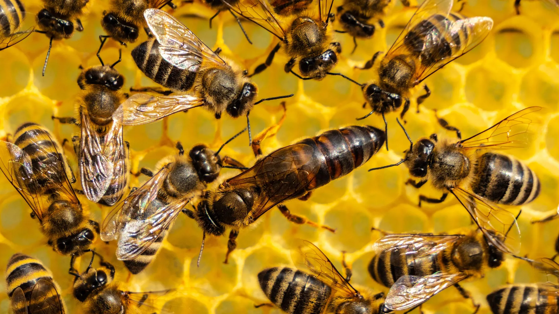
Researchers have discovered that future queens are raised inside specially designed nursery chambers built by young worker bees. These chambers provide unique wax, warmer conditions, and dedicated care that help guide a larva’s development into a healthy queen.

The findings, published in the journal Nature, show that the structures known as queen cells, sometimes called “royal cribs,” are much more than protective containers. They are carefully constructed environments that play a critical role in queen development. The research team also identified a previously unknown group of young worker bees called “queen cell builders” that appear specially suited for creating and maintaining these chambers.

“The old idea was relatively simple: take an egg, move it into a queen cell, feed it royal jelly, and you get a queen,” said Boris Baer, entomologist and director of the Center for Integrative Bee Research (CIBER) at the University of California, Riverside, whose laboratory contributed to the work. “What we found is that there’s an entire machinery behind this process. It’s much more sophisticated than we imagined.”

Queen Bees Need More Than Royal Jelly

Honeybee queens and worker bees start life in nearly the same way, emerging from almost identical eggs. Despite those similar beginnings, queens grow larger, develop more quickly, and can live much longer than workers. They also serve as the colony’s only egg-laying female, producing the next generation of bees.

For decades, researchers viewed royal jelly, a nutrient-rich substance fed to young larvae by worker bees, as the primary driver of this dramatic transformation.

The new study indicates that nutrition alone cannot explain what happens.

Using a combination of thermal imaging, behavioral monitoring, materials science techniques, and chemical analysis, the scientists examined the environments where queens are raised. They found major differences between queen cells and the familiar hexagonal chambers used to rear worker bees.

The Special Role of Queen Cells

Queen cells have a distinctive peanut-like shape and are built from wax that differs physically and chemically from ordinary hive wax. The material is less dense, more flexible, and better at retaining heat and moisture, creating favorable conditions for developing queens.

Researchers also found differences in the wax’s fatty acids and chemical signals, suggesting that queen cells provide a unique developmental setting.

To determine whether these chambers truly influence development, the team raised queen larvae in cells made either from queen wax or standard worker wax. Even when both groups received the same food, larvae raised in worker wax were more likely to die and ultimately developed into smaller queens.

The results suggest that the surrounding environment is just as important as diet in shaping a future queen.

Meet the Queen Cell Builders

The study also uncovered the worker bees responsible for creating and maintaining these royal nurseries.

Known as queen cell builders, these bees are generally younger than many other workers in the hive. While caring for developing queens, they maintain higher body temperatures and undergo physiological changes that appear linked to their specialized role.

The added warmth may help explain why queens develop so quickly. A queen bee reaches maturity in about 16 days, while worker bees require roughly 21 days. That faster development can be crucial when a colony urgently needs a new queen.

Rather than simply reusing existing wax, queen cell builders actively collect, modify, and enrich materials used in royal chambers. Their bodies also activate different biological pathways associated with wax production, effectively altering how they function while performing this task.

To see how these materials were gathered, researchers added trace amounts of graphite to ordinary honeycomb. Over time, darkened wax appeared inside queen cells, showing that workers were selectively collecting and transforming materials from elsewhere in the hive for use in queen development.

A Royal Court Inside the Hive

According to Baer, the process resembles something far more organized than a typical insect nursery.

The evidence points to a highly coordinated effort by the colony to produce its next ruler.

“You can think of it as something like Buckingham Palace,” he said. “There is a dedicated group of bees focused entirely on raising the queen, and if they don’t get it right, the colony cannot reproduce.”

The researchers observed the same pattern in both Asian and European honeybee species, suggesting this strategy evolved long ago and may be widespread among honeybees.

The project brought together experts in behavior, physiology, chemistry, materials science, and genomics. It was led by former UCR postdoctoral researchers Yu Fang and Yahya Al Naggar.

“In its collaborative nature, this project reflects the broader CIBER philosophy of bringing different disciplines together to tackle complex biological questions,” Baer said.

What the Discovery Means Beyond Bees

The findings could have implications that extend beyond honeybees. They suggest that development may be shaped not only by genetics and nutrition, but also by the physical and social environments organisms experience.

For years, queen bees appeared to offer one of biology’s simplest examples of development: special food creates a special insect. This research paints a much richer picture. A queen does not emerge from royal jelly alone. Instead, an entire colony works together to create the conditions needed for her success.

“This work highlights how much sophistication exists inside insect societies,” Baer said. “Honeybee colonies are not simply collections of individuals. They function as integrated biological systems capable of engineering their own environments.”

Share Button

Scientists open a million-year-old time capsule hidden beneath New Zealand

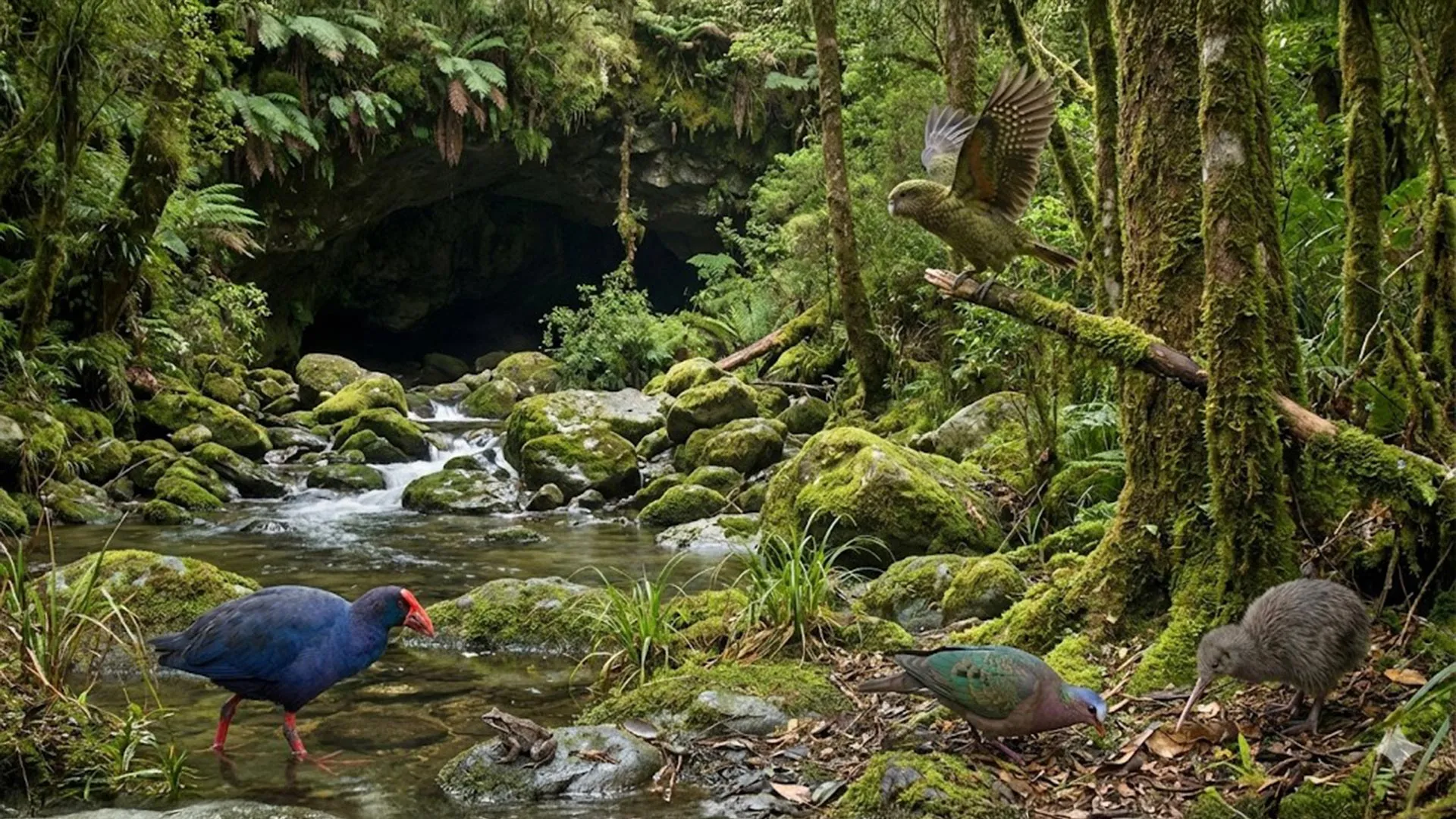
A remarkable fossil discovery inside a cave near Waitomo on New Zealand’s North Island is giving scientists an unprecedented look at a long vanished ecosystem. Researchers from Australia and New Zealand have uncovered the remains of ancient birds and frogs that lived around 1 million years ago, including a previously unknown relative of the iconic kākāpō.

The find marks the first time scientists have recovered a large collection of terrestrial vertebrate fossils from this period in New Zealand’s history. Preserved within the cave were fossils belonging to 12 bird species and four frog species, offering a rare snapshot of a world that existed hundreds of thousands of years before humans reached the islands.

The research, published in Alcheringa: An Australasian Journal of Palaeontology, suggests that New Zealand’s wildlife was already undergoing dramatic changes long before human settlement. Powerful volcanic eruptions and rapid climate shifts repeatedly reshaped habitats, driving extinctions and opening opportunities for new species to evolve.

Ancient Birds Lost to Time

Lead author Associate Professor Trevor Worthy of Flinders University says the fossils reveal a bird community unlike anything seen in New Zealand today.

“This is a newly recognized avifauna for New Zealand, one that was replaced by the one humans encountered a million years later,” says Associate Professor Worthy, from the College of Science and Engineering at Flinders University.

“This remarkable find suggests our ancient forests were once home to a diverse group of birds that did not survive the next million years.”

In biology, the term “avifauna” refers to the collection of bird species living in a particular place and time. The fossils indicate that the birds inhabiting New Zealand a million years ago were substantially different from those present when people eventually arrived.

The study involved paleontologists from Flinders University and Canterbury Museum, as well as volcanologists Joel Baker of the University of Auckland and Simon Barker of Victoria University of Wellington.

According to the researchers, approximately 33-50% of species disappeared during the million years before humans reached Aotearoa New Zealand.

Volcanoes and Climate Change Reshaped Ecosystems

Scientists believe these losses were largely caused by natural environmental upheaval.

“These extinctions were driven by relatively rapid climate shifts and cataclysmic volcanic eruptions,” says co-author Dr. Paul Scofield, Senior Curator of Natural History at Canterbury Museum.

The discovery helps fill one of the largest gaps in New Zealand’s fossil record.

“From our excavations at St Bathans in Central Otago over many years, we have a snapshot of life in Aotearoa between 20 and 16 million years ago. These new findings cast light on the 15 million year period from then to 1 million years ago, which is largely absent from New Zealand’s fossil record,” says Dr. Scofield.

“This wasn’t a missing chapter in New Zealand’s ancient history, it was a missing volume.”

Fossils are often compared to pages in Earth’s history book. In this case, researchers say they have uncovered an entire section of that story that was previously unknown.

A Possible Flying Ancestor of the Kākāpō

One of the most exciting discoveries is a newly identified parrot species called Strigops insulaborealis. It is an ancient relative of the kākāpō, one of New Zealand’s most famous birds.

Today, the kākāpō is the world’s only flightless parrot. It is also one of the heaviest parrots and is known for its unusual nighttime lifestyle. However, the newly discovered ancestor may have been very different.

Analysis of the fossilized bones suggests it had weaker legs than modern kākāpō. Because today’s birds rely heavily on their strong legs and climbing ability, researchers think the ancient species may have spent less time climbing and possibly retained the ability to fly.

Additional research will be needed to determine whether it truly could take to the air.

The cave also contained fossils from an extinct ancestor of the takahē, another distinctive New Zealand bird. Researchers also identified an extinct pigeon species closely related to Australia’s bronzewing pigeons.

“The shifting forest and shrubland habitats forced a reset of the bird populations,” adds Dr. Scofield.

“We believe this was a major driver for the evolutionary diversification of birds and other fauna in the North Island.”

Volcanic Ash Helps Date the Fossils

One reason the discovery is so important is that scientists can determine its age with unusual precision.

The fossils were trapped between two layers of volcanic ash preserved inside the cave. One ash layer came from an eruption about 1.55 million years ago. The second was produced by a massive eruption approximately 1 million years ago.

This natural geological sandwich provides clear age limits for the fossils.

Researchers say the younger eruption likely covered much of the North Island in meters of ash. While rain and erosion eventually removed much of that material, some remained protected inside caves.

The older ash layer also reveals something else remarkable. It shows that the fossil site is the oldest known cave on New Zealand’s North Island.

Rewriting New Zealand’s Natural History

Associate Professor Worthy says the fossils provide a crucial benchmark for understanding how New Zealand’s wildlife evolved.

The fossils “provide a critical, missing baseline for New Zealand’s natural history.”

For many years, scientists focused primarily on the ecological changes that occurred after humans arrived in New Zealand roughly 750 years ago. The new evidence shows that powerful natural forces had already been transforming the islands’ wildlife for hundreds of thousands of years.

“For decades, the extinction of New Zealand’s birds was viewed primarily through the lens of human arrival 750 years ago. This study proves that natural forces like super-volcanoes and dramatic climate shifts were already sculpting the unique identity of our wildlife over a million years ago.”

Share Button