Health officials warn little over half of 54-year-olds completed free at-home test kits.


Health officials warn little over half of 54-year-olds completed free at-home test kits.
Identical twins Nancy and Margo benefitted from the procedure while in the womb as part of a world-first medical trial.

Smartphones, electric vehicles, and countless portable electronics all depend on batteries. As demand for better energy storage grows, improvements in battery capacity, lifespan, and safety will play a major role in the future of electrification. One of the most promising technologies is the solid-state battery, which could allow smartphones to operate for several days on a single charge and give electric vehicles driving ranges up to three times greater than many current models.
Unlike conventional lithium-ion batteries, which use a liquid electrolyte between two solid electrodes, solid-state batteries replace the liquid with a solid electrolyte. This design offers several potential advantages, including higher energy density, improved safety, and longer battery life. But one stubborn problem has slowed commercial adoption. During charging, tiny tree-like structures called dendrites can grow from the lithium anode, pierce the solid electrolyte, and create internal short circuits.
Now, an interdisciplinary team at the Max Planck Institute for Sustainable Materials (MPI-SusMat) has identified exactly how these dendrites trigger fractures that ultimately lead to battery failure. Their findings were published in the journal Nature.
How Dendrites Crack Solid-State Batteries
Exactly how soft lithium dendrites manage to break through a hard ceramic electrolyte has long puzzled researchers.
“Although the electrodes and the forming dendrites consist of lithium metal, which is soft like a gummy bear, the dendrites are able to penetrate the ceramic electrolyte and lead to a short circuit,” says Dr. Yuwei Zhang, first author of the new publication and head of the group “Chemo-Mechanics of Battery Materials” at MPI-SusMat. “How can soft dendrites fracture the stiff solid ceramic? There are two hypotheses: either internal stress is built up inside the dendrites and induces mechanical fracture of the solid electrolyte. Or, electrons leak along the grain boundaries of the solid electrolyte promoting the formation of lithium nuclei that interconnect later.”
To determine which explanation was correct, the researchers used an advanced combination of sample preparation and materials characterization techniques. Every step was performed under vacuum and at cryogenic temperatures to eliminate interference from oxygen, water, or even the microscopes’ electron beams.
The team examined both the internal stress and the plastic deformation of lithium dendrites trapped inside cracks. Their analysis found no buildup of lithium ahead of the dendrite tip, ruling out one proposed mechanism.
“The soft lithium metal is able to penetrate the stiff ceramic electrolyte, like a continuous waterjet that penetrates a rock. We calculated that hydrostatic stress in the dendrite leads to brittle fracture of the solid electrolyte in the end,” says Zhang.
The researchers also confirmed their conclusions using phase field simulations and electron backscatter diffraction measurements.
New Strategies to Prevent Battery Failure
With a better understanding of how dendrites fracture solid electrolytes, the team is now investigating ways to stop or delay the process.
Potential solutions include making the solid electrolyte tougher so it resists cracking for longer, introducing microscopic voids that redirect dendrite growth and steer cracks away from vulnerable areas, or adding protective coatings to lithium electrodes to reduce dendrite formation in the first place.
The researchers say their work demonstrates the importance of understanding how materials behave at the microscopic level. Those insights could help transform solid-state batteries from a promising concept into a practical technology for future smartphones, electric vehicles, and other electronic devices.
The BBC join a crew based in Exeter as the UK sizzles in one of its longest-running heatwaves.

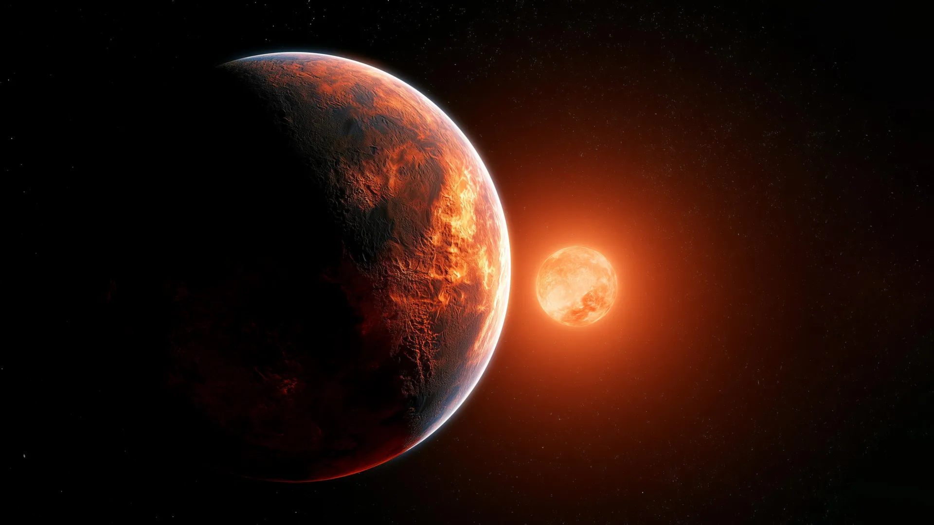
Since physicist Freeman Dyson first proposed the idea in 1960, the hypothetical “Dyson sphere” has become one of the most intriguing concepts in the search for extraterrestrial intelligence. Rather than a single solid shell, scientists now envision a Dyson “swarm” made up of countless orbiting structures that capture nearly all of a star’s energy.
While the concept has long been discussed in theory, an important question remains: if one actually existed, what would astronomers see? A new study by Amirnezam Amiri of the University of Arkansas, currently available as a pre-print on arXiv and scheduled for publication in Universe, explores exactly how these enormous structures might appear through modern telescopes. The research also identifies the types of stars most likely to host them.
Red Dwarfs and White Dwarfs Are Prime Targets
One of the strongest candidates is the red dwarf. These small, cool stars are the most common type in the Milky Way and consume their nuclear fuel so slowly that they can survive for trillions of years, far longer than the universe has existed so far.
Their relatively small size also makes them attractive from an engineering perspective. According to the study, a Dyson swarm could orbit a red dwarf at a distance of roughly 0.05 to 0.3 AU, requiring far less construction material than one built around a larger star like the Sun.
White dwarfs may be even more appealing. These dense stellar remnants are the leftover cores of Sun like stars that have exhausted their fuel and collapsed to only about 1% of their original size.
Because they are so compact, a Dyson swarm could orbit just a few million kilometers above the star’s surface, dramatically reducing the scale of the structure needed. White dwarfs also release energy at a steady rate for billions of years, making them reliable long term power sources.
How a Dyson Sphere Would Change a Star’s Appearance
Astronomers classify stars using the Hertzsprung-Russell (H-R) diagram, which plots stellar temperature against luminosity. A Dyson sphere would dramatically alter where a star appears on that chart.
Instead of allowing visible light to escape, the structure would absorb virtually all of the star’s radiation. Since energy cannot simply disappear, the same amount of energy would have to be emitted back into space, but as heat in the infrared portion of the spectrum. In effect, the megastructure would absorb starlight, use that energy for whatever purpose its builders intended, and then radiate the excess as infrared heat.
Although the star’s total energy output would remain unchanged, its apparent temperature would be much lower. Because H-R diagrams use bolometric luminosity (i.e. the luminosity over all of the spectra), the object would remain at the same luminosity but shift dramatically toward the cooler side of the diagram.
A Unique Infrared Signature
That temperature shift is one of the study’s most striking predictions. A typical red dwarf has a surface temperature of about 3000K. A surrounding Dyson sphere, however, could have an effective temperature as low as 50K, around two orders of magnitude colder.
No known natural stars occupy that region of the H-R diagram. Any object found there would immediately become an intriguing candidate for further investigation.
Another possible clue would be the absence of dust. Ordinary stars often display silicate emission associated with dusty disks. A Dyson swarm, by contrast, would consist of radiator panels rather than dust, giving it an unusually “clean” spectrum.
Looking for Strange Light Curves
The study also emphasizes that a true solid Dyson sphere is almost certainly impossible to build. Modern calculations indicate that even around relatively small stars, the amount of material required would be unrealistic.
Instead, an advanced civilization would likely construct a swarm of many independent solar collectors, leaving gaps between them or varying their density throughout the structure. As those components orbited the star, they could produce highly unusual, non natural variations in brightness that would stand out from the behavior of ordinary stars.
James Webb and the Hunt for Alien Megastructures
The James Webb Space Telescope is especially well suited to search for these hypothetical structures because it specializes in infrared observations. Older missions such as WISE are also contributing to the effort.
In May 2024, researchers from Project Hephaistos reported seven promising Dyson sphere candidates, all associated with red dwarfs, after examining a catalog of roughly 5 million stars. One candidate was later ruled out because a perfectly aligned supermassive black hole in the background explained the unusual signal.
That still leaves five candidates deserving closer study. While none have been confirmed as alien megastructures, Amiri’s work provides astronomers with another set of observational clues that could help distinguish genuine technosignatures from natural cosmic phenomena. If Dyson swarms do exist somewhere in the Milky Way, future infrared observations may finally reveal where they have been hiding.
People are being consulted about plans to stop vape companies using of enticing flavour descriptions that “attract” children into experimenting.
Identical twins Nancy and Margo benefitted from the procedure while in the womb as part of a world-first medical trial.
As a society, we take mothers for granted and raising children is a thankless job, says actor Kalki Koechlin.
LHS 3844b is an exoplanet just slightly larger than Earth that orbits the red dwarf star LHS 3884, located 48.5 light years from our solar system. Unlike Earth, it is tidally locked, meaning it rotates once on its axis in exactly the same amount of time it takes to orbit its star. As a result, one hemisphere experiences constant, blistering daylight while the other remains in permanent darkness so cold it approaches absolute zero (zero Kelvin).
At first glance, such an extreme environment seems completely inhospitable. Daytime temperatures can reach roughly 1,000 to 2,000 Kelvin, while the night side is so cold that particle motion effectively stops. Yet new research suggests these worlds may not be as hostile to life as they appear.
“Just looking at the extreme temperatures on the day and night sides — like 1,000-2,000 Kelvin on the day side and absolute zero on the night side — might lead one to conclude these exoplanets are too harsh for life. But,” says Daisuke Noto, a postdoctoral researcher in Hugo Ulloa’s Penn GEFLOW Lab at the University of Pennsylvania, “life might find a way.”
In a study published in Nature Communications, Noto and collaborators from the Japan Agency for Marine-Earth Science and Technology and Hokkaido University found that “such exoplanets may be more tolerant of sustaining life as ‘tidal locking’ can contribute to maintaining moderate thermal environments locally by distributing heat flux laterally.”
Why Tidally Locked Exoplanets Are So Common
The findings challenge a common assumption about planets that always show the same face to their stars. According to Noto, worlds with permanent day and night are actually much more common than planets like Earth, which experience a regular day and night cycle.
“Many celestial bodies like moons and planets that are very close to their parent stars are what we call tidally locked,” he explains. “Meaning, as they spin around on their axes and orbit around their parents, those rates/frequencies match, leading to the phenomena like us only seeing one side of our moon.”
This constant orientation creates a dramatic temperature contrast across the planet. Instead of focusing on surface conditions alone, the researchers wanted to understand what happens deep inside the planet, specifically within the mantle, the thick rocky layer between the crust and the core.
Recreating an Alien Planet in the Lab
Rather than relying only on computer simulations, the team built a physical laboratory model to mimic the interior of a tidally locked planet.
“Building an actual exoplanet in the lab wasn’t in the budget,” Noto jokes.
Instead, the researchers used a tabletop rectangular tank filled with viscous glycerol and tiny thermochromic liquid crystals that change color as temperatures shift. Similar experimental systems have long been used to study how heat moves through slow moving materials, making them useful stand ins for the rocky interiors of planets.
Unlike weather or ocean currents, which are strongly influenced by Earth’s rotation and gravity, convection inside a rocky mantle is driven mainly by differences in temperature and density. To reproduce those conditions, the team installed four thermostats around the tank to heat and cool different regions, creating temperature gradients similar to those expected between the permanently illuminated side, the permanently dark side, the surface, and the deep interior of a tidally locked exoplanet.
A Planetary Heat Engine
The experiments revealed a remarkably stable pattern. Hot material consistently rose beneath the day side, flowed across the upper region, cooled as it reached the night side, then sank before returning through the lower mantle. The result was one continuous circulation loop that behaved like a steady planetary heartbeat.
“It’s not chaotic like Earth’s mantle,” Noto says. “It’s slow and steady. Predictable. Kind of boring — but in a good way.”
The researchers also observed occasional mushroom shaped plumes rising from the heated base of the tank. Unlike volcanic hotspots on Earth, such as those beneath Hawaii or Iceland, these plumes remained fixed in one location rather than drifting over time.
Measurements of heat transport, known as Nusselt numbers, were comparable to those seen for Earth’s mantle. That finding suggests some tidally locked exoplanets could maintain localized geothermal environments that provide conditions favorable for life, particularly in more temperate mid latitudes.
What This Could Mean for Alien Life
The steady circulation pattern may influence more than just surface temperatures. Noto believes it could also affect the movement of a planet’s liquid core, potentially generating magnetic fields that differ from Earth’s familiar dipole field.
“That’s something we couldn’t test in this experiment,” he says, “but it’s an exciting direction for future work.”
Looking Beyond Other Worlds
Noto and Ulloa are continuing to develop similar laboratory models to investigate a wide range of geophysical processes. Earlier research from the Penn GEFLOW Lab explored how heat and mass move through confined spaces, providing new insight into the role of fluids in hydrothermal systems.
“We are planning on further extending the experimental methods to delve deeper into different systems on our planet in different contexts, the possibilities are, quite literally, out of this world,” says Noto.
Daisuke Noto is a postdoctoral researcher in the School of Arts & Sciences at the University of Pennsylvania.
Hugo Ulloa is an assistant professor in the Department of Earth and Environmental Science in Penn Arts & Sciences.
Other authors include Takehiro Miyagoshi and Takatoshi Yanagisawa of the Japan Agency for Marine-Earth Science and Technology; and Tomomi Terada and Yuji Tasaka of Hokkaido University.

Trees do not necessarily keep growing for as long as they keep photosynthesizing, according to a new study published in Science Advances. Researchers found that oak trees continue absorbing carbon dioxide well after their annual growth has ended, suggesting forests may store less carbon in wood than many climate models currently predict.
The discovery challenges a long standing assumption that higher rates of photosynthesis naturally lead to greater tree growth. If trees continue taking in carbon without turning much of it into new wood, less carbon may remain locked away over the long term.
Trees Keep Capturing Carbon After Growth Stops
Forests play a major role in slowing climate change because trees remove carbon dioxide (CO2) from the atmosphere and store much of it in their trunks, branches, and roots. Scientists have generally expected that rising atmospheric CO2 levels would boost photosynthesis, leading to faster growth and increased long term carbon storage.
The new findings suggest the relationship is more complicated. While trees may continue absorbing additional carbon, much of it does not necessarily become new wood. Instead, that carbon may be used to produce leaves, fuel short lived metabolic processes, or serve other functions, reducing the amount of carbon stored in forests compared with previous expectations.
The results could have important implications for climate forecasting.
“Right now, most models assume that if you have photosynthesis, you have growth. We find that’s not the case,” says lead author Mukund Palat Rao, an ecoclimatologist at Lamont-Doherty Earth Observatory, which is part of the Columbia Climate School. “Just because there is more photosynthesis might not necessarily mean more tree growth in the future.”
Why Photosynthesis and Growth Are Different
During photosynthesis, plants use sunlight to convert CO2 and water into sugars while releasing oxygen back into the atmosphere. The captured carbon remains inside the plant, but it is not all used to build wood.
Some of that carbon becomes woody tissue in the trunk, branches, and roots, where it can remain stored for decades, centuries, or even millennia. The rest supports the production of leaves and fruit, is temporarily stored as starch, or is converted into compounds released into the soil to nourish microbial communities, improve nutrient uptake, and help defend the tree against disease.
Because wood stores carbon for such long periods, understanding how much of the carbon captured through photosynthesis ultimately becomes woody biomass is critical for estimating how forests help slow climate change.
“Understanding how photosynthesis and growth are linked is very important from the perspective of understanding how forests will store carbon over long time scales,” says Rao.
Tracking Trees Across the United States
Scientists had previously suspected that carbon uptake and tree growth were not always synchronized, but there had been too few detailed observations to fully understand why.
To investigate, Rao and his colleagues combined several sources of data. They analyzed satellite imagery capable of detecting photosynthesis at 137 oak forest sites across the eastern United States and California. They also used instruments that measured CO2 levels in tree canopies every hour and sensors attached to tree trunks that tracked tiny changes in trunk size throughout the day. (Trees tend to expand at night as roots take up water, then shrink slightly in daytime as they transpire water, with the long-term trajectory adding up to growth.) The team also incorporated tree ring records and temperature data spanning 1950 through the present.
Together, these datasets provided daily measurements of photosynthesis, carbon uptake, and tree growth.
Trees Stop Growing Months Before Photosynthesis Ends
The researchers found a clear separation between growth and photosynthesis.
At eastern U.S. sites, oak trees typically grew from May through July but continued photosynthesizing into October. About 36 percent of their annual carbon assimilation occurred after growth had already stopped in late summer.
California oaks showed a different seasonal schedule but the same overall pattern. Growth generally occurred between December and April, then slowed during mid summer and ended by August even though photosynthesis continued. Roughly 26 percent of the trees’ yearly carbon uptake happened after growth had ceased.
According to Rao, the explanation is straightforward. Tree growth depends on internal water pressure, and that pressure drops quickly during hot, dry conditions.
“The moment you have dry and hot conditions, growth activity stops pretty instantly while photosynthesis seems to continue at a slightly decreased rate,” says Rao.
What Happens to the Extra Carbon?
Some of the carbon captured after growth ends is saved to help fuel growth when the next growing season begins. The remainder is used to produce new roots and leaves or is oxidized to keep living cells functioning through the winter.
Researchers still do not know exactly how much of that carbon eventually becomes long term woody biomass versus how much returns to the atmosphere over shorter time periods. However, the findings suggest that projections of forests growing larger and storing substantially more carbon in a warmer, CO2 rich world may need to be reconsidered.
The team also found that the disconnect between photosynthesis and growth became even stronger during years when local weather swung between unusually wet and unusually dry conditions. Because climate change is expected to increase this kind of variability in many regions, the pattern could become more common in the future.
Rao and his colleagues are now investigating whether similar patterns occur in other tree species, forest ecosystems, and climates. He expects the degree of separation between photosynthesis and growth will vary across different forests, but says many questions remain unanswered.
“I don’t really have answers yet,” he says. “There are many questions still left to address.”