Generative AI analyzes medical data faster than human research teams

In an early real world test of artificial intelligence in health research, scientists at UC San Francisco and Wayne State University discovered that generative AI could process enormous medical datasets far faster than traditional computer science teams — and in some cases produce even stronger results. Human experts had spent months carefully analyzing the same information.

To compare performance directly, researchers assigned identical tasks to different groups. Some teams relied entirely on human expertise, while others used scientists working with AI tools. The challenge was to predict preterm birth using data from more than 1,000 pregnant women.

Even a junior research pair made up of a UCSF master’s student, Reuben Sarwal, and a high school student, Victor Tarca, successfully developed prediction models with AI support. The system generated functioning computer code in minutes — something that would normally take experienced programmers several hours or even days.

The advantage came from AI’s ability to write analytical code based on short but highly specific prompts. Not every system performed well. Only 4 of the 8 AI chatbots produced usable code. Still, those that succeeded did not require large teams of specialists to guide them.

Because of this speed, the junior researchers were able to complete their experiments, verify their findings, and submit their results to a journal within a few months.

“These AI tools could relieve one of the biggest bottlenecks in data science: building our analysis pipelines,” said Marina Sirota, PhD, a professor of Pediatrics who is the interim director of the Bakar Computational Health Sciences Institute (BCHSI) at UCSF and the principal investigator of the March of Dimes Prematurity Research Center at UCSF. “The speed-up couldn’t come sooner for patients who need help now.”

Sirota is co-senior author of the study, published in Cell Reports Medicine on Feb. 17.

Why Preterm Birth Research Matters

Speeding up data analysis could improve diagnostic tools for preterm birth — the leading cause of newborn death and a major contributor to long term motor and cognitive challenges in children. In the United States, roughly 1,000 babies are born prematurely each day.

Researchers still do not fully understand what causes preterm birth. To investigate possible risk factors, Sirota’s team compiled microbiome data from about 1,200 pregnant women whose outcomes were tracked across nine separate studies.

“This kind of work is only possible with open data sharing, pooling the experiences of many women and the expertise of many researchers,” said Tomiko T. Oskotsky MD, co-director of the March of Dimes Preterm Birth Data Repository, associate professor in UCSF BCHSI, and co-author of the paper.

However, analyzing such a vast and complex dataset proved challenging. To tackle this, the researchers turned to a global crowdsourcing competition called DREAM (Dialogue on Reverse Engineering Assessment and Methods).

Sirota co-led one of three DREAM pregnancy challenges, focusing specifically on vaginal microbiome data. More than 100 teams worldwide participated, developing machine learning models designed to detect patterns linked to preterm birth. Most groups completed their work within the three month competition window. Yet it took nearly two years to consolidate the findings and publish them.

Testing AI on Pregnancy and Microbiome Data

Curious whether generative AI could shorten that timeline, Sirota’s group partnered with researchers led by Adi L. Tarca, PhD, co-senior author and professor in the Center for Molecular Medicine and Genetics at Wayne State University in Detroit, MI. Tarca had led the other two DREAM challenges, which focused on improving methods for estimating pregnancy stage.

Together, the researchers instructed eight AI systems to independently generate algorithms using the same datasets from the three DREAM challenges, without direct human coding.

The AI chatbots received carefully written natural language instructions. Much like ChatGPT, the systems were guided through detailed prompts designed to steer them toward analyzing the health data in ways comparable to the original DREAM participants.

Their objectives mirrored the earlier challenges. The AI systems analyzed vaginal microbiome data to identify signs of preterm birth and examined blood or placental samples to estimate gestational age. Pregnancy dating is almost always an estimate, yet it determines the type of care women receive as pregnancies progress. When estimates are inaccurate, preparing for labor becomes more difficult.

Researchers then ran the AI generated code using the DREAM datasets. Only 4 of the 8 tools produced models that matched the performance of the human teams, although in some cases the AI models performed better. The entire generative AI effort — from inception to submission of a paper — took just six months.

Scientists emphasize that AI still requires careful oversight. These systems can produce misleading results, and human expertise remains essential. However, by rapidly sorting through massive health datasets, generative AI may allow researchers to spend less time troubleshooting code and more time interpreting results and asking meaningful scientific questions.

“Thanks to generative AI, researchers with a limited background in data science won’t always need to form wide collaborations or spend hours debugging code,” Tarca said. “They can focus on answering the right biomedical questions.”

Authors: UCSF authors are Reuben Sarwal; Claire Dubin; Sanchita Bhattacharya, MS; and Atul Butte, MD, PhD. Other authors are Victor Tarca (Huron High School, Ann Arbor, MI); Nikolas Kalavros and Gustavo Stolovitzky, PhD (New York University); Gaurav Bhatti (Wayne State University); and Roberto Romero, MD, D(Med)Sc (National Institute of Child Health and Human Development (NICHD)).

Funding: This work was funded by the March of Dimes Prematurity Research Center at UCSF, and by ImmPort. The data used in this study was generated in part with support from the Pregnancy Research Branch of the NICHD.

Share Button

Puberty blockers trial paused over concerns from medicines watchdog

The medicines regulator is suggesting the minimum age limit for trial should be raised to 14.

Share Button

Multi-cancer blood test missed key goal in NHS trial

The company behind the test said there were positive signs that some of the most aggressive cancers could be prevented.

Share Button

Oxford breakthrough could make lithium-ion batteries charge faster and last much longer

A team at the University of Oxford has created an advanced technique that makes it possible to clearly see a vital component inside lithium-ion battery electrodes that scientists have struggled to track. The findings, published on February 17 in Nature Communications, could make battery manufacturing more efficient and help improve both charging speed and the overall lifespan of Li-ion batteries.

The research zeroed in on polymer binders used in the negative electrodes of lithium-ion batteries (anodes). These binders act like a glue that holds the electrode materials together. Even though they account for less than 5% of the electrode’s total weight, they strongly influence mechanical strength, electrical and ionic conductivity, and how long a battery can operate through repeated charge cycles.

Because binders are present in such small amounts and lack clear visual signatures, scientists have had difficulty determining exactly where they are located within the electrode. This has limited efforts to fine tune battery performance, since the way binders are distributed directly affects conductivity, structural stability, and long term durability.

Patent Pending Staining Technique Reveals Hidden Structure

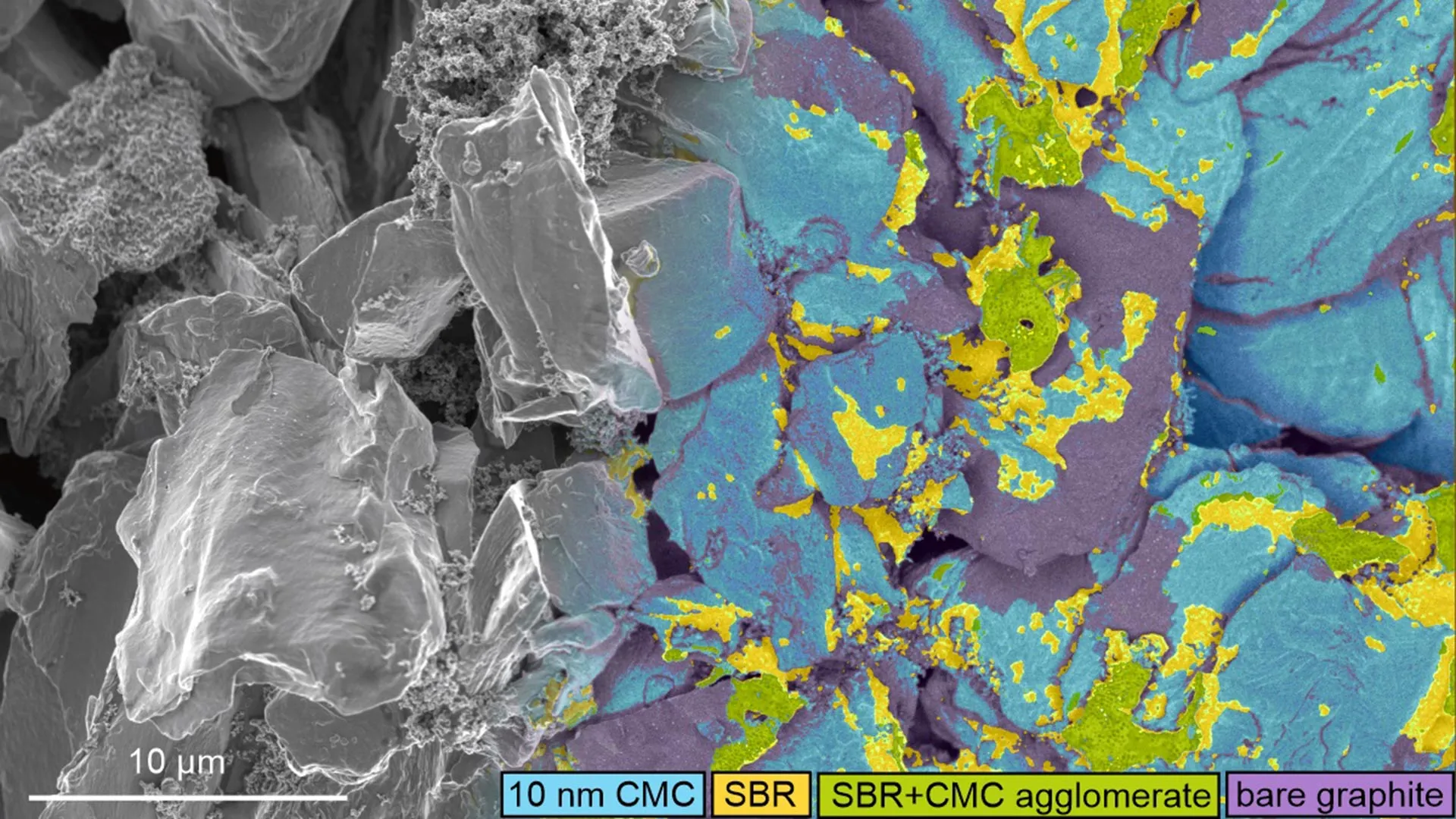
To overcome this obstacle, the researchers designed a patent-pending staining approach that attaches traceable silver and bromine markers to widely used cellulose- and latex-based binders in graphite- and silicon-based anodes. Once labeled, the binders can be detected because they emit characteristic X-rays (measured with energy-dispersive X-ray spectroscopy) or reflect high-energy electrons from the sample surface (measured with energy-selective backscattered electron imaging).

When viewed under an electron microscope, these signals provide detailed maps of where specific elements are located and what the electrode surface looks like. This allows scientists to analyze binder distribution with far greater precision than before.

Lead author Dr. Stanislaw Zankowski (Department of Materials, University of Oxford) said: “This staining technique opens up an entirely new toolbox for understanding how modern binders behave during electrode manufacturing. For the first time, we can accurately see the distribution of these binders not only generally (i.e., their thickness throughout the electrode), but also locally, as nanoscale binder layers and clusters, and correlate them with anode performance.”

The method works with standard graphite electrodes as well as advanced materials such as silicon or SiOx, making it relevant for both current lithium-ion batteries and next-generation designs.

Faster Charging and Longer Battery Life

By applying the new imaging tool, the team discovered that even subtle shifts in binder distribution can significantly change how efficiently a battery charges and how long it lasts. In testing, adjustments to slurry mixing and drying steps reduced the internal ionic resistance of experimental electrodes by as much as 40% — a major barrier to fast charging.

The researchers also captured detailed images of extremely thin layers of carboxymethyl cellulose (CMC) binder that coat graphite particles. The technique enabled clear detection of CMC layers just 10 nm thick and visualized structures spanning four orders of magnitude within a single image. The images revealed that what begins as a uniform CMC coating can break apart into uneven, patchy fragments during electrode processing, which may weaken battery performance and stability.

Co-author Professor Patrick Grant (Department of Materials, University of Oxford) said: “This multidisciplinary effort-spanning chemistry, electron microscopy, electrochemical testing, and modelling- has resulted in an innovative imaging approach that will help us to understand key surface processes that affect battery longevity and performance. This will drive forward advancements across a wide range of battery applications.”

The work was supported by the Faraday Institution’s Nextrode project and has already drawn significant interest from industry, including major battery producers and electric vehicle manufacturers.

Share Button

Air pollution linked to higher Alzheimer’s risk in 28 million older Americans

Older adults who live with higher levels of air pollution are more likely to develop Alzheimer’s disease, according to new research led by Yanling Deng of Emory University, U.S.A. The study was published February 17th in the open access journal PLOS Medicine.

Alzheimer’s is the most common type of dementia and affects roughly 57 million people worldwide. Scientists have long recognized air pollution as a risk factor for Alzheimer’s, as well as for chronic conditions such as hypertension, stroke, and depression. Because these conditions are also tied to dementia, researchers have questioned whether polluted air raises Alzheimer’s risk indirectly by contributing to those illnesses, or whether it harms the brain more directly. Another possibility was that existing health problems could make the brain more sensitive to pollution.

Large Medicare Study of 27.8 Million Older Adults

To explore these questions, the Emory team analyzed data from more than 27.8 million U.S. Medicare beneficiaries age 65 and older between 2000 and 2018. They compared levels of air pollution exposure with new cases of Alzheimer’s disease, while carefully considering the presence of other chronic health conditions.

The analysis revealed that people exposed to higher levels of air pollution had a greater likelihood of developing Alzheimer’s. The relationship was somewhat stronger among individuals who had previously suffered a stroke. In contrast, hypertension and depression did not meaningfully increase the pollution related risk.

Direct Brain Effects of Fine Particulate Pollution

Taken together, the results indicate that air pollution may raise Alzheimer’s risk mainly through direct effects on the brain rather than by triggering other chronic diseases. At the same time, a history of stroke appears to increase vulnerability, suggesting that certain individuals face compounded risks.

The findings also point to cleaner air as a potential strategy for lowering dementia rates and protecting cognitive health in aging populations.

The authors add, “In this large national study of older adults, we found that long-term exposure to fine particulate air pollution was associated with a higher risk of Alzheimer’s disease, largely through direct effects on the brain rather than through common chronic conditions such as hypertension, stroke, or depression.”

“Our findings suggest that individuals with a history of stroke may be particularly vulnerable to the harmful effects of air pollution on brain health, highlighting an important intersection between environmental and vascular risk factors.”

This work was supported by the National Institutes of Health (R01 AG074357 to KS and R01 ES034175 to YL).

Share Button

Scientists uncover oxygen-loving ancestor of all complex life

Scientists widely agree that complex life emerged after two very different microbes formed a close partnership. That merger eventually gave rise to plants, animals, and fungi, collectively known as eukaryotes. Yet one key question has lingered for years. How did these two organisms meet if one required oxygen to survive while the other was believed to thrive only in oxygen-free environments?

Researchers at The University of Texas at Austin now report evidence that may resolve that puzzle. Writing in the journal Nature, the team focused on a group of microbes called Asgard archaea, which are considered close relatives of the ancestors of complex life. Although most known Asgards live in deep-sea or other oxygen-poor environments, the new study shows that some members of this group can tolerate or even use oxygen. The discovery strengthens the long-standing theory that complex life evolved as predicted, likely in an environment where oxygen was present.

“Most Asgards alive today have been found in environments without oxygen,” explained Brett Baker an associate professor of marine science and integrative biology at UT. “But it turns out that the ones most closely related to eukaryotes live in places with oxygen, such as shallow coastal sediments and floating in the water column, and they have a lot of metabolic pathways that use oxygen. That suggests that our eukaryotic ancestor likely had these processes, too.”

The Great Oxidation Event and Early Eukaryotes

Baker’s team studies the genomes of Asgard archaea to identify new branches of the group and better understand how these microbes generate energy. Their latest findings align with what geologists and paleontologists have reconstructed about Earth’s early atmosphere.

More than 1.7 billion years ago, oxygen levels in the atmosphere were extremely low. Then oxygen concentrations rose sharply during what scientists call the Great Oxidation Event, eventually approaching levels similar to those today. Within a few hundred thousand years of this dramatic increase, the earliest known microfossils of eukaryotes appear in the fossil record. This close timing suggests that oxygen may have played a crucial role in the emergence of complex life.

“The fact that some of the Asgards, which are our ancestors, were able to use oxygen fits in with this very well,” Baker said. “Oxygen appeared in the environment, and Asgards adapted to that. They found an energetic advantage to using oxygen, and then they evolved into eukaryotes.”

Symbiosis and the Birth of Mitochondria

The prevailing model holds that eukaryotes arose when an Asgard archaeon formed a symbiotic relationship with an alphaproteobacterium. Over time, the two organisms became integrated into a single cell. The alphaproteobacterium eventually evolved into the mitochondria, the structure inside eukaryotic cells that produces energy.

In this study, researchers significantly expanded the known genetic diversity of Asgard archaea. They identified specific groups, including Heimdallarchaeia, that are especially closely related to eukaryotes but are relatively uncommon today.

“These Asgard archaea are often missed by low-coverage sequencing,” said co-author Kathryn Appler, a postdoctoral researcher at the Institut Pasteur in Paris, France. “The massive sequencing effort and layering of sequence and structural methods enabled us to see patterns that were not visible prior to this genomic expansion.”

Massive Genome Sequencing Effort

The work began with Appler’s Ph.D. research at The University of Texas Marine Science Institute in 2019, when she extracted DNA from marine sediments. The UT team and collaborators ultimately assembled more than 13,000 new microbial genomes. The project combined samples from multiple marine expeditions and required analyzing roughly 15 terabytes of environmental DNA.

From this extensive dataset, the researchers recovered hundreds of new Asgard genomes, nearly doubling the known genomic diversity of the group. By comparing genetic similarities and differences, they built an expanded Asgard archaea tree of life. The newly identified genomes also revealed previously unknown protein groups, doubling the number of recognized enzymatic classes within these microbes.

AI Analysis of Oxygen Metabolism Proteins

The team then examined Heimdallarchaeia more closely, comparing their proteins to those found in eukaryotes that are involved in energy production and oxygen metabolism. To do this, they used an artificial intelligence system called AlphaFold2 to predict the three-dimensional shapes of the proteins. Because a protein’s structure determines how it functions, this analysis provided important clues.

The results showed that several Heimdallarchaeia proteins closely resemble those used by eukaryotic cells for oxygen-based, energy-efficient metabolism. This structural similarity offers additional support for the idea that the ancestors of complex life were already adapted to using oxygen.

Other contributors to the study included former UT researchers Xianzhe Gong (currently at Shandong University in China), Pedro Leão (now at Radboud University in the Netherlands), Marguerite Langwig (now at the University of Wisconsin-Madison) and Valerie De Anda (currently at the University of Vienna). James Lingford and Chris Greening at Monash University in Australia, along with Kassiani Panagiotou and Thijs Ettema at Wageningen University in the Netherlands, also participated in the research.

Funding was provided in part by the Gordon and Betty Moore and Simons Foundations, the National Natural Science Foundation of China and the National Health and Medical Research Council of Australia.

Share Button

Sepsis warning after woman’s quadruple amputation

Manjit Sangha, from Penn, near Wolverhampton, says her life drastically change in one weekend.

Share Button

Cats may hold clues for human cancer treatment

The household cat could hold the key to understanding certain types of cancer, such as breast cancer.

Share Button

Single vaccine could protect against all coughs, colds and flus, researchers say

A Stanford University team have tested their nasal spray vaccine in animals but still need to do human clinical trials.

Share Button

Scientists discover the body’s hidden “off switch” for inflammation

Researchers at University College London have identified a biological process that helps the body shut down inflammation once it is no longer needed. The discovery could pave the way for new treatments for chronic diseases that affect millions of people around the world.

Inflammation is an essential defense mechanism that protects us from infection and injury. However, if it continues unchecked, it can contribute to serious conditions including arthritis, heart disease, and diabetes. Until now, scientists did not clearly understand how the body transitions from an active immune attack to a healing phase.

Fat Derived Molecules That Calm the Immune System

The study, published in Nature Communications, found that small fat-based molecules known as epoxy-oxylipins act as natural regulators of the immune response. These molecules help prevent the buildup of specific immune cells called intermediate monocytes*, which are associated with chronic inflammation — linked to tissue damage, illness and disease progression.

To explore this process, researchers conducted a carefully controlled experiment in healthy volunteers. Participants received a small injection of UV-killed E. coli bacteria in the forearm. This triggered a temporary inflammatory response — pain, redness, heat and swelling — similar to what occurs after infection or injury.

Volunteers were divided into two groups: prophylactic arm and therapeutic arm.

At different stages, participants were given a drug called GSK2256294. This medication blocks an enzyme known as soluble epoxide hydrolase (sEH), which normally breaks down epoxy-oxylipins.

In the prophylactic arm, 24 volunteers participated — 12 received the drug and 12 received placebo (placebo). They were treated two hours before inflammation began to test whether boosting epoxy-oxylipins early could prevent harmful immune changes.

In the therapeutic arm, another 24 volunteers — 12 treated and 12 untreated (placebo) — received the drug four hours after inflammation had started. This approach reflected how treatment would occur in real world settings once symptoms appear.

Boosting Protective Lipids Reduced Harmful Immune Cells

In both groups, blocking sEH increased levels of epoxy-oxylipins. Participants who received the drug experienced faster pain resolution and had significantly lower levels of intermediate monocytes in both blood and tissue — the immune cells linked to chronic inflammation and disease. Notably, the medication did not meaningfully change visible symptoms such as redness or swelling.

Further investigation showed that one specific epoxy-oxylipin, 12,13-EpOME, works by suppressing a protein signaling pathway known as p38 MAPK, which drives monocyte transformation. Laboratory experiments and additional testing in volunteers who received a p38 blocking drug confirmed this mechanism.

First author Dr. Olivia Bracken (UCL Department of Ageing, Rheumatology and Regenerative Medicine) said: “Our findings reveal a natural pathway that limits harmful immune cell expansion and helps calm inflammation more quickly.

“Targeting this mechanism could lead to safer treatments that restore immune balance without suppressing overall immunity.

“With chronic inflammation ranked as a major global health threat, this discovery opens a promising avenue for new therapies.”

Corresponding author Professor Derek Gilroy (UCL Division of Medicine) said: “This is the first study to map epoxy-oxylipin activity in humans during inflammation.

“By boosting these protective fat molecules, we could design safer treatments for diseases driven by chronic inflammation.”

He added: “This was an entirely human-based study with direct relevance to autoimmune diseases, as we used a drug already suitable for human use — one that could be repurposed to treat flares in chronic inflammatory conditions, an area currently bereft of effective therapies.”

Scientists chose to investigate epoxy-oxylipins because previous animal research suggested they can reduce inflammation and pain. However, their role in human biology had not been clearly defined. Unlike well known inflammatory signals such as histamine and cytokines, epoxy-oxylipins belong to a lesser studied pathway that researchers believed might help naturally quiet the immune system.

Next Steps for Arthritis and Heart Disease Research

The findings open the possibility of clinical trials to test sEH inhibitors as treatments for diseases such as rheumatoid arthritis and cardiovascular disease.

Dr. Bracken said: “For instance, rheumatoid arthritis is a condition in which the immune system attacks the cells that line your joints. sEH inhibitors could be trialled alongside existing medications to investigate if they can help prevent or slow down joint damage incurred by the condition.”

Dr. Caroline Aylott, Head of Research Delivery at Arthritis UK, said: “The pain of arthritis can affect how we move, think, sleep and feel, along with our ability to spend time with loved ones. Pain is incredibly complex and is affected by many different factors. We also know that everybody’s pain is different.

“That is why it is important that we invest in research like this, that helps us understand what causes and influences people’s experience of pain.

“We are excited to see the results of this study which has found a natural process that could stop inflammation and pain. We hope in the future that this will lead to new pain management options for people with arthritis.”

The study was funded by Arthritis UK and included researchers from UCL, King’s College London, University of Oxford, Queen Mary University of London, and the National Institute of Environmental Health Sciences, USA.

Notes

*Intermediate monocytes are white blood cells that help fight infection and repair tissue. In short bursts, they help coordinate the immune response and support recovery, but if they persist or grow in excess, they keep the immune system switched on, leading to chronic inflammation.

Share Button