Scientists unearth a 112-million-year-old time capsule filled with ancient insects

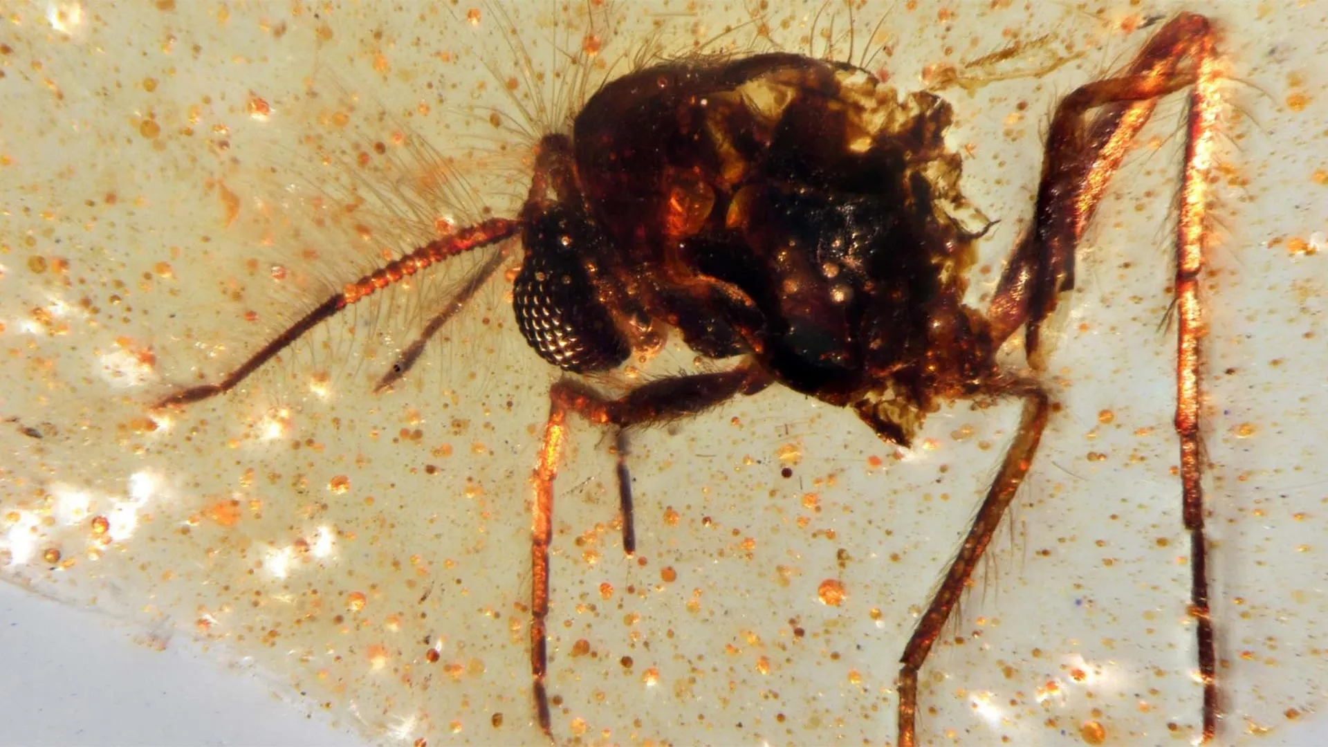
Scientists have uncovered the first South American amber deposits containing preserved insects in a quarry in Ecuador, according to a study published in Communications Earth & Environment. The discovery captures a vivid picture of a 112-million-year-old forest that once thrived on the ancient supercontinent Gondwana and opens new doors for exploring a long-overlooked prehistoric ecosystem.

Amber (fossilized tree resin) has been found in samples dating back as far as 320 million years, but it became far more common between 120 million and 70 million years ago, during the Cretaceous era (143.1 million to 66 million years ago). These ancient resins sometimes contain “bio-inclusions” — trapped remains of plants or animals — that offer rare, detailed glimpses of life forms such as insects and flowers that are not usually preserved as fossils. Until now, nearly all known major amber deposits were located in the Northern Hemisphere, leaving scientists with limited insight into what Southern Hemisphere ecosystems looked like during the time when the continents were beginning to separate from Gondwana.

To investigate, Xavier Delclòs and his research team examined amber and surrounding rock samples collected from the Genoveva quarry in Ecuador. The amber, dated to roughly 112 million years ago, belongs to the Hollín Formation, a sedimentary layer that stretches across Ecuador’s Oriente Basin. The team identified two distinct kinds of amber: one formed underground near the roots of resin-producing plants, and another that developed in the open air. Among 60 samples of the latter, the researchers found 21 bio-inclusions representing five insect orders, including Diptera (flies), Coleoptera (beetles), and Hymenoptera (a group that includes ants and wasps), as well as a fragment of spider web. In addition, the rock surrounding the amber contained numerous plant fossils such as spores, pollen, and other botanical traces.

According to the researchers, the characteristics of the fossils indicate that the amber originated in a warm, humid forest filled with dense vegetation and resin-producing trees in southern Gondwana. They emphasize that this rare find provides a crucial new resource for understanding life and biodiversity during this key period in Earth’s history.

Share Button

Report shows ‘stark’ gender gap in HIV prevention

A meeting will discuss how to tackle what organisers say is a growing inequality in HIV prevention.

Share Button

MIT’s “stealth” immune cells could change cancer treatment forever

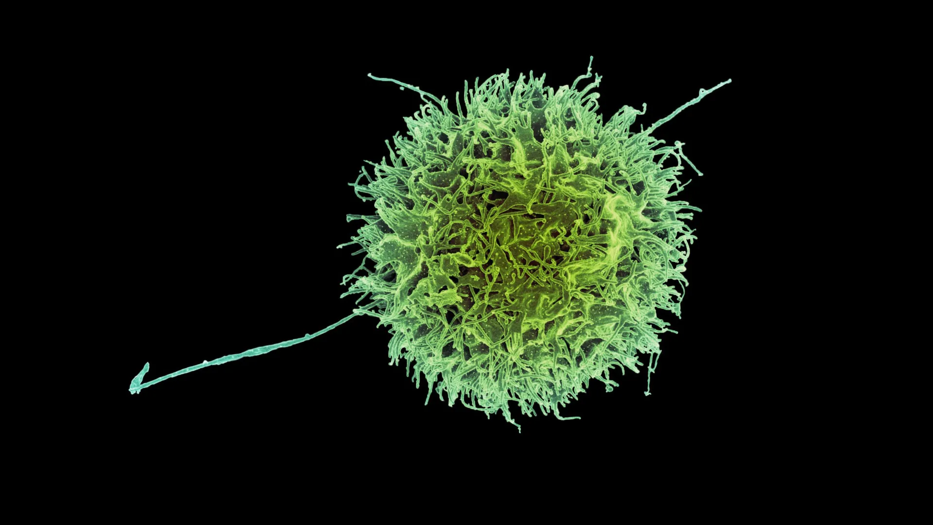
Scientists have created a new and more advanced form of immune-based cancer therapy using engineered cells known as CAR-NK (natural killer) cells. Like CAR-T cells, these modified immune cells can be programmed to recognize and attack cancer, but they rely on a different type of immune cell that naturally targets abnormal or infected cells.

A team from MIT and Harvard Medical School has now developed a more effective way to engineer CAR-NK cells that dramatically reduces the chance of the body’s immune system rejecting them. Immune rejection has been one of the biggest limitations of cell-based therapies, often weakening their effectiveness.

This innovation could also make it possible to produce “off-the-shelf” CAR-NK treatments that are available immediately after diagnosis, rather than waiting weeks for custom-engineered cells. Traditional CAR-NK and CAR-T manufacturing methods typically require several weeks to complete before patients can begin treatment.

“This enables us to do one-step engineering of CAR-NK cells that can avoid rejection by host T cells and other immune cells. And, they kill cancer cells better and they’re safer,” says Jianzhu Chen, an MIT professor of biology, a member of the Koch Institute for Integrative Cancer Research, and one of the senior authors of the study. 

In tests using mice with humanized immune systems, the newly engineered cells successfully destroyed most cancer cells while avoiding attack from the host’s own immune defenses.

Rizwan Romee, an associate professor of medicine at Harvard Medical School and Dana-Farber Cancer Institute, is also a senior author of the paper, which was published in Nature Communications. The study’s lead author is Fuguo Liu, a postdoctoral researcher at the Koch Institute and a research fellow at Dana-Farber.

Evading the immune system

Natural killer (NK) cells are a vital part of the body’s built-in immune defense, responsible for identifying and destroying cancerous and virus-infected cells. They eliminate these threats through a process called degranulation, which releases a protein known as perforin. This protein punctures the membrane of target cells, leading to their death.

To produce CAR-NK cells for treatment, doctors typically collect a blood sample from the patient. NK cells are then extracted and engineered to express a specialized protein called a chimeric antigen receptor (CAR), which is designed to target specific markers found on cancer cells.

Once modified, the cells must multiply in the lab for several weeks before there are enough to be infused back into the patient. The same general process is used for CAR-T cell therapies, some of which have already been approved to treat blood cancers like lymphoma and leukemia. CAR-NK therapies, however, are still being tested in clinical trials.

Because growing enough personalized CAR-NK cells takes time and the patient’s cells may not always be healthy enough for reliable use, scientists have been exploring an alternative: creating NK cells from healthy donors. These donor-derived cells could be mass-produced and stored for rapid use. The challenge, however, is that the recipient’s immune system often identifies donor cells as foreign and destroys them before they can attack the cancer.

In their latest research, the MIT team aimed to solve this problem by helping NK cells “hide” from immune detection. Their experiments showed that removing surface proteins known as HLA class 1 molecules allowed NK cells to avoid attack from T cells in the host’s immune system. These proteins normally act as identity markers that tell the immune system whether a cell belongs to the body.

To make use of this insight, the researchers added a sequence of siRNA (short interfering RNA) that silences the genes responsible for producing HLA class 1 proteins. Along with this genetic tweak, they introduced the CAR gene itself and another gene that encodes either PD-L1 or single-chain HLA-E (SCE), both of which help strengthen the NK cells’ cancer-fighting abilities.

All of these genetic components were combined into a single DNA construct, which allowed the team to efficiently convert donor NK cells into immune-evasive CAR-NK cells. Using this method, they engineered cells that target CD-19, a protein commonly found on malignant B cells in lymphoma patients.

NK cells unleashed

The researchers tested these CAR-NK cells in mice with a human-like immune system. These mice were also injected with lymphoma cells.

Mice that received CAR-NK cells with the new construct maintained the NK cell population for at least three weeks, and the NK cells were able to nearly eliminate cancer in those mice. In mice that received either NK cells with no genetic modifications or NK cells with only the CAR gene, the host immune cells attacked the donor NK cells. In these mice, the NK cells died out within two weeks, and the cancer spread unchecked.

The researchers also found that these engineered CAR-NK cells were much less likely to induce cytokine release syndrome — a common side effect of immunotherapy treatments, which can cause life-threatening complications.

Because of CAR-NK cells’ potentially better safety profile, Chen anticipates that they could eventually be used in place of CAR-T cells. For any CAR-NK cells that are now in development to target lymphoma or other types of cancer, it should be possible to adapt them by adding the construct developed in this study, he says.

The researchers now hope to run a clinical trial of this approach, working with colleagues at Dana-Farber. They are also working with a local biotech company to test CAR-NK cells to treat lupus, an autoimmune disorder that causes the immune system to attack healthy tissues and organs.

The research was funded, in part, by Skyline Therapeutics, the Koch Institute Frontier Research Program through the Kathy and Curt Marble Cancer Research Fund and the Elisa Rah Memorial Fund, the Claudia Adams Barr Foundation, and the Koch Institute Support (core) Grant from the National Cancer Institute.

Share Button

12,000-year-old rock art found in Arabia reveals a lost civilization

Recent research has revealed the crucial role of early human groups who settled in northern Arabia soon after the intensely dry period known as the Last Glacial Maximum (LGM). These communities followed the return of seasonal water sources and left behind an extraordinary record of their presence through monumental rock art.

An international team of archaeologists, coordinated by the Heritage Commission of the Saudi Ministry of Culture, uncovered the findings through the Green Arabia Project. The team included experts from the Max Planck Institute of Geoanthropology, KAUST (King Abdullah University of Science and Technology), University College London, Griffith University, and several other institutions.

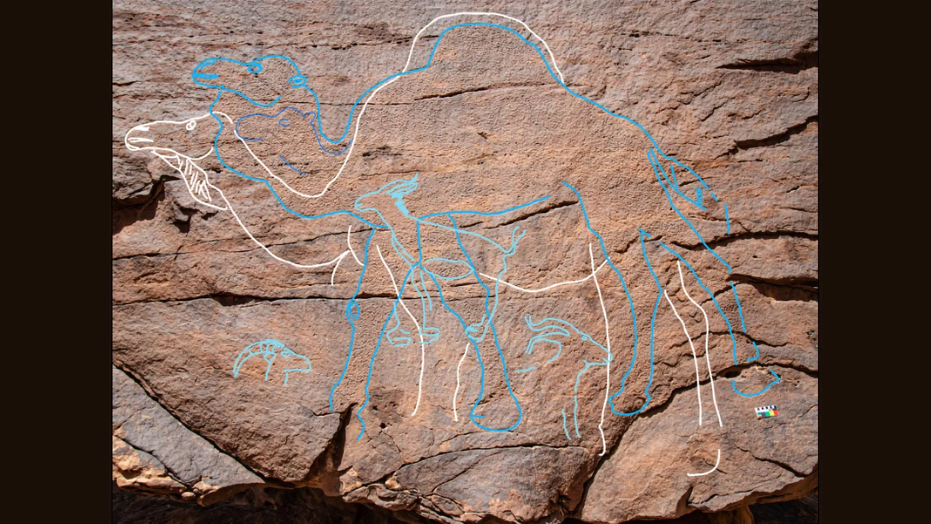
Researchers documented over 60 rock art panels featuring 176 individual engravings across three previously unexplored locations: Jebel Arnaan, Jebel Mleiha, and Jebel Misma, situated along the southern edge of the Nefud Desert in northern Saudi Arabia.

The engravings, which depict animals such as camels, ibex, equids, gazelles, and aurochs, include 130 highly detailed and life-sized figures, some reaching up to 3 meters long and more than 2 meters tall.

Dating to between 12,800 and 11,400 years ago, the engravings correspond to a time when temporary lakes and rivers reappeared after centuries of extreme aridity.

Sediment analysis confirmed the existence of these ancient water sources, which would have provided essential support for human groups venturing into the desert interior and allowed them to survive in this challenging environment.

“These large engravings are not just rock art – they were probably statements of presence, access, and cultural identity,” said lead author, Dr. Maria Guagnin from Max Planck Institute of Geoanthropology.

Dr. Ceri Shipton, co-lead author from the Institute of Archaeology, University College London, said: “The rock art marks water sources and movement routes, possibly signifying territorial rights and intergenerational memory.”

Unlike previously known sites where engravings were hidden in crevices, the Jebel Mleiha and the Jebel Arnaan panels were etched onto towering cliff faces, some up to 39 meters high, in visually commanding locations.

One panel would have required ancient artists to climb and work precariously on narrow ledges, underscoring the sheer effort and significance of the imagery.

Artefacts including Levantine-style El Khiam and Helwan stone points, green pigment, and dentalium beads suggest long-distance connections to Pre-Pottery Neolithic (PPN) populations in the Levant region.

However, the scale, content, and placement of the Arabian engravings set them apart.

“This unique form of symbolic expression belongs to a distinct cultural identity adapted to life in a challenging, arid environment,” said Dr. Faisal Al-Jibreen, from the Heritage Commmission, Saudi Ministry of Culture.

“The project’s interdisciplinary approach has begun to fill a critical gap in the archaeological record of northern Arabia between the LGM and the Holocene, shedding light on the resilience and innovation of early desert communities,” said Michael Petraglia, lead of the Green Arabia project.

The study ‘Monumental rock art illustrates that humans thrived in the Arabian Desert during the Pleistocene-Holocene transition’ has been published in Nature Communications.

Share Button

For the first time, scientists pinpoint brain cells linked to depression

Scientists at McGill University and the Douglas Institute have discovered that two distinct types of brain cells show changes in people experiencing depression.

Published in Nature Genetics, the research provides new clues that could guide the creation of treatments focused on these specific cells. It also enhances scientific understanding of depression, a condition that affects more than 264 million people globally and is one of the leading causes of disability.

“This is the first time we’ve been able to identify what specific brain cell types are affected in depression by mapping gene activity together with mechanisms that regulate the DNA code,” said senior author Dr. Gustavo Turecki, a professor at McGill, clinician-scientist at the Douglas Institute and Canada Research Chair in Major Depressive Disorder and Suicide. “It gives us a much clearer picture of where disruptions are happening, and which cells are involved.”

Rare brain bank enables breakthrough

The team conducted their work using post-mortem brain tissue from the Douglas-Bell Canada Brain Bank, one of the few collections worldwide that includes donations from people with psychiatric conditions.

Through advanced single-cell genomic analysis, the researchers examined RNA and DNA from thousands of individual brain cells to determine which ones behaved differently in people with depression and which DNA sequences might explain these variations. The study analyzed tissue from 59 individuals who had depression and 41 who did not.

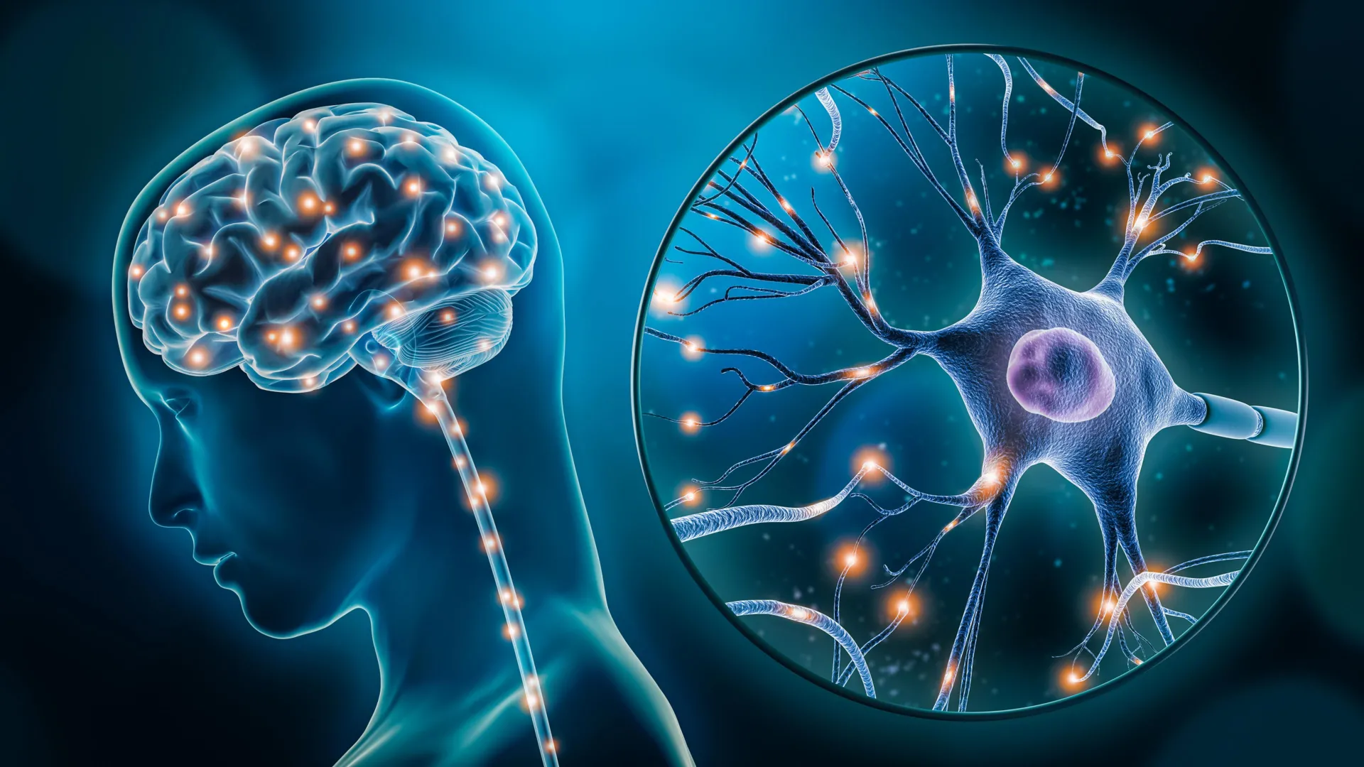
They discovered that gene activity was altered in two types of brain cells: a class of excitatory neurons responsible for mood and stress regulation, and a subtype of microglia, the immune cells that manage inflammation in the brain. In both cell types, many genes were expressed differently in people with depression, pointing to possible disruptions in vital neural systems.

By identifying the specific cells affected, the research deepens understanding of the biological foundation of depression and helps dispel outdated views of the condition.

“This research reinforces what neuroscience has been telling us for years,” Turecki said. “Depression isn’t just emotional, it reflects real, measurable changes in the brain.”

Looking ahead, the scientists intend to explore how these cellular changes influence brain function and whether targeting them could lead to more effective treatments.

About the study

“Single-nucleus chromatin accessibility profiling identifies cell types and functional variants contributing to major depression” by Anjali Chawla and Gustavo Turecki et al., was published in Nature Genetics.

The study was funded by Canadian Institutes of Health Research, Brain Canada Foundation, Fonds de recherche du Québec – Santé and Healthy Brains, Healthy Lives initiative at McGill University.

Share Button

A facelift at 28? Why young people are turning to plastic surgery

Gone are the days when facelifts were for the ageing wealthy. Now younger people are going under the knife.

Share Button

William visibly moved as widow tells of husband’s suicide

Prince William’s foundation is giving £1m to set up a suicide prevention network.

Share Button

‘Marginalised and menopausal’ women given spotlight

Women from ethnic minority communities typically experience perimenopause symptoms earlier and for longer.

Share Button

How 20 minutes of nature can boost your health

Spending just 20 minutes in nature can lower blood pressure, heart rate and stress levels.

Share Button

A hidden “backup heater” that helps burn fat and boost metabolism

Scientists at Washington University School of Medicine in St. Louis have uncovered a new way that brown fat, a type of fat that burns energy, can boost the body’s metabolism. This process allows cells to consume more fuel and generate heat, improving overall metabolic health. Conducted in mice, the research points to new possibilities for using brown fat to address metabolic conditions such as insulin resistance and obesity.

The findings were published Sept. 17 in Nature.

Brown fat is unique because it turns energy (calories) from food into heat. Unlike white fat, which stores energy, or muscle, which uses it immediately, brown fat helps keep the body warm in cold environments. Exposure to cold can increase the amount of brown fat, and scientists have long suggested that activating it could support weight loss by increasing calorie burning.

“The pathway we’ve identified could provide opportunities to target the energy expenditure side of the weight loss equation, potentially making it easier for the body to burn more energy by helping brown fat produce more heat,” said senior author Irfan Lodhi, PhD, a professor of medicine in the Division of Endocrinology, Metabolism & Lipid Research at WashU Medicine. “Boosting this kind of metabolic process could support weight loss or weight control in a way that is perhaps easier to maintain over time than traditional dieting and exercise. It’s a process that basically wastes energy — increasing resting energy expenditure — but that’s a good thing if you’re trying to lose weight.”

A back-up heater in brown fat

Until now, scientists understood brown fat’s heat production mainly through mitochondria, the energy centers of cells. Mitochondria in brown fat can shift from making fuel to generating heat through a molecule called uncoupling protein 1. However, studies have shown that mice lacking this protein can still burn energy and produce heat, suggesting another system at work.

The new research identifies peroxisomes, small structures within cells that process fats, as an alternative heat source in brown fat. When exposed to cold, these peroxisomes multiply. This effect was even stronger in mice whose mitochondria lacked uncoupling protein 1, suggesting that peroxisomes can step in when mitochondria lose their ability to produce heat.

Lodhi and his team discovered that peroxisomes burn fuel and release heat through a process involving a protein called acyl-CoA oxidase 2 (ACOX2). Mice that lacked ACOX2 in their brown fat were less able to tolerate cold, showed lower body temperatures after exposure to cold, and had poorer insulin sensitivity. When fed high-fat diets, they also gained more weight than typical mice.

In contrast, mice genetically engineered to make unusually high amounts of ACOX2 in brown fat showed increased heat production, better cold tolerance and improved insulin sensitivity and weight control when fed the same high-fat diet.

Using a fluorescent heat sensor they developed, the researchers found that when ACOX2 metabolized certain fatty acids, brown fat cells got hotter. They also used an infrared thermal imaging camera to show that mice lacking ACOX2 produced less heat in their brown fat.

While human bodies can manufacture these fatty acids, the molecules also are found in dairy products and human breast milk and are made by certain gut microbes. Lodhi said this raises the possibility that a dietary intervention based on these fatty acids — such as a food, probiotic or “nutraceutical” intervention — could boost this heat-production pathway and the beneficial effects it appears to have. He and his colleagues also are investigating possible drug compounds that could activate ACOX2 directly.

“While our studies are in mice, there is evidence to suggest this pathway is relevant in people,” Lodhi said. “Prior studies have found that individuals with higher levels of these fatty acids tend to have lower body mass indices. But since correlation is not causation, our long-term goal is to test whether dietary or other therapeutic interventions that increase levels of these fatty acids or that increase activity of ACOX2 could be helpful in dialing up this heat production pathway in peroxisomes and helping people lose weight and improve their metabolic health.”

This work was supported by the National Institutes of Health (NIH), grant numbers R01DK133344, R01DK115867, R01DK132239, GM103422, T32DK007120, S10 OD032315, DK020579 and DK056341; and by the FP7 funded European Infrafrontier-I3 project. The content is solely the responsibility of the authors and does not necessarily represent the official views of the NIH.

Lodhi and Liu are named on a provisional patent application filed by Washington University related to targeting ACOX2 activation as a treatment for obesity and related metabolic diseases.

Share Button