Families affected by a rare inherited disease say they have had to “scramble” for information.


Families affected by a rare inherited disease say they have had to “scramble” for information.

Physicians rely on nuclear medicine scans, like SPECT scans, to watch the heart pump, track blood flow and detect diseases hidden deep inside the body. But today’s scanners depend on expensive detectors that are difficult to make.
Now, scientists led by Northwestern University and Soochow University in China have built the first perovskite-based detector that can capture individual gamma rays for SPECT imaging with record-breaking precision. The new tool could make common types of nuclear medicine imaging sharper, faster, cheaper and safer.
For patients, that could mean shorter scan times, clearer results and lower doses of radiation.
The study was published on Aug. 30 in the journal Nature Communications.
“Perovskites are a family of crystals best known for transforming the field of solar energy,” said Northwestern’s Mercouri Kanatzidis, the study’s senior author. “Now, they are poised to do the same for nuclear medicine. This is the first clear proof that perovskite detectors can produce the kind of sharp, reliable images that doctors need to provide the best care for their patients.”
“Our approach not only improves the performance of detectors but also could lower costs,” said co-corresponding author Yihui He, a professor at Soochow University. “That means more hospitals and clinics eventually could have access to the best imaging technologies.”
Kanatzidis is a Charles E. and Emma H. Morrison Professor of Chemistry at Northwestern’s Weinberg College of Arts and Sciences and a senior scientist at Argonne National Laboratory. Yihui He is a former postdoctoral fellow from Kanatzidis’ laboratory.
Nuclear medicine, like SPECT (single-photon emission computing tomography) imaging, works like an invisible camera. Physicians implant a tiny, safe, short-lived radiotracer in a specific part of a patient’s body. The tracer emits gamma rays, which pass outward through tissues and eventually hit a detector outside of the body. Each gamma ray is like a pixel of light. After collecting millions of these pixels, computers can construct a 3D image of working organs.
Today’s detectors, which are either made from cadmium zinc telluride (CZT) or sodium iodide (NaI), have several disadvantages. CZT detectors are incredibly expensive, sometimes reaching into the price range of hundreds of thousands to millions of dollars for a whole camera. Because CZT crystals are brittle and prone to cracking, these detectors also are difficult to manufacture. While cheaper than CZT detectors, NaI detectors are bulky and produce blurrier images — like taking a photo through a foggy window.
To overcome these issues, the scientists turned to perovskite crystals, a material that Kanatzidis has studied for more than a decade. In 2012, his group built the first solid-film solar cells made from perovskites. Then, in 2013, Kanatzidis discovered that single perovskite crystals were highly promising for detecting X-rays and gamma rays. This breakthrough, enabled by his group’s growth of high-quality single crystals, sparked a worldwide surge of research and effectively launched a new field in hard radiation detection materials.
“This work demonstrates how far we can push perovskite detectors beyond the laboratory,” Kanatzidis said. “When we first discovered in 2013 that perovskite single crystals could detect X-rays and gamma rays, we could only imagine their potential. Now, we’re showing that perovskite-based detectors can deliver the resolution and sensitivity needed for demanding applications like nuclear medicine imaging. It’s exciting to see this technology moving closer to real-world impact.”
Building on this foundation, Kanatzidis and He led the crystal growth, surface engineering and device design for the new study. By carefully growing and shaping these crystals, the researchers created a pixelated sensor — just like the pixels in a smartphone camera — that delivers record-breaking clarity and stability.
Leading the design and development of the prototype gamma-ray detector, He developed the camera’s pixelated architecture, optimized the multi-channel readout electronics and carried out the high-resolution imaging experiments that validated the device’s capabilities. He, Kanatzidis and their team demonstrated that perovskite-based detectors can achieve record energy resolutions and unprecedented single-photon imaging performance, paving the way for practical integration into next-generation nuclear medicine imaging systems.
“Designing this gamma-ray camera and demonstrating its performance has been incredibly rewarding,” He said. “By combining high-quality perovskite crystals with a carefully optimized pixelated detector and multi-channel readout system, we were able to achieve record-breaking energy resolution and imaging capabilities. This work shows the real potential of perovskite-based detectors to transform nuclear medicine imaging.”
In experiments, the detector was able to differentiate among gamma rays of different energies with the best resolution reported thus far. It also sensed extremely faint signals from a medical radiotracer (technetium-99m) commonly used in clinical practice and distinguished incredibly fine features, producing crisp images that could separate tiny radioactive sources spaced just a few millimeters apart. The detector also remained highly stable, collecting nearly all the tracer’s signal without loss or distortion. Because these new detectors are more sensitive, patients potentially could require shorter scan times or smaller doses of radiation.
Northwestern spinout company Actinia Inc. is commercializing this technology — working with partners in the medical device field to bring it out of the lab and into hospitals. Because they are easier to grow and use simpler components, perovskites offer a far less expensive alternative to CZT and NaI detectors without sacrificing quality. Perovskite-based detectors also offer a realistic pathway to imaging using a lower dose of a radiotracer than can be used with a NaI detector but at a price that ensures widespread patient access.
“Demonstrating that perovskites can deliver single-photon gamma-ray imaging is a milestone,” He said. “It shows these materials are ready to move beyond the laboratory and into technologies that directly benefit human health. From here, we see opportunities to refine the detectors further, scale up production and explore entirely new directions in medical imaging.”
“High-quality nuclear medicine shouldn’t be limited to hospitals that can afford the most expensive equipment,” Kanatzidis said. “With perovskites, we can open the door to clearer, faster, safer scans for many more patients around the world. The ultimate goal is better scans, better diagnoses and better care for patients.”
The study, “Single photon γ-ray imaging with high energy and spatial resolution perovskite semiconductor for nuclear medicine,” was supported by the Defense Threat Reduction Agency (award number HDTRA12020002), the Consortium for Interaction of Ionizing Radiation with Matter University Research Alliance, the National Key R&D Program of China (award number 2021YFF0502600), the National Natural Science Foundation of China (award number U2267211) and the Jiangsu Natural Science Foundation (award number BK20240822).
Music can evoke powerful memories and offers a sense of normality for people dealing with illness or death.

The official number of exoplanets — planets outside our solar system — tracked by NASA has reached 6,000. Confirmed planets are added to the count on a rolling basis by scientists from around the world, so no single planet is considered the 6,000th entry. The number is monitored by NASA’s Exoplanet Science Institute (NExScI), based at Caltech’s IPAC in Pasadena, California. There are more than 8,000 additional candidate planets awaiting confirmation, with NASA leading the world in searching for life in the universe.
“This milestone represents decades of cosmic exploration driven by NASA space telescopes — exploration that has completely changed the way humanity views the night sky,” said Shawn Domagal-Goldman, acting director, Astrophysics Division, NASA Headquarters in Washington. “Step by step, from discovery to characterization, NASA missions have built the foundation to answering a fundamental question: Are we alone? Now, with our upcoming Nancy Grace Roman Space Telescope and Habitable Worlds Observatory, America will lead the next giant leap — studying worlds like our own around stars like our Sun. This is American ingenuity, and a promise of discovery that unites us all.”
The milestone comes 30 years after the first exoplanet was discovered around a star similar to our Sun, in 1995. (Prior to that, a few planets had been identified around stars that had burned all their fuel and collapsed.) Although researchers think there are billions of planets in the Milky Way galaxy, finding them remains a challenge. In addition to discovering many individual planets with fascinating characteristics as the total number of known exoplanets climbs, scientists are able to see how the general planet population compares to the planets of our own solar system.
For example, while our solar system hosts an equal number of rocky and giant planets, rocky planets appear to be more common in the universe. Researchers have also found a range of planets entirely different from those in our solar system. There are Jupiter-size planets that orbit closer to their parent star than Mercury orbits the Sun; planets that orbit two stars, no stars, and dead stars; planets covered in lava; some with the density of Styrofoam; and others with clouds made of gemstones.
“Each of the different types of planets we discover gives us information about the conditions under which planets can form and, ultimately, how common planets like Earth might be, and where we should be looking for them,” said Dawn Gelino, head of NASA’s Exoplanet Exploration Program (ExEP), located at the agency’s Jet Propulsion Laboratory in Southern California. “If we want to find out if we’re alone in the universe, all of this knowledge is essential.”
Searching for other worlds
Fewer than 100 exoplanets have been directly imaged, because most planets are so faint they get lost in the light from their parent star. The other four methods of planet detection are indirect. With the transit method, for instance, astronomers look for a star to dim for a short period as an orbiting planet passes in front of it.
To account for the possibility that something other than an exoplanet is responsible for a particular signal, most exoplanet candidates must be confirmed by follow-up observations, often using an additional telescope, and that takes time. That’s why there is a long list of candidates in the NASA Exoplanet Archive (hosted by NExScI) waiting to be confirmed.
“We really need the whole community working together if we want to maximize our investments in these missions that are churning out exoplanets candidates,” said Aurora Kesseli, the deputy science lead for the NASA Exoplanet Archive at IPAC. “A big part of what we do at NExScI is build tools that help the community go out and turn candidate planets into confirmed planets.”
The rate of exoplanet discoveries has accelerated in recent years (the database reached 5,000 confirmed exoplanets just three years ago), and this trend seems likely to continue. Kesseli and her colleagues anticipate receiving thousands of additional exoplanet candidates from the ESA (European Space Agency) Gaia mission, which finds planets through a technique called astrometry, and NASA’s upcoming Nancy Grace Roman Space Telescope, which will discover thousands of new exoplanets primarily through a technique called gravitational microlensing.
Future exoplanets
At NASA, the future of exoplanet science will emphasize finding rocky planets similar to Earth and studying their atmospheres for biosignatures — any characteristic, element, molecule, substance, or feature that can be used as evidence of past or present life. NASA’s James Webb Space Telescope has already analyzed the chemistry of over 100 exoplanet atmospheres.
But studying the atmospheres of planets the size and temperature of Earth will require new technology. Specifically, scientists need better tools to block the glare of the star a planet orbits. And in the case of an Earth-like planet, the glare would be significant: The Sun is about 10 billion times brighter than Earth — which would be more than enough to drown out our home planet’s light if viewed by a distant observer.
NASA has two main initiatives to try overcoming this hurdle. The Roman telescope will carry a technology demonstration instrument called the Roman Coronagraph that will test new technologies for blocking starlight and making faint planets visible. At its peak performance, the coronagraph should be able to directly image a planet the size and temperature of Jupiter orbiting a star like our Sun, and at a similar distance from that star. With its microlensing survey and coronagraphic observations, Roman will reveal new details about the diversity of planetary systems, showing how common solar systems like our own may be across the galaxy.
Additional advances in coronagraph technology will be needed to build a coronagraph that can detect a planet like Earth. NASA is working on a concept for such a mission, currently named the Habitable Worlds Observatory.
More about ExEP, NExScI
NASA’s Exoplanet Exploration Program is responsible for implementing the agency’s plans for the discovery and understanding of planetary systems around nearby stars. It acts as a focal point for exoplanet science and technology and integrates cohesive strategies for future discoveries. The science operations and analysis center for ExEP is NExScI, based at IPAC, a science and data center for astrophysics and planetary science at Caltech. JPL is managed by Caltech for NASA.

UNSW engineers have made a significant advance in quantum computing: they created ‘quantum entangled states’ – where two separate particles become so deeply linked they no longer behave independently – using the spins of two atomic nuclei. Such states of entanglement are the key resource that gives quantum computers their edge over conventional ones.
The research was published on Sept. 18 in the journal Science, and is an important step towards building large-scale quantum computers – one of the most exciting scientific and technological challenges of the 21st century.
Lead author Dr Holly Stemp says the achievement unlocks the potential to build the future microchips needed for quantum computing using existing technology and manufacturing processes.
“We succeeded in making the cleanest, most isolated quantum objects talk to each other, at the scale at which standard silicon electronic devices are currently fabricated,” she says.
The challenge facing quantum computer engineers has been to balance two opposing needs: shielding the computing elements from external interference and noise, while still enabling them to interact to perform meaningful computations. This is why there are so many different types of hardware still in the race to be the first operating quantum computer: some are very good for performing fast operations, but suffer from noise; others are well shielded from noise, but difficult to operate and scale up.
The UNSW team has invested on a platform that – until today – could be placed in the second camp. They have used the nuclear spin of phosphorus atoms, implanted in a silicon chip, to encode quantum information.
“The spin of an atomic nucleus is the cleanest, most isolated quantum object one can find in the solid state,” says Scientia Professor Andrea Morello, UNSW School of Electrical Engineering & Telecommunications.
“Over the last 15 years, our group has pioneered all the breakthroughs that made this technology a real contender in the quantum computing race. We already demonstrated that we could hold quantum information for over 30 seconds – an eternity, in the quantum world – and perform quantum logic operations with less than 1% errors.
“We were the first in the world to achieve this in a silicon device, but it all came at a price: the same isolation that makes atomic nuclei so clean, makes it hard to connect them together in a large-scale quantum processor.”
Until now, the only way to operate multiple atomic nuclei was for them to be placed very close together inside a solid, and to be surrounded by one and the same electron.
“Most people think of an electron as the tiniest subatomic particle, but quantum physics tells us that it has the ability to ‘spread out’ in space, so that it can interact with multiple atomic nuclei,” says Dr Holly Stemp, who conducted this research at UNSW and is now a postdoctoral researcher at MIT in Boston.
“Even so, the range over which the electron can spread is quite limited. Moreover, adding more nuclei to the same electron makes it very challenging to control each nucleus individually.”
Making atomic nuclei talk through electronic ‘telephones’
“By way of metaphor one could say that, until now, nuclei were like people placed in a sound-proof room,” Dr Stemp says.
“They can talk to each other as long as they are all in the same room, and the conversations are really clear. But they can’t hear anything from the outside, and there’s only so many people who can fit inside the room. This mode of conversation doesn’t ‘scale’.
“With this breakthrough, it’s as if we gave people telephones to communicate to other rooms. All the rooms are still nice and quiet on the inside, but now we can have conversations between many more people, even if they are far away.”
The ‘telephones’ are, in fact, electrons. Mark van Blankenstein, another author on the paper, explains what’s really going on at the sub-atomic level.
“By their ability to spread out in space, two electrons can ‘touch’ each other at quite some distance. And if each electron is directly coupled to an atomic nucleus, the nuclei can communicate through that.”
So how far apart were the nuclei involved in the experiments?
“The distance between our nuclei was about 20 nanometers – one thousandth of the width of a human hair,” says Dr Stemp.
“That doesn’t sound like much, but consider this: if we scaled each nucleus to the size of a person, the distance between the nuclei would be about the same as that between Sydney and Boston!”
She adds that 20 nanometers is the scale at which modern silicon computer chips are routinely manufactured to work in personal computers and mobile phones.
“You have billions of silicon transistors in your pocket or in your bag right now, each one about 20 nanometers in size. This is our real technological breakthrough: getting our cleanest and most isolated quantum objects talking to each other at the same scale as existing electronic devices. This means we can adapt the manufacturing processes developed by the trillion-dollar semiconductor industry, to the construction of quantum computers based on the spins of atomic nuclei.”
A scalable way forward
Despite the exotic nature of the experiments, the researchers say these devices remain fundamentally compatible with the way all current computer chips are built. The phosphorus atoms were introduced in the chip by the team of Professor David Jamieson at the University of Melbourne, using an ultra-pure silicon slab supplied by Professor Kohei Itoh at Keio University in Japan.
By removing the need for the atomic nuclei to be attached to the same electron, the UNSW team has swept aside the biggest roadblock to the scale-up of silicon quantum computers based on atomic nuclei.
“Our method is remarkably robust and scalable. Here we just used two electrons, but in the future we can even add more electrons, and force them in an elongated shape, to spread out the nuclei even further,” Prof. Morello says.
“Electrons are easy to move around and to ‘massage’ into shape, which means the interactions can be switched on and off quickly and precisely. That’s exactly what is needed for a scalable quantum computer.”

When German physicist Wilhelm Röntgen discovered X-rays in the late 1800s while experimenting with cathode ray tubes, it was a breakthrough that transformed science and medicine. So much so that the basic concept remains in use today. But a team of researchers at Sandia National Laboratories believes they’ve found a better way, harnessing different metals and the colors of light they emit.
“It’s called colorized hyperspectral X-ray imaging with multi-metal targets, or CHXI MMT for short,” said project lead Edward Jimenez, an optical engineer. Jimenez has been working with materials scientist Noelle Collins and electronics engineer Courtney Sovinec to create X-rays of the future.
“With this new technology, we are essentially going from the old way, which is black and white, to a whole new colored world where we can better identify materials and defects of interest,” Collins said.
The team found they could achieve this using tiny, patterned samples of varied metals such as tungsten, molybdenum, gold, samarium and silver.
The Basics of X-ray Creation
To understand the concept, one must understand the basics of X-ray creation. Traditional X-rays are generated by bombarding a single metal target, or anode, with high-energy electrons. Those X-rays are channeled into a beam and directed at a subject or material. Denser tissues, like bone, absorb more X-rays, while less dense tissues, like muscles and organs allow more to pass through. A detector records the pattern, creating an image.
While X-ray technology has advanced over time, the basic concept remains the same, which limits resolution and clarity.
A New Type of X-Ray Image
The Sandia team set out to solve that limitation by making the X-ray focal spot smaller. The smaller the spot, the sharper the image.
They achieved this by designing an anode with metal dots patterned to be collectively smaller than the beam, effectively reducing the focal point.
But the team decided they wanted to push the limits and took the concept a step further.
“We chose different metals for each dot,” Sovinec said. “Each metal emits a particular ‘color’ of X-ray light. When combined with an energy discriminating detector, we can count individual photons, which provide density information, and measure the energy of each photon. This allows us to characterize the elements of the sample.”
The result is colorized images with what the team calls revolutionary image clarity and a better understanding of an object’s composition.
“We get a more accurate representation of the shape and definition of that object, which is going to allow us to make unprecedented measurements and unprecedented observations,” Jimenez said.
Far-reaching applications
The team sees this as a major advancement for X-ray technology with a wide range of uses, from airport security and quality control to nondestructive testing and advanced manufacturing.
They also hope its impact will improve medical diagnostics.
“With this technology, you can see even slight differences between materials,” Jimenez said. “We hope this will help better identify things like cancer and more effectively analyze tumor cells. In mammography you are trying to catch something before it grows. In breast tissue, it’s hard to identify the different dots, but with colorization you have a sharper beam and higher resolution image that increases the system’s capability to detect a microcalcification. It’s really exciting to be a part of that.”
“From here we will continue to innovate,” Collins said. “We hope to identify threats faster, diagnose diseases quicker and hopefully create a safer, healthier world.”
The team was recently awarded an R&D 100 award for their technology. They were among six winners from Sandia. Click for R&D 100 Submission video with soundbites.
As its popularity grows, research suggests creatine may also benefit short-term memory, mood and focus.
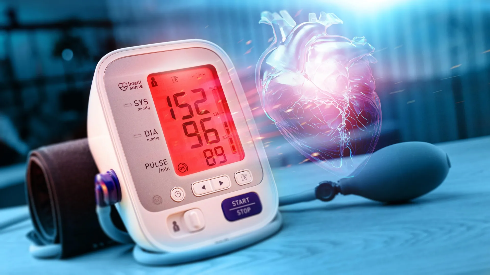
Few people with high blood pressure were using salt substitutes, even though they are a simple and effective way to lower sodium intake and manage blood pressure, according to preliminary research presented at the American Heart Association’s Hypertension Scientific Sessions 2025. The meeting is the premier scientific exchange focused on recent advances in basic and clinical research on high blood pressure and its relationship to cardiac and kidney disease, stroke, obesity and genetics.
High blood pressure occurs when the force of blood flowing through the blood vessels is consistently too high. High blood pressure can lead to other serious events such as heart attack and stroke. Using data from 2017 to 2020, 122.4 million (46.7%) adults in the U.S. had high blood pressure and it contributed to more than 130,000 deaths. Too much sodium and too little potassium in the diet are risk factors for high blood pressure.
“Overall, less than 6% of all U.S. adults use salt substitutes, even though they are inexpensive and can be an effective strategy to help people control blood pressure, especially people with difficult-to-treat high blood pressure,” said lead study author Yinying Wei, M.C.N., R.D.N., L.D., and Ph.D. candidate in the departments of applied clinical research and hypertension section, cardiology division, at UT Southwestern Medical Center in Dallas. “Health care professionals can raise awareness about the safe use of salt substitutes by having conversations with their patients who have persistent or hard-to-manage high blood pressure.”
Salt substitutes are products that replace some or all of the sodium with potassium. Potassium salt tastes similar to regular salt, except when heated it can have a bitter aftertaste. Many foods contain some sodium in their natural state, however, the largest amount of sodium comes from processed and packaged foods and meals prepared at restaurants. The American Heart Association recommends consuming no more than 2,300 mg of sodium a day, with an ideal limit of less than 1,500 mg per day for most adults, especially for those with high blood pressure. For most people, cutting back by 1,000 mg a day can improve blood pressure and heart health.
This study is the first to examine long-term trends in salt substitute use among a nationally representative sample of U.S. adults. Using data from the National Health and Nutrition Examination Survey (NHANES) from 2003 to 2020, researchers analyzed the use of products that replace salt with potassium-enriched or other alternative salts.
The investigation focused on people with high blood pressure, and an additional analysis was conducted among adults eligible to use salt substitutes, including people with normal kidney function and those not taking medications or supplements that affect blood potassium levels. Some salt substitutes contain potassium, and they can raise blood potassium to dangerous levels in people with kidney disease or those taking certain medications or potassium supplements. Excessive potassium can lead to irregular heart rhythms. People with high blood pressure who are thinking about switching from regular salt to a salt substitute should first consult with a health care professional.
The analysis found:
“Salt substitute use remained uncommon over the last two decades including among people with high blood pressure,” Wei said. “Even among individuals with treated and poorly managed or untreated high blood pressure, most continued to use regular salt.”
“This study highlights an important and easy missed opportunity to improve blood pressure in the U.S. — the use of salt substitutes,” said Amit Khera, M.D., M.Sc., FAHA, an American Heart Association volunteer expert. “The fact that use of salt substitutes remains so low and has not improved in two decades is eye-opening and reminds patients and health care professionals to discuss the use of these substitutes, particularly in visits focused on high blood pressure.” Khera, who was not involved in this study, is a professor of medicine, clinical chief of cardiology and director of preventive cardiology at UT Southwestern Medical Center in Dallas.
The study has several limitations. First, information about salt substitute use was self-reported, so there may have been underreporting or misclassification. In addition, all types of salt substitutes were included in the analysis, therefore, the analysis could not specifically separate potassium-enriched salt from other types of salt substitutes. Finally, the survey data did not capture how much salt substitute the participants used.
“Future research should explore why salt substitute-use remains low by investigating potential barriers, such as taste acceptance, cost and limited awareness among both patients and clinicians,” said Wei. “These insights may help guide more targeted interventions.”
Study details, background and design:
Research Highlights:

Researchers led by Hiroshi Ohno at the RIKEN Center for Integrative Medical Sciences (IMS) in Japan have discovered why smoking tobacco helps people suffering from ulcerative colitis, a chronic disease typified by inflammation of the large intestine. Published in the scientific journal Gut, the study shows that smoking produces metabolites that encourage bacteria from the mouth to grow in the large intestines where they trigger an immune response. These findings imply that protection against ulcerative colitis can be achieved through prebiotics like hydroquinone or probiotic therapy with bacteria like Streptococcus mitis, thus eliminating the need to smoke and all the associated risks for other diseases.
Inflammatory bowel disease comes in two main varieties, Crohn’s disease and ulcerative colitis. Although both cause chronic abdominal pain, diarrhea, fatigue and weight loss, their causes and the exact type and location of the inflammation differ. Along with these differences is a mystery that has puzzled doctors and scientists for over 40 years; smoking increases the risk of Crohn’s disease but somehow protects against ulcerative colitis. As both diseases are related to gut inflammation — which is an immune response — and gut immunity depends in part on the types of bacteria in the gut, Ohno and his team at RIKEN IMS set out to investigate whether the differential effects of smoking on these diseases can be explained by gut bacteria.
The researchers used a combination of human clinical data and experiments with mice to reach their conclusions. Among those with ulcerative colitis, they found that smokers had certain bacteria usually found in the mouth, such as Streptococcus, growing in the gut, specifically in the colonic mucosa that cover the inner lining of the intestines. This phenomenon did not occur in ex-smokers. Thus, while these bacteria normally pass all the way through the digestive system as we swallow saliva throughout the day, smoking somehow allows them to settle down in the gut mucosa.
The next question was why? The researchers also examined gut metabolites — small substances produced in the gut when food is broken down and processed by the body and gut bacteria. They found that levels of several gut metabolites were higher in smokers with ulcerative colitis than in ex-smokers with colitis. In mice, the researchers found that one of these metabolites, called hydroquinone, promoted the growth of Streptococcus in the gut mucosa. So, smoking-related metabolites like hydroquinone allow mouth bacteria like Streptococcus to flourish in the mucus layer that covers the inner lining of the intestines. But how do these bacteria help reduce inflammation? And why don’t they help in Crohn’s disease?
The researchers then went back to the oral bacteria that they had discovered was growing in the gut mucosa of smokers with ulcerative colitis, and isolated 10 strains from the saliva of smokers. When they treated mouse models of Crohn’s disease and ulcerative colitis with each of these 10 strains for five days, they found that giving the mice Streptococcusmitis had almost the same effect as smoking. Inflammation was reduced in mice with ulcerative colitis and exacerbated in mice with Crohn’s disease.
Analysis showed that S. mitis triggered the emergence of helper Th1 cells, which are an important part of the gut’s immune response to invaders. In Crohn’s disease this likely worsens the condition because the original inflammation is actually caused by these same helper Th1 cells. But in colitis, the Th1 cells fight against an initial Th2-immune response, and this ends up reducing inflammation.
As smoking poses high risks for cancer, heart disease, and many other illnesses, it is not a sustainable treatment for ulcerative colitis. “Our results indicate the relocation of bacteria from the mouth to the gut, particularly those of the Streptococcus genus, and the subsequent immune response in the gut, is the mechanism through which smoking helps protect against the disease,” says Ohno. “Logically, direct treatment with this kind of bacteria, or indirect treatment with hydroquinone, is thus likely to mimic the beneficial effects of smoking but avoid all the negative effects.”

Lund University archaeologists have revealed details of late medieval artillery from the wreck of the royal Danish-Norwegian flagship, Gribshunden. The shipwreck is the only known example of its kind from the medieval period — as both ship and weapons are nearly identical to those of the early Spanish and Portuguese explorers. The new study tells the story of how early modern maritime adventurers were equipped to start the process of dominance and colonisation across the world.
“Diving on this late medieval royal shipwreck is of course exciting. However, the greatest satisfaction is when we can actually put the pieces of the puzzle together later on; combining Martin’s castle expertise with Kay’s deep understanding of artillery,” explains Brendan Foley, the marine archeologist behind the study, who worked closely with fellow LU archaeologist Martin Hansson and medieval artillery expert Kay Douglas Smith.
Gribshunden, the flagship of the Danish-Norwegian King Hans, sank mysteriously in 1495 off the coast of Ronneby, Sweden. The wreck is internationally significant as the world’s best-preserved ship from the Age of Exploration — a proxy for the vessels of Christopher Columbus and Vasco da Gama.
Ocean-going ships like Gribshunden and the artillery they carried were critical technologies for European explorers after 1492. The voyages to America and into the Indian Ocean via the Cape of Good Hope led to European colonization around the world. Gribshunden is a rare archaeological resource, as it is the most complete example yet discovered of a late medieval carvel warship.
The ship carried 50 or more small calibre guns firing lead shot with an iron core. They were intended for anti-personnel use at close range, with tactics designed to injure or kill the enemy ships’ personnel, followed by boarding to capture the vessel. Led by Lund University Professor Nicolo Dell’Unto, the Lund University team recreated the guns from 3D models of the artifacts
A Danish ‘floating castle’
Gribshunden was built near Rotterdam between 1483-84. King Hans of Denmark and Norway had taken possession of the ship by spring 1486. The high cost of building and equipping these ships meant Gribshunden probably absorbed about 8% of the Danish national budget in 1485.
Hans utilized his flagship differently from other monarchs; he personally sailed on it frequently, using it not for exploration, but to solidify his grasp on his kingdom. It was his floating castle, enabling royal travel to Sweden and all around the Danish realm including Gotland and especially Norway. The king used this vessel in ways similar to a terrestrial royal fortification. This included several soft power functions: economic, diplomatic, social, cultural, and administrative. Underpinning all of these was the obvious hard power of the ship’s martial purpose embodied by the guns and other weapons carried aboard.
Evidence of explosion
Gribshunden served the crown for a decade before sinking while the king was en route from Copenhagen to a political summit in Sweden, where he expected to unify the entire Nordic region in a new Kalmar Union. Historical documents including eyewitness accounts relate that while Hans was ashore in Ronneby, an explosion and fire claimed the ship while it was anchored off the town.
Among the 22-lead artillery shots from Gribshunden, several are flattened on one or two sides. This may be a result of the explosion that sank the vessel. Shot stored in the hold near the gunpowder ricocheted inside the ship.
No Nordic expansion into North America
So, given the existence of these warships, why didn’t Denmark compete in expanding to the Americas? Denmark and Norway shared the long Viking and medieval Nordic history of exploration and settlement in the west, with colonies in Iceland and Greenland, and settlements in North America. Coupled with adoption of this new enabling technology, Hans might have successfully competed with the Iberian rulers in global exploration and expansion to the Americas.
However, Hans’ primary concern was consolidating rule over the Baltic region. In pursuit of that goal, Hans himself sailed on Gribshunden into the Atlantic on several royal visits, and to Kalmar on the ship’s final voyage.
One reason for Denmark’s inattention to the Americas might have been a 1493 papal bull signed by Pope Alexander VI. This granted Spain rights to the Americas, and a treaty between Spain and Portugal ceded the Indian Ocean to the latter. Prior to the Reformation, the threat of excommunication for ignoring the papal ‘Inter Caetera’ was very real.
Summary of the publication:
More about the study:
The study was written by archaeologists Brendan Foley and Martin Hansson, with English medieval artillery expert Kay Douglas Smith. The project is conducted in collaboration with Blekinge Museum, Vikingeskibsmueet, and Ronneby municipality. The research was funded by grants from the Swedish Research Council (Vetenskapsrådet), Crafoordska Stifltelsen, Huckleberry Foundation (USA), and with support from Blekinge Museum and the Lund University Department of Archaeology and Ancient History.
The Gribshunden artillery artifacts are exhibited and curated at the Blekinge Museum facility in Rosenholm, with select artifacts on temporary display in Kallvattenkuren in Ronneby, and in Museet for Søfart in Helsingør, Denmark. Plans are underway to create a dedicated Gribshunden museum in Ronneby, where the artillery and other objects from the wreck may ultimately find a permanent home.
