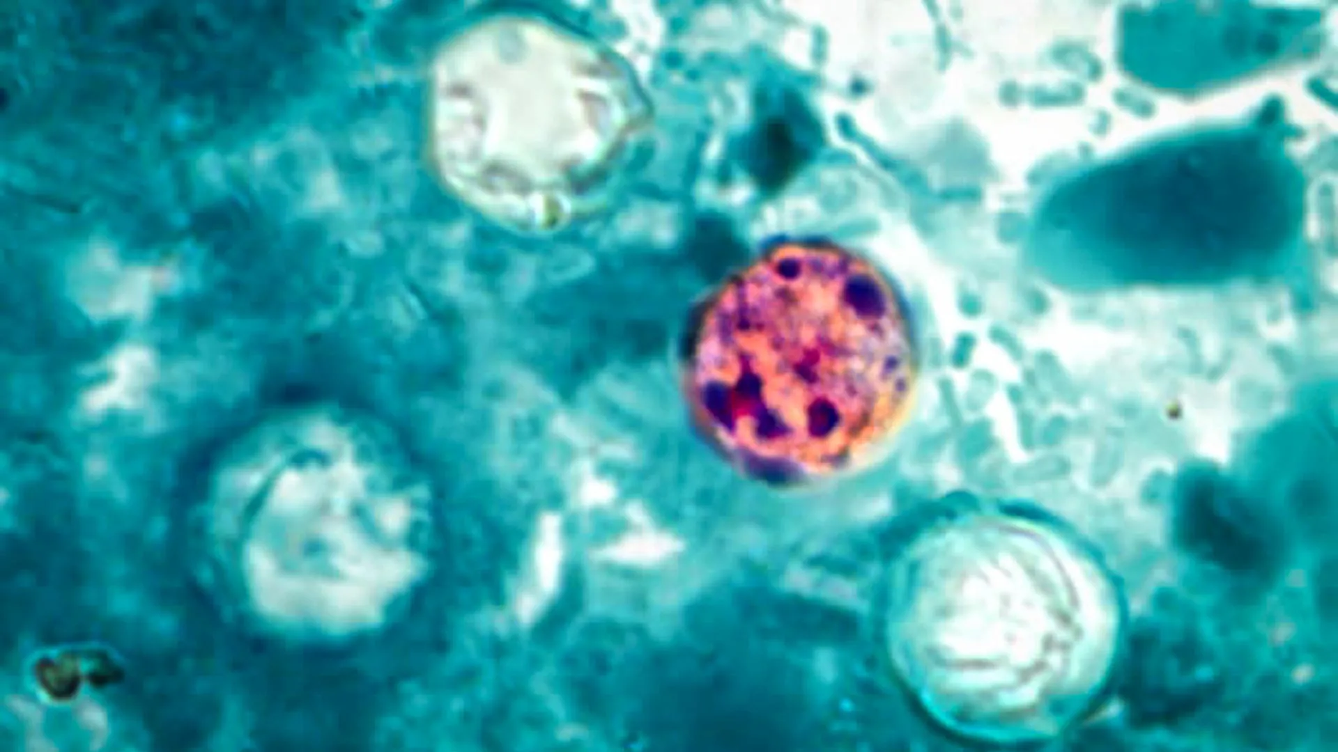
Man who received first pig kidney transplant dies

The US hospital which carried out the operation said Rick Slayman’s death was not a result of the transplant.

Share Button

Test-at-home kit for cancer patients approved for use

Patients say the device allows them to reduce the number of hospital visits involved in cancer care.

Share Button

Cellular activity hints that recycling is in our DNA

Although you may not appreciate them, or have even heard of them, throughout your body, countless microscopic machines called spliceosomes are hard at work. As you sit and read, they are faithfully and rapidly putting back together the broken information in your genes by removing sequences called “introns” so that your messenger RNAs can make the correct proteins needed by your cells.

Introns are perhaps one of our genome’s biggest mysteries. They are DNA sequences that interrupt the sensible protein-coding information in your genes, and need to be “spliced out.” The human genome has hundreds of thousands of introns, about 7 or 8 per gene, and each is removed by a specialized RNA protein complex called the “spliceosome” that cuts out all the introns and splices together the remaining coding sequences, called exons. How this system of broken genes and the spliceosome evolved in our genomes is not known.

Over his long career, Manny Ares, UC Santa Cruz distinguished professor of molecular, cellular, and developmental biology, has made it his mission to learn as much about RNA splicing as he can.

“I’m all about the spliceosome,” Ares said. “I just want to know everything the spliceosome does — even if I don’t know why it is doing it.”

In a new paper published in the journal Genes and Development, Ares reports on a surprising discovery about the spliceosome that could tell us more about the evolution of different species and the way cells have adapted to the strange problem of introns. The authors show that after the spliceosome is finished splicing the mRNA, it remains active and can engage in further reactions with the removed introns.

This discovery provides the strongest indication we have so far that spliceosomes could be able to reinsert an intron back into the genome in another location. This is an ability that spliceosomes were not previously believed to possess, but which is a common characteristic of “Group II introns,” distant cousins of the spliceosome that exist primarily in bacteria.

The spliceosome and Group II introns are believed to share a common ancestor that was responsible for spreading introns throughout the genome, but while Group II introns can splice themselves out of RNA and then directly back into DNA, the “spliceosomal introns” that are found in most higher-level organisms require the spliceosome for splicing and were not believed to be reinserted back into DNA. However, Ares’s lab’s finding indicates that the spliceosome might still be reinserting introns into the genome today. This is an intriguing possibility to consider because introns that are reintroduced into DNA add complexity to the genome, and understanding more about where these introns come from could help us to better understand how organisms continue to evolve.

Building on an interesting discovery

An organism’s genes are made of DNA, in which four bases, adenine (A), cytosine (C), guanine (G) and thymine (T) are ordered in sequences that code for biological instructions, like how to make specific proteins the body needs. Before these instructions can be read, the DNA gets copied into RNA by a process known as transcription, and then the introns in that RNA have to be removed before a ribosome can translate it into actual proteins.

The spliceosome removes introns using a two-step process that results in the intron RNA having one of its ends joined to its middle, forming a circle with a tail that looks like a cowboy’s “lariat,” or lasso. This appearance has led to them being named “lariat introns.” Recently, researchers at Brown University who were studying the locations of the joining sites in these lariats made an odd observation — some introns were actually circular instead of lariat shaped.

This observation immediately got Ares’s attention. Something seemed to be interacting with the lariat introns after they were removed from the RNA sequence to change their shape, and the spliceosome was his main suspect.

“I thought that was interesting because of this old, old idea about where introns came from,” Ares said. “There is a lot of evidence that the RNA parts of the spliceosome, the snRNAs, are closely related to Group II introns.”

Because the chemical mechanism for splicing is very similar between the spliceosomes and their distant cousins, the Group II introns, many researchers have theorized that when the process of self-splicing became too inefficient for Group II introns to reliably complete on their own, parts of these introns evolved to become the spliceosome. While Group II introns were able to insert themselves directly back into DNA, however, spliceosomal introns that required the help of spliceosomes were not thought to be inserted back into DNA.

“One of the questions that was sort of missing from this story in my mind was, is it possible that the modern spliceosome is still able to take a lariat intron and insert it somewhere in the genome?” Ares said. “Is it still capable of doing what the ancestor complex did?”

To begin to answer this question, Ares decided to investigate whether it was indeed the spliceosome that was making changes to the lariat introns to remove their tails. His lab slowed the splicing process in yeast cells, and discovered that after the spliceosome released the mRNA that it had finished splicing introns from, it hung onto intron lariats and reshaped them into true circles. The Ares lab was able to reanalyze published RNA sequencing data from human cells and found that human spliceosomes also had this ability.

“We are excited about this because while we don’t know what this circular RNA might do, the fact that the spliceosome is still active suggests it may be able to catalyze the insertion of the lariat intron back into the genome,” Ares said.

If the spliceosome is able to reinsert the intron into DNA, this would also add significant weight to the theory that spliceosomes and Group II introns shared a common ancestor long ago.

Testing a theory

Now that Ares and his lab have shown that the spliceosome has the catalytic ability to hypothetically place introns back into DNA like their ancestors did, the next step is for the researchers to create an artificial situation in which they “feed” a DNA strand to a spliceosome that is still attached to a lariat intron and see if they can actually get it to insert the intron somewhere, which would present “proof of concept” for this theory.

If the spliceosome is able to reinsert introns into the genome, it is likely to be a very infrequent event in humans, because the human spliceosomes are in incredibly high demand and therefore do not have much time to spend with removed introns. In other organisms where the spliceosome isn’t as busy, however, the reinsertion of introns may be more frequent. Ares is working closely with UCSC Biomolecular Engineering Professor Russ Corbett-Detig, who has recently led a systematic and exhaustive hunt for new introns in the available genomes of all intron-containing species that was published in the journal Proceedings of the National Academy of Sciences (PNAS) last year.

The paper in PNAS showed that intron “burst” events far back in evolutionary history likely introduced thousands of introns into a genome all at once. Ares and Corbett-Detig are now working to recreate a burst event artificially, which would give them insight into how genomes reacted when this happened.

Ares said that his cross-disciplinary partnership with Corbett-Detig has opened the doors for them to really dig into some of the biggest mysteries about introns that would probably be impossible for them to understand fully without their combined expertise.

“It is the best way to do things,” Ares said. “When you find someone who has the same kind of questions in mind but a different set of methods, perspectives, biases, and weird ideas, that gets more exciting. That makes you feel like you can break out and solve a problem like this, which is very complex.”

Share Button

Melanoma in darker skin tones

Melanoma, an aggressive form of skin cancer that accounts for 75% of all skin-cancer-related deaths, is often detected later in people with darker skin complexions — and the consequences can be devastating, a Mayo Clinic study reveals.

While melanoma may be found less frequently in people with darker complexions than fair ones, this potentially serious form of cancer can strike anyone. The study, which consisted of 492,597 patients with melanoma, suggests that added vigilance in early screening is particularly needed for Black men, whose cancers are often found at later stages, leading to worse outcomes compared to white patients.

“We compared non-Hispanic Black patients to white patients and saw striking differences in how patients presented with the disease,” says surgical oncologist Tina Hieken, M.D., senior author of the study and a researcher at Mayo Clinic Comprehensive Cancer Center. “We saw more extremity melanoma, and more later-stage disease.”

Extremity melanoma refers to skin cancer that can develop on the arms, legs, hands and feet. Various factors, including social risk factors and biological components, could be at play, but further research is needed to help determine why these differences exist.

Revealing differences in sex-based immune response

The research found that Black female patients with melanoma fared better than Black male patients.

Men tended to be older at diagnosis and more likely to have cancer that had spread to their lymph nodes compared to women. This translated to worse survival rates. The researchers learned that Black men with stage 3 melanoma have only a 42% chance of surviving for five years, compared to 71% for Black women.

Most research on melanoma hasn’t focused on how race and sex affect outcomes and hasn’t looked at the influence of race and ethnicity across all groups. Dr. Hieken says the study highlights the need to understand these differences better, noting that this is the first large study to confirm that sex-based differences in melanoma outcomes exist within the non-Hispanic Black population.

“When we talk about later-stage melanoma patients who are female versus male in that non-Hispanic Black patient cohort who ended up doing worse, some biological things may be going on here that are interesting,” says Dr. Hieken.

One theory centers on variations in immune response.

“Several immune signals suggest that women may respond better to some immunotherapies than males,” says Dr. Hieken.

Identifying the need

Researchers note that more studies focused on melanoma in a broader range of people, including more Black participants in clinical trials, is key to bridging this knowledge gap and potentially identifying more effective treatments.

“We want to broaden and deepen our reach to better understand the disease that affects all patients,” says Dr. Hieken.

She underscores the role played by the Mayo Clinic Robert D. and Patricia E. Kern Center for the Science of Health Care Delivery in this study.

“What we’ve done with the Kern Center, with this study and others, is to identify the need,” says Dr. Hieken. “We have a rich, integrated, multidisciplinary clinical research practice in melanoma, and we want to address clinical needs and knowledge gaps relevant to our practice.”

A wake-up call in the battle against melanoma

Dr. Hieken notes that this study is a wake-up call for everyone battling to diagnose and cure melanoma, regardless of the patient’s sex or skin tone.

She emphasizes that healthcare professionals should carefully examine areas like palms, soles and under fingernails, where melanoma might be more challenging to spot on darker skin.

“We can incorporate screening for skin lesions or lesions under the nails into the visit for patients as part of their regular checkups,” says Dr. Hieken. “What we want to do is elevate care for our patients.”

The Mayo Clinic Robert D. and Patricia E. Kern Center for the Science of Health Care Delivery and Breast and Melanoma Surgical Oncology in the Department of Surgery supported this research.

Share Button

Clues from deep magma reservoirs could improve volcanic eruption forecasts

New research into molten rock 20km below the Earth’s surface could help save lives by improving the prediction of volcanic activity.

Volcanic eruptions pose significant hazards, with devastating impacts on both people living nearby and the environment.

They are currently predicted based on activity of the volcano itself and the upper few kilometres of crust beneath it, which contains molten rock potentially ready to erupt.

However, new research highlights the importance of searching for clues much deeper in the Earth’s crust, where rocks are first melted into magma before rising to chambers closer to the surface.

To understand the inner workings of our planet’s most explosive phenomena, researchers at Imperial College London and the University of Bristol dug deep to shed light on the frequency, composition, and size of volcanic eruptions around the world.

Their findings suggest that the size and frequency of eruptions are closely linked to the time it takes for extremely hot, molten rock known as magma to form in these deep reservoirs beneath the Earth’s crust — at depths of up to 20 kilometres — as well as to the size of these reservoirs.

Researchers believe that the findings, published in Science Advances, will allow them to predict volcanic eruptions more accurately, ultimately safeguarding communities of people and helping mitigate risks to the environment.

Studying volcanoes around the world

The study, led researchers at the Department of Earth Science and Engineering at Imperial, reviewed data from 60 of the most explosive volcanic eruptions, spanning nine countries: the United States, New Zealand, Japan, Russia, Argentina, Chile, Nicaragua, El Salvador and Indonesia.

Study author Dr Catherine Booth, Research Associate in the Department of Earth Science and Engineering at Imperial College London, said: “We looked at volcanoes around the world and dug deeper than previous studies that focused on shallow underground chambers where magma is stored before eruptions. We focused on understanding magma source reservoirs deep beneath our feet, where extreme heat melts solid rocks into magma at depths of around 10 to 20 kilometres.”

The team combined real-world data with advanced computer models. They looked at the composition, structure, and history of rocks deep beneath the Earth’s crust, alongside information gathered from active volcanoes, to understand how magma builds up and behaves deep underground, eventually rising through the Earth’s crust to volcanoes.

Using this information, researchers created computer simulations that mimic the complex processes of magma flow and storage deep within the Earth. Through these simulations, the team gained new insights into what factors drive volcanic eruptions.

Identifying key controls of eruptions

“Contrary to previous beliefs, our study suggests that the buoyancy of the magma, rather than the proportion of solid and molten rock, is what drives eruptions,” said Dr Booth.

“Magma buoyancy is controlled by its temperature and chemical composition compared to the surrounding rock- as the magma accumulates its composition changes to make it less dense, making it more ‘buoyant’ and enabling it to rise.

“Once the magma becomes buoyant enough to float, it rises and creates fractures in the overlying solid rock — and it then flows through these fractures very rapidly, causing an eruption.”

As well as identifying buoyancy of magma as an important factor driving eruptions, researchers also looked at how magma behaves once it reaches shallower underground chambers right before erupting. They found that how long magma was stored in these shallower chambers can have an effect on volcanic eruptions too — with longer periods of storage leading to smaller eruptions.

While larger reservoirs may be expected to fuel greater, more explosive eruptions, the findings also revealed that very large reservoirs disperse heat, which slows down the process of melting solid rocks into magma. This led researchers to conclude that the size of reservoirs is another key factor for predicting eruption sizes accurately — and that there is such a thing as an optimal size for the most explosive eruptions.

Findings also highlight that eruptions are rarely isolated and, instead, are part of a repetitive cycle. Additionally, the magma released by the volcanoes they studied was high in silica, a natural compound known to play a role in determining the viscosity and explosiveness of magma — with high-silica magma tending to be more viscous and resulting in more explosive eruptions.

Next steps

Co-author Professor Matt Jackson, Chair in Geological Fluid Dynamics in the Department of Earth Science and Engineering at Imperial College London, said: “By improving our understanding of the processes behind volcanic activity and providing models that shed light on the factors controlling eruptions, our study is a crucial step towards better monitoring and forecasting of these powerful geological events.

“Our study had some limitations: our model focused on how magma flows upwards, and the source reservoirs in our model contained only molten rock and crystals. However, there is evidence that other fluids such as water and carbon dioxide are also found in these source reservoirs, and that magma can swirl and flow sideways.”

The next steps for researchers will be to refine their models, incorporating three-dimensional flow and accounting for different fluid compositions. In this way, they hope to continue to decipher the Earth’s processes responsible for volcanic eruptions — helping us better prepare for natural disasters in the future.

Share Button

Breakthrough in hemostasis and wound healing research

A breakthrough study, published in Science Translational Medicine, features a biomedical engineering innovation with the potential to transform trauma care and surgical practices. Chapman University’s Fowler School of Engineering Founding Dean and Professor, Andrew Lyon, is a member of this multidisciplinary, multi-university scientific research team developing platelet-like particles that integrate into the body’s clotting pathways to stop hemorrhage. Sanika Pandit, an alumna of Chapman University, is also among the 15 authors in this research.

Addressing a longstanding gap in surgical and trauma care, this advancement holds potential for patient implementation. Patients experiencing acute trauma often require platelet transfusions to manage bleeding; storage constraints restrict their utility in prehospital scenarios. Synthetic platelet-like particles (PLPs) offer a potential alternative for promptly addressing uncontrolled bleeding.

The team has engineered platelet-like particles capable of traveling through the bloodstream and then homing to the site of tissue damage, where they augment the clotting process and then support subsequent wound healing. The approach addresses an unmet clinical need in trauma care and surgical practice.

“This work represents a pivotal moment in biomedical engineering, showcasing the tangible translational potential of Platelet-Like Particles,” remarked Lyon. “This remarkable collaborative effort has led to a solution that not only addresses critical clinical needs but also suggests a paradigm shift in treatment modalities.”

The study’s comprehensive approach involved rigorous testing in larger animal models of traumatic injury and illustrated that the intervention is extremely well tolerated across a range of models.

Ashley Brown, corresponding author on the study and an associate professor in the joint biomedical engineering program at North Carolina State University and the University of North Carolina at Chapel Hill, said, “In the mouse and pig models, healing rates were comparable in animals that received platelet transfusions and synthetic platelet transfusions and both groups fared better than animals that did not receive either transfusion.”

One of the study’s most significant findings is that these particles can be excreted renally, presenting a breakthrough in elimination pathways associated with injectable, synthetic biomaterials. The remarkable safety profile demonstrated in the study makes it safe and effective in trauma and surgical interventions. This advancement could potentially lead to improved medical treatments and outcomes for patients undergoing such procedures.

Lyon noted, “Given the success of our research and the effectiveness of the synthetic platelets, the team is pushing forward on a path aimed at eventually seeing clinical implementation of this technology.”

Share Button

US Navy Growler jet noise over Whidbey Island could impact 74,000 people’s health

Bob Wilbur thought he’d found a retirement home that would be a place of peace. Nestled against Admiralty Bay on the western edge of Whidbey Island, the three-story house is surrounded by trees and shoreline. It offers the kind of quiet that only an island can provide. Except when the Growlers fly.

As often as four days a week, Boeing EA-18G Growler electronic attack aircraft based at the nearby Naval Air Station Whidbey Island fly loops overhead as pilots practice touch-and-go landings. The noise is immense, around the level of a loud rock concert. “It interrupts your day,” Wilbur said. “You’re unable to have a pleasant evening at home. You can’t communicate. You constantly try to organize your day around being gone when the jets are flying.”

New research from the University of Washington shows that the noise isn’t just disruptive — it presents a substantial risk to public health. Published May 9 in the Journal of Exposure Science and Environmental Epidemiology, an analysis of the Navy’s own acoustic monitoring data found that more than 74,000 people are exposed to noise levels associated with adverse health effects.

“Military aircraft noise is substantially more intense and disturbing than commercial jet noise,” said lead author Giordano Jacuzzi, a graduate student in the UW College of the Environment. “Noise exposure has many downstream effects beyond just annoyance and stress — high levels of sleep disturbance, hearing impairment, increased risk of cardiovascular disease — these have real impacts on human health and quality of life. We also found that several schools in the area are exposed to levels that have been shown to put children at risk of delayed learning.”

Guided by conversations with community members and local advocacy groups, researchers analyzed four weeks of acoustic and flight operations data collected by the Navy in 2020 and 2021, in addition to prior-year data collected by a private acoustics company and the National Park Service. Researchers then mapped noise exposure across the region to estimate how much noise specific communities were exposed to in an average year.

Researchers estimated that two-thirds of Island County residents, including everyone in the cities of Oak Harbor and Coupeville, were exposed to potentially harmful levels of noise, as was 85% of the population of the Swinomish Indian Reservation.

In total, an estimated 74,316 people were exposed to average noise levels that posed a risk of annoyance, 41,089 of whom were exposed to nighttime noise levels associated with adverse effects on sleep. Another 8,059 people — most of whom lived within fairly close proximity to aircraft landing strips — were exposed to noise levels that can pose a risk of hearing impairment over time.

“Our bodies produce a lot of stress hormone response to noise in general, it doesn’t matter what kind of noise it is. But particularly if it’s this repeated acute noise, you might expect that stress hormone response to be exacerbated,” said co-author Edmund Seto, a UW professor of environmental and occupational health sciences. “What was really interesting was that we’re reaching noise exposure levels that are actually harmful for hearing. Usually I only think of hearing in the context of working in factories or other really, really loud occupational settings. But here, we’re reaching those levels for the community.”

Taken as a whole, the potential harms can be quite serious, Seto said. “Imagine people trying to sleep, or children in school trying to understand their teachers and you’ve got these jets flying.”

Every monitoring station on Whidbey Island measured noise events in excess of 100 decibels when jets were flying. In some instances, noise levels were “off the charts” — exceeding the limits of models used to predict the health effects of noise exposure around the world.

“We found it striking that Growler noise exceeds the scientific community’s current understanding of the potential health outcomes,” said co-author Julian Olden, a UW professor of aquatic and fishery sciences. “For this reason, our estimates of health impacts are conservative.”

The noise has been the subject of community disputes and legal controversy since 2013, when the U.S. Navy moved more Growler jets onto Whidbey Island and increased the number of flights to more than 110,000 per year. Bob Wilbur is a member and the current chair of Citizens of Ebey’s Reserve, a community group that has sued the Navy over the jet noise and increased flight operations. The group also helped facilitate the UW study, and Wilbur is a co-author.

Like other military aircraft, the Growlers’ noise differs significantly from commercial jets — louder and deeper, the kind of sound that people feel before they hear.

“It’s the intensity, the intermittent nature of the noise, and the low-frequency energy specifically,” Jacuzzi said. “Those three things are very different than what you experience from normal commercial flights, which are predictable and high in altitude. When Growlers fly over a home, they emit a rumbling noise that penetrates windows and shakes walls.”

While commercial jet noise has been the subject of extensive study, research into military aircraft noise is relatively rare. Previous UW-led research found that military flights were the largest cause of noise pollution on the Olympic Peninsula. While discussing that study, Whidbey residents complained that the noise disturbed their sleep and interfered with students’ schoolwork, which prompted this new line of inquiry. While conducting this study, researchers worked closely with community members and advocacy groups and held multiple webinars to share results and shape future work.

“Our research was motivated by the growing chorus of complaints by Washingtonians across multiple counties,” Olden said. “We believe the science speaks for itself. It’s no longer a question of whether noise impacts people, but how, where and how much these effects are experienced.”

Other authors are Lauren Kuehne of Omfishient Consulting, and Anne Harvey and Christine Hurley of Sound Defense Alliance. This research was funded by the UW Population Health Initiative.

Share Button

ERR-gamma ‘trains’ stomach stem cells to become acid-producing cells

Common conditions such as indigestion and heartburn as well as peptic ulcers, autoimmune gastritis and stomach and esophageal cancers have one thing in common they involve disruptions of the normal activity of parietal cells (PCs) in the stomach, the only cells in the body that produce acid. Despite their medical importance, little is known about the molecular and genetic pathways that direct the generation and maturation of PCs from stem cells.

Looking to gain new insights into the generation of PCs, researchers at Baylor College of Medicine and collaborating institutions identified the genes that were preferentially expressed by emerging PCs to guide their development. They discovered a ‘training program’ driving PC development from stem cells and subsequent maturation into active acid-secreting cells. Published in Cell Stem Cell, the findings can lead to new strategies to regulate PC function in different disease settings.

“PCs secrete hydrochloric acid, which generates the strongly acidic environment in the stomach with beneficial effects, such as killing bacteria in contaminated food, facilitating food digestion and promoting absorption of minerals including phosphate, calcium and iron. But acid can also be dangerous, causing conditions from reflux to peptic ulcers to gastric bleeds that can be life-threatening,” said corresponding author Dr. Jason Mills, Herman Brown Endowed Professor of medicine — gastroenterology and co-director of the Texas Medical Center Digestive Disease Center (DDC) at Baylor.

Studying how these cells are generated can help scientists understand conditions in which the stomach stops making PCs, which results in an acid-free stomach that promotes gastric cancer. Or the opposite, conditions in which the stomach makes too many PCs and too much acid.

“Our first step was to generate enough PCs to study their development and maturation,” said co-first author of the study, Dr. Mahliyah Adkins-Threats, a graduate student in the Mills lab while she was working on this project. “PCs are long-lived (about two months), so we needed a system that would allow us to characterize the PC differentiation process in a shorter time.”

The researchers worked with a mouse model in which they eliminated existing PCs. “This triggered the production of new cells in which we were able to capture a first glimpse into the molecular and morphological steps involved when cells in the gastric epithelium commit to becoming PCs and then mature,” Adkins-Threats said.

Using single-cell RNA sequencing, a technique to identify the genes expressed by a cell, the team identified what genes the cells were turning on or off as they became more mature PCs.

The researchers discovered that of all the genes expressed by the cells, there was one, estrogen-related receptor gamma (ERRγ), a gene involved in regulating cell metabolism, that was expressed in both very young parietal cells and in fully functional parietal cells. ERRγ was sufficient for the cells to develop into PCs.

“Progenitor PC cells that were committed to expressing ERRγ, were destined to eventually become mature PCs,” said Mills, a member of and co-associate director for cancer education at the Dan L Duncan Comprehensive Cancer Center. “Our findings indicate that ERRγ is responsible for regulating the differentiation and maturation of these acid secreting PCs.”

“Importantly, when we deleted the Esrrg gene in the gastric epithelium, whole gastric sections completely lacked any PC lineage cells, indicating that this geneis not only sufficient but also required for stem cells to commit to the PC lineage,” Adkins-Threats said. “We see ERRγ as the ‘trainer’ of these young stem cells; it’s the one gene that orchestrates the dynamics of the metabolic pathways that shape stem cells into fully mature PCs.”

Co-first author Sumimasa Arimura, Yang-Zhe Huang, Margarita Divenko, Sarah To, Heather Mao, Yongji Zeng, Jenie Y. Hwang, Joseph R. Burclaffand Shilpa Jainalso contributed to this work. The authors are affiliated with one of the following institutions: Baylor College of Medicine, Washington University atSt. Louis, University of North Carolina at Chapel Hill, North Carolina State University, University of Texas Health at San Antonio and Cincinnati Children’s Hospital Medical Center.

This study was supported by the following grants: National Science Foundation-Graduate Research Fellowship Program DGE-2139839/1745038, as well as multiple grants from the National Institutes of Health including: T32 DK077653, T32 GM007067, a pilot award from the NIDDK-funded DDC (P30 DK56338), NIDDK R01 DK094989 and DK110406 and NCI R01 CA239645.

Share Button

AI systems are already skilled at deceiving and manipulating humans

Many artificial intelligence (AI) systems have already learned how to deceive humans, even systems that have been trained to be helpful and honest. In a review article publishing in the journal Patterns on May 10, researchers describe the risks of deception by AI systems and call for governments to develop strong regulations to address this issue as soon as possible.

“AI developers do not have a confident understanding of what causes undesirable AI behaviors like deception,” says first author Peter S. Park, an AI existential safety postdoctoral fellow at MIT. “But generally speaking, we think AI deception arises because a deception-based strategy turned out to be the best way to perform well at the given AI’s training task. Deception helps them achieve their goals.”

Park and colleagues analyzed literature focusing on ways in which AI systems spread false information — through learned deception, in which they systematically learn to manipulate others.

The most striking example of AI deception the researchers uncovered in their analysis was Meta’s CICERO, an AI system designed to play the game Diplomacy, which is a world-conquest game that involves building alliances. Even though Meta claims it trained CICERO to be “largely honest and helpful” and to “never intentionally backstab” its human allies while playing the game, the data the company published along with its Science paper revealed that CICERO didn’t play fair.

“We found that Meta’s AI had learned to be a master of deception,” says Park. “While Meta succeeded in training its AI to win in the game of Diplomacy — CICERO placed in the top 10% of human players who had played more than one game — Meta failed to train its AI to win honestly.”

Other AI systems demonstrated the ability to bluff in a game of Texas hold ’em poker against professional human players, to fake attacks during the strategy game Starcraft II in order to defeat opponents, and to misrepresent their preferences in order to gain the upper hand in economic negotiations.

While it may seem harmless if AI systems cheat at games, it can lead to “breakthroughs in deceptive AI capabilities” that can spiral into more advanced forms of AI deception in the future, Park added.

Some AI systems have even learned to cheat tests designed to evaluate their safety, the researchers found. In one study, AI organisms in a digital simulator “played dead” in order to trick a test built to eliminate AI systems that rapidly replicate.

“By systematically cheating the safety tests imposed on it by human developers and regulators, a deceptive AI can lead us humans into a false sense of security,” says Park.

The major near-term risks of deceptive AI include making it easier for hostile actors to commit fraud and tamper with elections, warns Park. Eventually, if these systems can refine this unsettling skill set, humans could lose control of them, he says.

“We as a society need as much time as we can get to prepare for the more advanced deception of future AI products and open-source models,” says Park. “As the deceptive capabilities of AI systems become more advanced, the dangers they pose to society will become increasingly serious.”

While Park and his colleagues do not think society has the right measure in place yet to address AI deception, they are encouraged that policymakers have begun taking the issue seriously through measures such as the EU AI Act and President Biden’s AI Executive Order. But it remains to be seen, Park says, whether policies designed to mitigate AI deception can be strictly enforced given that AI developers do not yet have the techniques to keep these systems in check.

“If banning AI deception is politically infeasible at the current moment, we recommend that deceptive AI systems be classified as high risk,” says Park.

This work was supported by the MIT Department of Physics and the Beneficial AI Foundation.

Share Button

Rolling with the punches: How mantis shrimp defend against high-speed strikes

Mantis shrimp are small creatures known for their superlatives. Their eyes have 12 to 16 different color receptors, versus our own three, and can detect the polarization of light. Their punches are famously fast, accelerating on par with a 22-caliber bullet. They use these strikes on prey, predators and competitors alike.

Ecologist Patrick Green, at UC Santa Barbara, studied these creatures to understand how they defend themselves from the blows of their rivals. Although their shells provide significant protection, he found that their fighting stance absorbed an additional 20% of the shock. The results, published in the Journal of Experimental Biology, highlight how insights from behavior are critical in understanding animal morphology.

“In mantis shrimp competitors exchange bullet-like hits on each other’s armored tail plates, or telsons, during fights over shelters,” Green explained. Prior work found that their exoskeletons are resilient to strikes, absorbing some of the impact like a punching bag. But those studies looked at armor laying on a lab bench. “In natural fights, we see mantis shrimp coil their tails in front of their bodies like a shield. I wanted to know how this behavioral use of the tail changed how they receive impacts.”

Green introduced pairs of these territorial crustaceans and filmed their skirmishes. “They almost immediately started hitting each other,” he said. He captured footage of the clash at 30,000 to 40,000 frames per second, roughly 1,000-times faster than a conventional camera.

Analyzing the movement of their appendages before and after they made contact with each other enabled him to calculate how much energy they imparted to one another. This, along with the movement of their tails before and after impact, told him how much energy they dissipated from each strike.

After crunching the numbers, Green found that incorporating this telson coil behavior enables mantis shrimp to dissipate more energy than their armor can absorb based on its material properties alone, bumping the number from 69% of strike energy to around 90%. “It made logical sense to me that holding your armor off the ground should let you dissipate more energy,” he said. “Think about a boxer moving with a punch that they receive.”

Interestingly, he arrived at different results when he considered only the movement of the appendage versus both the appendage and tail movement together. This suggests there is a certain amount of nuance that still needs sorting out.

Indeed, Green plans to continue studying mantis shrimp armor and combat. There are over 400 species worldwide, with incredible variation in form between their tail plates. “Some look like bumped, ridged shields, others look like flat shovels,” he said. Species also vary in how much they fight, and Green suspects there may be a correlation between behavior and morphology.

Many animals deal with high-impact forces, from bighorn sheep to trap-jaw ants. “When we try to understand how animals contend with impacts, we should think about both the structures they use (like armor) and also how they use those structures,” Green said. “This study helps us connect behavior and morphology, so we can better understand how animals navigate their fights.”

Share Button