Metagenomics offers a new way of detecting infections in intensive care patients.


Metagenomics offers a new way of detecting infections in intensive care patients.
New guidance says trans patients should be cared for on single-sex hospital wards or bays that align with their biological sex.

Even when a person is fully unconscious under general anesthesia, their brain may still be doing something remarkable: processing language, recognizing patterns, and even anticipating which words will come next.
Researchers at Baylor College of Medicine have discovered that the brain retains surprisingly sophisticated abilities during anesthesia, despite the absence of conscious awareness. The findings, published in the journal Nature, challenge traditional assumptions about the relationship between consciousness and thinking. They could also provide new insights into how the brain processes memories and language, potentially leading to advances in technologies that help people communicate after neurological injuries.
“Our findings show that the brain is far more active and capable during unconsciousness than previously thought,” said Dr. Sameer Sheth, professor and Cullen Foundation Endowed chair of neurosurgery and a McNair Scholar at Baylor. “Even when patients are fully anesthetized, their brains continue to analyze the world around them.”
Recording Brain Activity During General Anesthesia
To investigate what happens inside the unconscious brain, Sheth, who is also a neurosurgeon at Baylor St. Luke’s Medical Center, and his colleagues studied patients undergoing epilepsy surgery while under general anesthesia.
These procedures offered an unusual opportunity to examine the hippocampus, a brain structure deeply involved in learning and memory. Located within the brain’s temporal lobe, the hippocampus helps organize information and plays an important role in forming new memories.
The researchers used advanced recording devices called Neuropixels probes to monitor electrical signals from hundreds of individual neurons in the hippocampus. These tiny probes can detect activity from many nerve cells simultaneously, providing a detailed picture of how groups of neurons respond to information.
According to the researchers, this technology had not previously been used to investigate this particular brain region in this way. It allowed the team to examine how the hippocampus responds to sounds and language while patients are unconscious and unable to report what they experience.
The first experiment involved playing a series of repeating tones, occasionally interrupted by a sound that differed from the others.
Although the patients were under anesthesia, neurons in their hippocampus responded differently to the unexpected sounds. Even more surprisingly, the brain cells became better at distinguishing these unusual tones as the experiment continued.
This improvement suggested that the hippocampus was showing signs of neural plasticity, the brain’s ability to change its responses through experience. In other words, some of the basic processes associated with learning appeared to remain active even without conscious awareness.
The Unconscious Brain Can Process Language and Predict Words
The researchers then tested whether the unconscious brain could handle something more complicated than simple sounds.
They played short stories to the anesthetized patients while continuing to record activity from neurons in the hippocampus.
The results revealed a surprising level of language processing. Distinct patterns of neural activity contained information about different categories of words, including nouns, verbs, and adjectives. This suggested that the hippocampus was responding to meaningful features of language rather than simply detecting that sounds were occurring.
But the most intriguing discovery went a step further.
The researchers found that neural activity also carried information that could be used to predict upcoming words in a sentence. Even though the patients were unconscious, their brains appeared to anticipate how the language unfolding around them might continue.
“The brain appears to anticipate what comes next in a story, even without conscious awareness,” said Sheth, who is also Director of The Gordon and Mary Cain Pediatric Neurology Research Foundation Laboratories within the Duncan Neurological Research Institute at Texas Children’s Hospital.
This ability is related to a process known as predictive coding. Rather than passively receiving information, the brain continually uses patterns and previous experience to anticipate what might happen next. When listening to someone speak, for example, the brain can use the words already heard to form expectations about what is likely to follow.
Scientists have typically studied this kind of sophisticated prediction in people who are awake and paying attention. Finding evidence of it during general anesthesia suggests that at least some of the underlying neural processes can operate independently of conscious awareness.
“This kind of predictive coding is something we associate with being awake and attentive, yet it’s happening here in an unconscious state,” said Dr. Benjamin Hayden, professor of neurosurgery at Baylor.
What Does This Discovery Mean for Consciousness?
The findings raise important questions about what consciousness actually contributes to the brain’s ability to process information.
Consciousness is generally associated with subjective awareness, including the experience of hearing, thinking, and understanding the world around us. Yet the study suggests that some neural processes involved in interpreting language and predicting future information can continue even when that awareness is absent.
This does not necessarily mean that the anesthetized patients consciously understood the stories being played to them. Instead, it indicates that the brain can carry out surprisingly complex aspects of language processing without a person being aware of doing so.
The distinction could help researchers better understand how consciousness emerges.
One possibility is that awareness depends not simply on whether individual brain regions remain active, but on how information is coordinated and shared across larger networks of brain regions.
Under this view, a structure such as the hippocampus might continue analyzing information even when the broader communication between brain regions needed for conscious experience is disrupted.
A Surprising Connection Between the Brain and Artificial Intelligence
The discovery also highlights an intriguing similarity between human brain activity and artificial intelligence (AI).
Large language models, the technology behind many modern AI chatbots, generate text by using patterns learned from enormous amounts of language data to predict what should come next.
The researchers observed a related predictive ability in the hippocampus, where neural signals reflected information about upcoming words even during unconsciousness.
Although biological brains and AI models operate in fundamentally different ways, both can use patterns in language to anticipate future information. Studying these similarities may help scientists better understand how prediction works in natural and artificial systems.
The findings could also have practical implications for brain-computer interfaces, which use signals recorded from the brain to communicate with external devices.
One potential application involves speech prosthetics, technologies designed to help people communicate when injuries or neurological conditions have impaired their ability to speak.
If scientists can reliably identify meaningful language signals in the brain, those signals might eventually be used to support communication technologies for patients who have lost speech abilities.
“Can we use these signals to deploy and run a speech prosthetic for some of the parts of the brain that are damaged by stroke or injury? These are questions that we can now consider in relation to this part of the brain,” said Dr. Vigi Katlowitz, first author and a neurosurgery resident with Baylor.
Scientists Are Rethinking the Limits of the Unconscious Brain
Despite the striking results, the researchers emphasize that several questions remain unanswered.
The experiments were conducted under one particular type of general anesthesia, so the findings cannot automatically be extended to other anesthetic methods or states of unconsciousness.
It is also unclear whether similar language processing occurs during sleep or coma, two conditions that differ significantly from medically induced unconsciousness.
Another important limitation is that the researchers focused on the hippocampus. Further studies will be needed to determine whether comparable abilities persist in other brain regions and how these areas interact when consciousness is absent.
Nevertheless, the results provide compelling evidence that unconsciousness does not necessarily mean the brain has stopped performing sophisticated information processing.
Instead, the brain may continue analyzing patterns, responding to unexpected events, and anticipating language even when a person has no conscious awareness of the surrounding world.
“This work pushes us to rethink what it means to be conscious,” said Sheth. “The brain is doing much more behind the scenes than we fully understand.”

Nearly five decades of satellite observations reveal a troubling transformation in the Arctic, where sea ice is shrinking in both summer and winter, and much of the remaining ice is becoming younger and thinner.
The Arctic Ocean is covered by a vast expanse of frozen seawater that expands and contracts with the seasons. Ice typically builds up throughout the long, dark winter, reaching its greatest extent in March before retreating during the warmer months to a September minimum. This seasonal cycle has continued for thousands of years, but satellite measurements reveal that the amount of ice covering the ocean has declined substantially at both ends of the cycle.
In 2026, Arctic sea ice reached its annual minimum extent on September 12 (above), covering approximately 4.6 million square kilometers (1.78 million square miles). According to NASA and the National Snow and Ice Data Center (NSIDC), this tied the minimums recorded in 2008, 2010, and 2025 for the 10th-lowest level since satellite monitoring began.
The maps below illustrate how Arctic sea ice has changed over time, comparing September conditions since 1990 (left), when ice coverage is typically at its lowest, with the following March (right), when it approaches its annual peak. The sequence continues through March 2026.
Decades of Arctic Sea Ice Loss
Satellites have continuously tracked the seasonal expansion and retreat of Arctic sea ice since late 1978, providing scientists with nearly five decades of observations. Those measurements reveal a long-term decline that accelerated during the 2000s, leading to a succession of exceptionally low ice levels.
One particularly striking pattern is that every summer from 2007 through 2026 ranks among the 20 lowest September sea ice extents in the satellite record. The smallest extent ever observed occurred in September 2012.
However, the rate of summer ice loss has not been consistent. During the past decade, September sea ice coverage has remained comparatively stable rather than continuing its earlier rapid decline. Scientists emphasize that short-term weather conditions can strongly influence how much ice disappears in any given summer.
For instance, increased cloud cover over the Arctic during recent years has reduced the amount of sunlight reaching the ice, helping to limit additional melting. NASA researchers have identified this as one factor contributing to the recent slowdown. Even with that temporary stabilization, however, September ice coverage remains far below its historical average.
The Arctic Is Losing Its Oldest, Thickest Ice
The changes extend beyond the shrinking area covered by sea ice. Scientists have also documented a major shift in the ice itself, which is becoming increasingly young and thin.
Historically, much of the Arctic contained multiyear ice, which survives at least one summer melting season. Because this ice can persist and accumulate over multiple years, it generally becomes thicker and more resilient than ice that forms during a single winter.
Satellite observations now show that multiyear ice has declined significantly. As a result, the Arctic is increasingly covered by thinner first-year ice, which forms during the cold months but is more susceptible to melting when summer returns.
The decline is also evident during winter, when Arctic sea ice normally reaches its greatest coverage. In March 2026, the annual maximum statistically tied the March 2025 maximum for the lowest winter peak in the satellite record.
Could the Arctic Become Nearly Ice-Free?
Natural climate patterns influence Arctic sea ice from year to year. One example is the Arctic Oscillation, a pattern of atmospheric circulation that affects temperatures, winds, and ice conditions across the region.
However, natural fluctuations cannot fully account for the persistent decline recorded by satellites since 1978. Rising global temperatures, together with natural climate variability, have contributed to the growing loss of Arctic sea ice.
Looking ahead, scientists project that the Arctic Ocean could become essentially free of sea ice during the summer at least once before the middle of the 21st century. Such an event would mark a profound change in a region that has maintained extensive seasonal ice cover for millennia.
How Satellites Track Arctic Ice Through Clouds
Scientists measure Arctic sea ice coverage using satellites equipped with passive microwave sensors. These instruments detect microwave energy naturally emitted by Earth’s surface, allowing researchers to distinguish frozen seawater from the surrounding open ocean.
Sea ice emits more microwave energy than liquid water, producing a recognizable contrast in satellite observations. Unlike visible-light imaging, microwave measurements can penetrate cloud cover, making it possible to monitor the Arctic consistently even when storms or persistent clouds obscure the surface.
The continuous satellite record began in 1978 with NASA’s Nimbus-7 spacecraft. Additional observations came from the Defense Meteorological Satellite Program (DMSP), beginning in 1987, and NASA’s Aqua satellite, which provided measurements between 2002 and 2011.
In 2025, researchers began incorporating observations from the Japan Aerospace Exploration Agency’s GCOM-W satellite, continuing the long-running effort to measure changes in the planet’s polar ice.
The satellite maps also contain a white circular region at the North Pole known as the “pole hole.” This represents an area beyond the historical coverage of the satellite sensors, where direct measurements were unavailable. When calculating total sea ice extent, scientists assume this region is covered by ice.
NASA Earth Observatory images by Michala Garrison, using data from the National Snow and Ice Data Center.
Fewer than 800 Scots have received jabs for weight loss on the NHS despite them being approved for use three years ago, BBC News has found.
Robbie Butler says a new ED at Altnagelvin hospital in Derry is a priority but he does not have the money to pay for it.
Only five of Scotland’s 14 health boards currently offer the treatment specifically for weight loss, it has emerged.
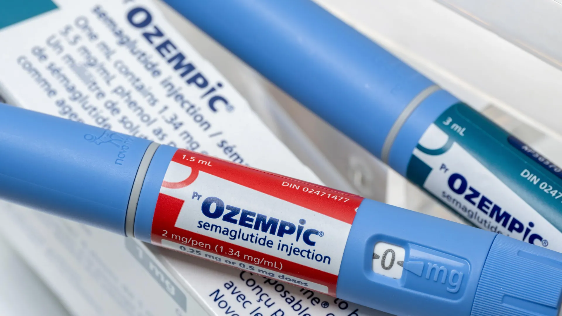
A large study led by Rutgers researchers has identified a possible vision risk associated with widely used diabetes and weight loss medications, including Ozempic and Wegovy. The findings suggest that people taking these drugs may face a slightly higher risk of developing a rare eye condition that can cause sudden and potentially permanent vision loss.
The condition, known as ischemic optic neuropathy, occurs when blood flow to the optic nerve is reduced. Because the optic nerve carries visual information from the eye to the brain, damage to it can lead to significant vision problems.
Study Links GLP-1 Drugs to Rare Vision Loss
The study, published in Annals of Internal Medicine, analyzed information from a large database of US adults ages 18 to 65 with Type 2 diabetes. Researchers focused on patients who had recently started taking medications to manage their condition, including a class of drugs known as glucagon-like peptide-1 receptor agonists, or GLP-1s.
These medications include semaglutide (sold as Ozempic and Wegovy) and tirzepatide (sold as Mounjaro and Zepbound). In addition to helping control blood sugar and manage weight, GLP-1 medications can reduce the risks of certain cardiovascular and kidney problems. However, scientists are still investigating their potential side effects as their use becomes increasingly widespread.
The researchers found that GLP-1 use was associated with a modest increase in the likelihood of ischemic optic neuropathy. Although the condition was uncommon, its potential consequences make the finding important.
“Although ischemic optic neuropathy was rare, it is clinically important because it can involve sudden vision loss,” said Chintan Dave, a core faculty member of the Center for Pharmacoepidemiology and Treatment Science at Rutgers Institute for Health, Health Care Policy and Aging Research. “The increase in risk was small — approximately three to four additional cases per 10,000 patients treated with GLP-1s over 18 months, but clinicians and patients should be aware of this potential association.”
While the absolute risk remains low, researchers say the growing popularity of GLP-1 medications makes continued safety monitoring especially important. These drugs are now widely prescribed to people both with and without Type 2 diabetes.
Who May Be Most at Risk?
The study also identified patterns among patients who developed the eye condition. Cases were more common among men and adults in the older portion of the study population.
“Most patients in our study who had ischemic optic neuropathy were either men or men and women aged 50 to 65,” said Dave, an associate professor at the Ernest Mario School of Pharmacy and the lead author of the study. “Because ischemic optic neuropathy can present with sudden vision loss, which may be permanent, early recognition of symptoms and prompt ophthalmologic evaluation may be especially important for patients at higher baseline risk.”
More than 80% of ischemic optic neuropathy cases are estimated to involve the non-arteritic anterior form of the condition. This occurs when blood supply to the front portion of the optic nerve becomes insufficient, potentially damaging the nerve and interfering with vision.
The resulting vision loss is often permanent. According to Dave, approximately one-third of affected patients experience some recovery, usually during the first six months. Even when improvement occurs, some degree of visual impairment typically remains.
Rather than causing complete blindness, the condition usually affects part of the vision in one eye. Patients may notice sudden blurriness, dimming, or missing areas in their field of view.
Do Weight Loss Drugs Actually Cause Vision Loss?
Despite the association, the researchers caution that their findings do not establish that GLP-1 medications directly cause optic nerve damage.
Other factors, including differences in patients’ underlying health conditions, could help explain the increased risk observed in the study. Additional research will be necessary to determine whether the medications themselves contribute to the condition.
For now, the findings highlight the importance of recognizing sudden vision changes and seeking prompt medical evaluation, particularly among people who may already be at greater risk.
The research team also included Kamika Reynolds and Kimberly O’Malley of the Institute for Health, along with Jason Roy of the Rutgers School of Public Health.
We asked experts how safe it is for children and teenagers to take protein supplements.
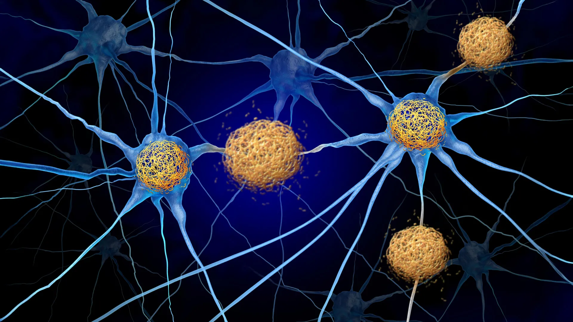
For decades, researchers have invested billions of dollars in Alzheimer’s treatments designed to target amyloid beta, a peptide that forms harmful deposits in the brain. But a biochemist at the University of California, Santa Cruz, believes scientists may have overlooked another potential contributor to the disease: a smaller, closely related peptide that was long considered harmless.
In a new commentary published in ChemBioChem, UC Santa Cruz professor Jevgenij Raskatov highlights research suggesting that this lesser-known peptide, called P3, can form microscopic clumps and threadlike structures known as fibrils. Evidence also suggests that P3 may interact with amyloid beta (Aβ), potentially influencing how it accumulates in the brain and how toxic it becomes. These findings raise the possibility that P3 contributes to the nerve cell damage associated with Alzheimer’s disease.
“The P3 peptide is, most likely, not the innocent bystander it was commonly thought to be. There’s still more research to be done. But this could turn Alzheimer’s research on its head,” said Raskatov, whose laboratory investigates amyloid peptides in hopes of identifying ways to prevent their harmful effects and develop better Alzheimer’s treatments. “P3 is a distinct aggregating peptide that is itself potentially neurotoxic and may be contributing to Alzheimer’s disease.”
Why Alzheimer’s Treatments Have Had Limited Success
Alzheimer’s is the most widespread neurodegenerative disease, affecting approximately 35 million people worldwide and generating annual costs exceeding $800 billion. The number of people living with the condition is expected to double by 2050.
Despite decades of intensive research, developing effective treatments has proved extraordinarily difficult. More than 400 clinical trials have investigated potential Alzheimer’s drugs, with most focusing on amyloid beta. Many of these efforts have failed, while others have produced only modest benefits. Some treatments have also been associated with serious complications, including brain hemorrhages and strokes.
Amyloid beta originates from a much larger protein embedded in cell membranes called the Amyloid Precursor Protein (APP). Two enzymes, β-secretase and γ-secretase, cut this protein in sequence, releasing amyloid beta peptides of different lengths.
Researchers have concentrated particularly on two versions, Aβ40 and Aβ42, which contain 40 and 42 amino acids, respectively. Aβ42 has attracted the greatest attention because it is especially prone to sticking together and forming toxic aggregates. As a result, it has remained a central target of Alzheimer’s drug development for decades.
Existing medications, including cholinesterase inhibitors and N-methyl-D-aspartate (NMDA) receptor antagonists, can temporarily ease certain symptoms but do not stop the disease from progressing. More recently, antibody treatments such as Lecanemab and Donanemab have been approved to help remove amyloid beta from the brain.
However, Raskatov argues that the benefits achieved so far remain limited, underscoring the need to investigate other biological processes involved in Alzheimer’s.
“Progress has been extremely slow, and the current state of the art in Alzheimer’s therapy leaves much to be desired,” he said. “We need fundamentally new approaches to the problem.”
The Overlooked Peptide That May Also Damage Brain Cells
Unlike amyloid beta, P3 forms when Amyloid Precursor Protein is processed through a different sequence of molecular cuts. In this pathway, an enzyme called α-secretase acts first, followed by γ-secretase.
Raskatov has proposed calling this peptide “Amyloid α,” or Aα, to better distinguish it from amyloid beta and reflect its ability to form aggregates.
For years, scientists largely assumed that P3 was incapable of forming amyloid deposits, was not toxic to cells, and would remain soluble in water. Under that interpretation, the peptide would simply dissolve rather than accumulate in the brain.
These assumptions were never adequately verified, according to Raskatov. Nevertheless, they helped establish the view that P3 was unlikely to matter in Alzheimer’s disease, leaving it largely absent from research efforts.
Raskatov, a specialist in peptide chemistry, and researchers in his laboratory decided to test those long-standing beliefs.
Over the past five years, the team has published three major studies demonstrating that P3 can form amyloid deposits at least as readily as amyloid beta. In fact, their experiments showed that P3 produces these aggregates more rapidly.
The researchers also found evidence that P3 itself may be harmful to neurons, although its toxicity appears lower than that of amyloid beta.
According to Raskatov, an independent research group in the United Kingdom has confirmed and expanded upon his team’s findings. Other laboratories are now beginning to investigate how P3 and amyloid beta interact, including whether their relationship affects the accumulation and toxicity of these peptides.
Together, the results challenge the long-held assumption that P3 is biologically harmless and irrelevant to Alzheimer’s. However, further research is needed to establish whether and how it contributes to the disease in patients.
Challenging Decades of Alzheimer’s Research
For much of the past several decades, amyloid beta has dominated scientific explanations of Alzheimer’s disease. David Teplow, an emeritus professor of neurology at UCLA and a prominent Alzheimer’s researcher, said the peptide has long been regarded as a central cause of the condition.
But an independent evaluation of Raskatov’s research led Teplow to conclude that scientists are beginning to reconsider this narrow focus.
“This reevaluation has far-reaching consequences for both basic science and clinical research into the causes and treatment of Alzheimer’s disease,” said Teplow, a founding editorial board member of the Journal of Molecular Neuroscience and the American Journal of Neurodegenerative Disease, as well as editor-in-chief of Progress in Molecular Biology and Translational Science.
Raskatov has also encountered a troubling problem while examining how other scientists have interpreted his team’s discoveries.
He identified at least four papers published in respected, peer-reviewed journals that cited his laboratory’s research as evidence that P3 was harmless and unable to form amyloid deposits. Yet his team’s experiments had demonstrated precisely the opposite.
“This is exactly the opposite of what we have actually shown,” Raskatov said. “We remain in the dark on how this sort of grand confusion may have come about. Clearly, there is more work ahead of us.”
The emerging evidence suggests that understanding Alzheimer’s may require looking beyond amyloid beta alone. By examining P3 and its interactions with other amyloid peptides, researchers hope to gain a more complete picture of the processes that damage brain cells and potentially identify new directions for treatment.
