‘Families deserve clarity about the qualifications and training of those caring for their children’


‘Families deserve clarity about the qualifications and training of those caring for their children’
Physical therapists have long emphasized the importance of strength training, especially for older adults. According to experts, strength training isn’t just for working out; it can also help build independence by keeping older folks safe and stable in their daily activities. If you’re looking for an easy (and affordable) way to strength train at home, this set of five resistance bands may be it. While the pink and blue packs go for close to $17, the multicolor set is just $12, making it 33% off.
Advertisement
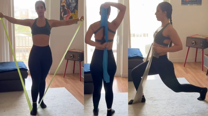
Each color band offers a different level of resistance, from three to 20 pounds. The bands are made of thermoplastic elastomer (TPE), a stretchy, latex-free material that blends qualities of rubber and plastic. Each band measures 59 inches long and 6 inches wide, making them extra accessible to hold and use. Reviewers who have tried many bands compliment the durability and quality of this set, with many saying it’s the best set they’ve tried.
Advertisement
“What I really like is that these are both wide and long. What that means is that they are long enough to give you the stretch necessary for physical therapy and wide enough that you can hold them tightly,” one user wrote. “These are far superior to the ones that the PT facility I went to uses.”
Because the bands are light and are easy to hold, they’re loved by older reviewers for making movement more accessible. A 69-year-old reviewer says the bands are “good even when I’m watching TV to work out with them,” joking that he’s a enjoying them as a “grumpy old man!”
Advertisement
“I bought these for my elderly mother to add a bit of resistance for her exercises,” one reviewer wrote. “…Being in her mid 70s she is work on stabilizing herself and adding some upper body strength, too. While anyone could get benefit from these, I believe this is the perfect end user for this type (beginner and elderly).”
Many reviewers who bought the bands for their older parents say they ended up buying a set for themselves. “I bought these for my mother. They are super easy for her to use. She is 88 years old and she loves them,” Jeff wrote. “They are super awesome and we really loved them.”
Shopper Newton calls the bands “incredibly helpful” for both building strength and “recovering safely” at home. “They’ve allowed me to start my muscle recovery process at home, progressing step by step at my own pace,” Newton says. “…I also like that they can be adjusted to fit different needs and levels.”
Advertisement
“I bought these for my elderly mother to add a bit of resistance for her exercises. They provide a wide range of ‘weight’ and start with as little as 5lbs. She’s able to comfortably use the red one (the second lightest), so this set will be all she needs. Being in her mid 70s she is work on stabilizing herself and adding some upper body strength, too. While anyone could get benefit from these, I believe this is the perfect end user for this type (beginner and elderly).” — Jaych79
“Wide, long, and easy for an elderly woman with arthritic hands to use. Decent selection of resistance options, though probably not suitable for non-rehab type workouts” — Joshua Noble
“What I really like is that these are both wide and long. What that means is that they are long enough to give you the stretch necessary for physical therapy and wide enough that you can hold them tightly. These are far superior to the ones that the PT facility I went to uses.” — Catfish Jeff
“These resistance bands have been incredibly helpful for me. They’ve allowed me to start my muscle recovery process at home, progressing step by step at my own pace. They are very strong and durable, yet comfortable to use. I also like that they can be adjusted to fit different needs and levels. Overall, a great tool for anyone looking to build strength or recover safely at home.” — Newton
“Fast delivery, love the package that tells you the strength of each one, individually packaged. Just started with them, but you could tell the durability. My age it’s good even when I’m watching TV to work out with them. 69-year-old army veteran. Enjoying grumpy old man!” — Roy
“I bought these for my mother. They are super easy for her to use. She is 88 years old and she loves them. They are super awesome and we really loved them. Yes, I even use them some myself. Great product. I highly recommend it.” — Jeff

Amazon
,
Amazon
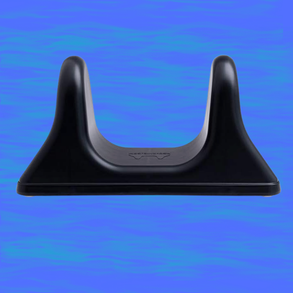
HuffPost contributor Megan Lasher swears by Pso-Rite’s muscle release tool for releasing back pain after long days on their feet. “You simply lie faced-down on the tool, situating it so the points of the U sit right above your hip joints, and wait as the muscle slowly begins to release,” they wrote. “In my experience, using the tool creates the type of cathartic pain you’d experience with a deep tissue massage. There’s a pinch as you settle into place, and a slow release the longer you lay on the prongs of the U. The full effect sets in after about a minute for me personally, and lasts for a few days (but I rarely go so long between sessions.)”
Amazon
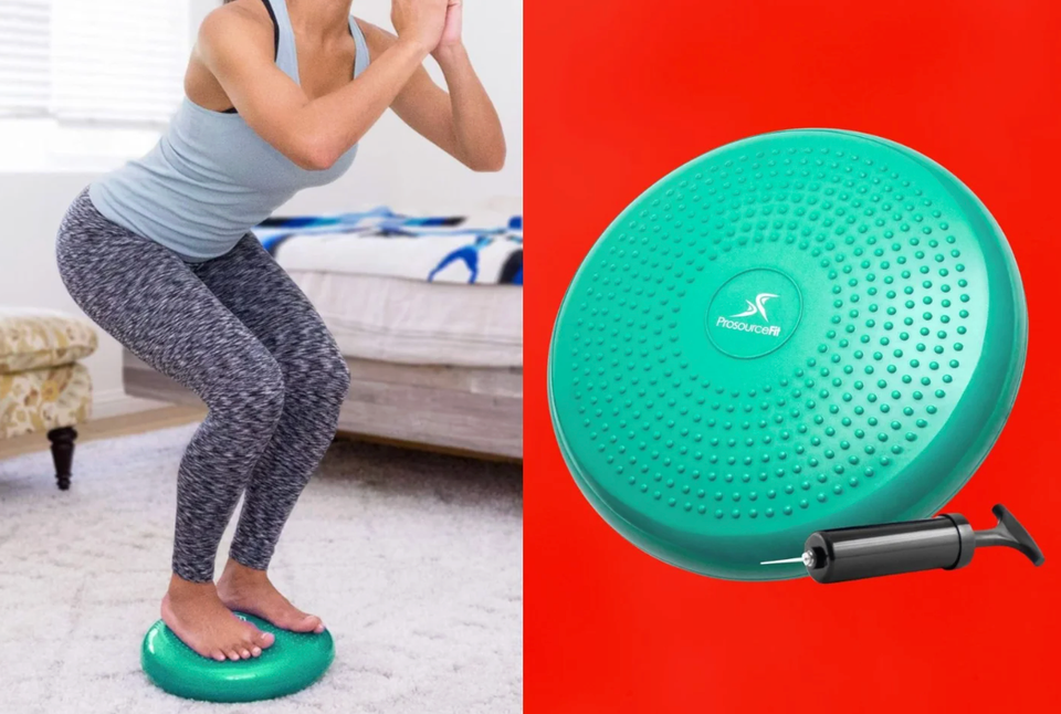
“These discs can be helpful when used in the right context,” Annalise Calo, physical therapist at WAVE Physical Therapy & Pilates in Ohio said of balancing tools like the ProsourceFit disc. It’s a 15-inch inflatable cushion that can be used to perform a number of balancing and core-strengthening exercises. ”[They] move as your weight shifts, helping to improve proprioception (awareness of your position in space) and reaction time,” Calo said.
Amazon
Advertisement
The Real Deal: We use deal trackers and commerce experience to sift through “fake” hike-and-drop deals and other deceptive sales tactics. Products will usually be rated at least 4 stars with a minimum 15% discount. (And when there’s an exception, we’ll tell you why.)
Advertisement
Denmark’s team doctor said an ICD implanted into the footballer’s chest responded as it should after he collapsed on Sunday.
Kimberley Wilson gives tips on how to manage your emotions.
A third of the weight loss from obesity jabs can come from muscle, say experts.
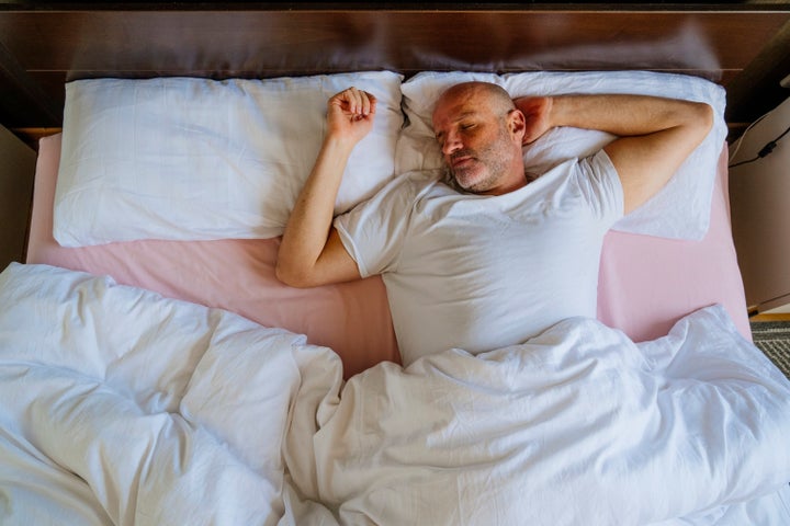
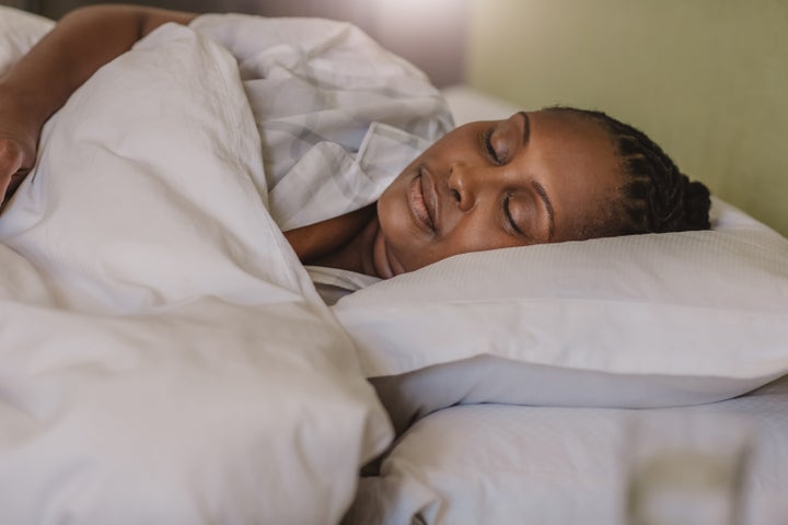
If you want to age well, the amount of sleep you get matters. A new study suggests there’s a “Goldilocks” time frame you want to aim for ― and it’s slightly less than eight hours.
Analyzing biological clocks throughout the human body, researchers found that too few hours of sleep (anything less than six hours) may speed biological aging in the brain, lung, heart and immune system. But too much sleep (more than eight hours) also accelerates aging in nearly every organ in the body, according to study, published in the journal Nature last month.
Advertisement
If you’re looking to age minimally, the “sweet spot” is between 6.4 and 7.8 hours of sleep per day.
Getting either too much or too little sleep was also associated with a range of physical health conditions, including obesity, type 2 diabetes, hypertension, coronary artery disease and gastritis. Insufficient and excessive sleep durations were also significantly associated with depression and anxiety disorders.
The study supports “the idea that sleep is important in maintaining organ health within a coordinated brain-body network, including metabolic balance and a healthy immune system,” Junhao Wen, the lead researcher and an assistant professor of radiology at Columbia University Vagelos College of Physicians and Surgeons, said in a press release.
Wen and his colleagues used advanced statistical models called “aging clocks” to figure out the amount of sleep that’s associated with accelerated aging.
Advertisement
While most aging clocks estimate biological aging across the entire body, organs age at different rates. Wen and his team developed organ-specific aging clocks that may provide patients with more personalized and precise insights into their health.
“In the liver, for example, we have an aging clock built with protein data, an aging clock of metabolic data, and an aging clock of imaging data,” Wen said.
The hypothesis is that different organs, even within the same person, age at different rates, Wen told The Washington Post.
Advertisement
This doesn’t mean that sleep duration alone is aging our organs faster or slower, but too much or little sleep may be markers of poorer overall health in the body.
KARRASTOCK via Getty Images
One limitation for the study? Wen said more research is needed involving people of Asian and African descent, since the data for sleepers was taken from U.K. Biobank data, which is heavily weighted toward people of white European ancestry.
Advertisement
Research has long confirmed that chronically under-sleeping can lead to a reduced lifespan and a range of health issues: hypertension, cardiovascular disease, stroke. But those who love to sleep may wonder: Why might too much sleep be a problem?
“Chronically long sleep time is associated with many health issues because oversleeping is often a byproduct of an underlying disease such as a sleep disorder like sleep apnea, inflammatory conditions, cancer, neurodegenerative disorder, and poor mental health,” said Dr. Chelsie Rohrscheib, a neuroscientist and researcher at Wesper, a company that designs at-home tools for diagnosing sleep disorders like sleep apnea.
Some studies have shown that long-sleepers have markers of inflammation, which is associated with diseases like cardiovascular disease and cancer, said Rohrscheib, who is not affiliated with this current study.
Advertisement
“The other answer is that long sleeping increases a sedentary lifestyle and reduces the total amount of physical activity achieved each day,” Rohrscheib told HuffPost.
Goodboy Picture Company via Getty Images
The study also found that women may need a little more sleep than men. The difference is relatively small, though, said Rohrscheib: typically about 10 to 20 additional minutes per night.
Advertisement
The exact reasons for this remains unclear, but sleep researchers believe it may be related to hormonal differences and fluctuations throughout life, particularly during the luteal phase of the menstrual cycle, pregnancy and menopause.
“Others have proposed that women may experience greater emotional and cognitive demands, which could increase the need for sleep to support essential brain functions such as memory consolidation, emotional regulation, and overnight recovery,” she said.
Dr. Chris Winter, a a neurologist and host of the podcast “Sleep Unplugged,” said the study’s most interesting finding was its suggested optimal sleep range of 6.4 to 7.8 hours per night ― slightly below the eight hours many of us have been taught is the sleep “magic number.”
Advertisement
“The range is essentially an average of seven hours of sleep, not eight,” Winter, who was not affiliated with the study, told HuffPost. “I would love to dislodge from the public’s collective mentality about sleep that ‘eight hours’ of sleep is ideal for everyone, or even attainable for everyone. It’s not.”
Winter’s takeaway is that we should aim for a consistent seven hours or so on average, though if eight works for you, more power to you. A person’s need for sleep varies greatly based on age, lifestyle and genetics.
“If you struggle to get eight consistently, this is not something to overcome to optimize your health,” he said. “It may simply be that your body favors a number below what you are shooting for, yet still well within the range of optimized.”
Advertisement
Not all fruit and veg is equal for getting nutrients called flavanols, say researchers.
Androgenetic alopecia (AGA) is the most common type of hair loss, affecting millions of men and women around the world. It is often known as male or female pattern hair loss, and it usually develops gradually as hair follicles shrink over time. As follicles become smaller, they produce thinner, shorter hairs until growth may slow dramatically or stop.
Current treatments, including finasteride and minoxidil, can help some people, but they are not ideal for everyone. Finasteride works by targeting hormones involved in follicle shrinkage, while minoxidil is commonly used on the scalp to encourage growth. However, some patients worry about unwanted effects, including sexual side effects linked to finasteride or scalp irritation associated with minoxidil. Because of this, many people continue to look for options that feel safer, more natural, or more comprehensive.
Ancient Root Meets Modern Hair Science
A new scientific review suggests that Polygonum multiflorum, a root long used in traditional Chinese medicine, may deserve serious attention as a potential therapy for androgenetic alopecia. The herb has been used for more than 1,000 years and has traditionally been associated with “blacken hair and nourish essence.”
What makes the review especially interesting is that the plant does not appear to act through only one biological route. Instead, researchers report that Polygonum multiflorum may influence several processes involved in hair loss and regrowth at the same time.
In androgenetic alopecia, a hormone called dihydrotestosterone plays a major role. It can gradually shrink hair follicles, making it harder for them to keep producing strong, healthy hair. According to the review, Polygonum multiflorum may help reduce the impact of this hormone, protecting follicles from one of the major drivers of pattern hair loss.
A Multi Path Approach to Hair Regrowth
The review also describes several other possible benefits. Polygonum multiflorum may help prevent follicle cells from dying too early, which is important because healthy follicles depend on active, living cells to maintain the hair growth cycle. It may also turn on key biological signals involved in regeneration, including Wnt and Shh pathways.
These pathways are important because they help control how cells grow, communicate, and repair tissue. In hair follicles, they are closely linked to the shift from resting phases into active growth. When these signals are stronger, follicles may be more likely to reenter a growth state.
The herb may also improve blood flow to the scalp. Better circulation can help bring oxygen and nutrients to follicles, supporting the environment needed for healthier hair growth. This is one reason researchers see Polygonum multiflorum as potentially broader than conventional treatments that focus on a single target.
“Our analysis bridges ancient wisdom and modern science,” said Han bixian, the first author of a review on the topic recently published in the Journal of Holistic Integrative Pharmacy. “What surprised us was how consistently historical texts — from the Tang Dynasty onward — described effects that align perfectly with today’s understanding of hair biology. Modern studies now confirm that this isn’t folklore; it’s pharmacology.”
From Traditional Records to Laboratory Evidence
The review brings together several kinds of evidence, including laboratory research, clinical reports, and historical herbal records. Those older records are not being treated as proof by themselves. Instead, researchers are comparing traditional claims with modern biological findings to see where they overlap.
That overlap appears to be one of the main reasons for renewed interest in Polygonum multiflorum. The review suggests that the herb may do more than slow hair loss. By acting on growth factors and signaling pathways, it may help create conditions that support regeneration.
This is an important distinction. Many hair loss treatments are designed mainly to preserve existing hair or slow further thinning. A treatment that actively supports regrowth through multiple mechanisms could offer a different kind of approach, especially for people who have not responded well to existing options.
Safety Depends on Proper Preparation
The review also emphasizes that preparation matters. In traditional Chinese medicine, Polygonum multiflorum is typically processed before use. This step is considered important because processing can affect both safety and biological activity.
“When properly processed — a key step in traditional preparation — the herb shows a favorable safety profile, making it more acceptable to patients wary of side effects like sexual dysfunction or scalp irritation linked to current medications,” This article highlights.
That point is especially relevant because natural products are not automatically risk free. Herbs can contain powerful compounds, and their effects may vary depending on preparation, dose, and product quality. The review presents processed Polygonum multiflorum as a more acceptable option for some patients, but it does not suggest that people should self treat without guidance.
More Clinical Testing Is Still Needed
Although the findings are promising, the researchers stress that stronger clinical evidence is still needed. Much of the current support comes from laboratory studies, historical records, and limited clinical observations. Large, carefully designed human trials would be necessary to confirm how well Polygonum multiflorum works for androgenetic alopecia and how safe it is across different groups of patients.
Still, the review points to a larger idea with growing scientific importance. Traditional remedies may contain biologically active compounds that can inspire new treatments when they are studied with modern methods. In the case of Polygonum multiflorum, centuries of use are now being examined through the lens of hormone biology, cell survival, growth signaling, and scalp circulation.
For people dealing with hair loss, the research offers a hopeful but cautious message. A root used for more than a millennium may not replace today’s treatments yet, but it could help guide the next generation of hair regrowth therapies.

A century old idea from Erwin Schrödinger has taken a major step forward, thanks to new research into how humans perceive differences between colors.
A team led by Los Alamos scientist Roxana Bujack used geometry to build a mathematical definition of color perception based on hue, saturation, and lightness. Their results, presented at a visualization science conference, formalize Schrödinger’s model of color and show that these familiar color qualities are built into the structure of color perception itself.
“What we conclude is that these color qualities don’t emerge from additional external constructs such as cultural or learned experiences but reflect the intrinsic properties of the color metric itself,” Bujack said. “This metric geometrically encodes the perceived color distance — that is, how different two colors appear to an observer.”
Completing Schrödinger’s Color Puzzle
By defining these perceptual attributes more rigorously, the researchers have supplied a missing piece in Schrödinger’s long standing vision for a closed mathematical model of color. The goal was to define hue, saturation, and lightness using only the geometric property of highest color similarity.
Human color vision is based on three types of cone cells, which are centered around red, blue, and green. That gives color spaces three dimensions, allowing scientists to organize and compare colors mathematically.
In the 19th century, mathematician Bernhard Riemann proposed that perceptual color spaces are not flat or straight, but curved. In the 1920s, Schrödinger built on that idea by defining hue, saturation, and lightness within a Riemannian model of color perception, using a metric that describes how people perceive color differences.
Fixing a Century Old Mathematical Gap
Schrödinger’s definitions have shaped color science for roughly 100 years. But while the Los Alamos team was developing algorithms for scientific visualization, they found that the mathematics behind the model had important weaknesses.
The biggest problem involved the neutral axis, the line of grays that runs from black to white. Schrödinger’s definitions of hue, saturation, and lightness depend on where a color sits in relation to that axis, yet he never formally defined the axis itself.
That omission created a serious gap. Without a precise definition of the neutral axis, the entire construction was formally incomplete. The team’s most important advance was finding a way to define the neutral axis using only the geometry of the color metric.
To accomplish that, the researchers had to move beyond the traditional Riemannian model. That shift represents a major mathematical advance for visualization science.
A Better Model of How Colors Change
The team also corrected two other important issues in the older framework.
One involved the Bezold- Brücke effect, a phenomenon in which changing light intensity can make a color appear to shift in hue. The researchers addressed this by using the shortest path in their geometric model of color perception rather than relying on a simple straight line.
They also used the shortest path in a non-Riemannian space to account for diminishing returns in color perception, another effect that had not been fully captured by the older approach.
Why Color Perception Matters
The research was presented at the Eurographics Conference on Visualization and builds on a broader Los Alamos project on color perception. That project also produced a groundbreaking 2022 paper in the Proceedings of the National Academy of Sciences.
A more precise model of color perception could have wide value in fields that depend on accurate color, including photography, video, visualization, and related technologies. It could also improve the way scientists create and interpret visual data.
Scientific visualization plays an important role in helping researchers understand complex information. Better color models can support more effective analysis across many areas, including national security sciences.
The team’s work now provides a foundation for future color modeling in non-Riemannian space.
Funding: This work was supported by the Laboratory Directed Research and Development program at Los Alamos and by the National Nuclear Security Administration’s Advanced Simulation and Computing program.
Fuelled by social media, the market for children’s skincare is booming. Experts fear for the long-term impact on girls