Fatty liver breakthrough: A common vitamin shows promise

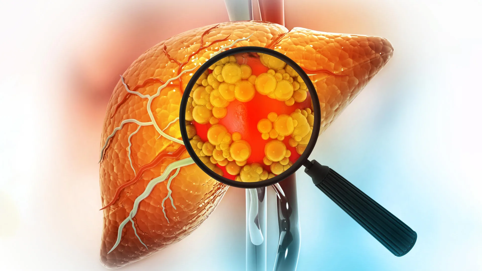
Metabolic-associated fatty liver disease (MASLD) impacts roughly 30% of people globally and has long lacked effective, targeted therapies. Now, researchers have uncovered a key genetic factor that worsens the condition. Even more surprising, the most effective way to target this factor may be an already approved and widely available treatment: vitamin B3.

An international research team led by Professor Jang Hyun Choi at UNIST, working with Professor Hwayoung Yun at Pusan National University (PNU) and Professor Neung Hwa Park at Ulsan University Hospital (UUH), has identified microRNA-93 (miR-93) as a central regulator in MASLD. This marks the first time this molecule has been clearly linked to how the disease develops and progresses.

How miR-93 Disrupts Liver Function

MiR-93 is a small RNA molecule found in liver cells that controls the activity of certain genes. The researchers discovered that levels of miR-93 are unusually high in both people with fatty liver disease and in animal models. Their analysis showed that miR-93 drives fat buildup, inflammation, and scarring in the liver by suppressing SIRT1, a gene that plays a key role in managing fat metabolism inside liver cells.

To better understand its role, the team used gene editing to stop the production of miR-93 in mice. These animals showed significantly less fat accumulation in the liver, along with improved insulin sensitivity and better overall liver function. In contrast, mice engineered to produce excess miR-93 experienced more severe metabolic problems in the liver.

Vitamin B3 Emerges as a Potential Treatment

The researchers then screened 150 FDA-approved drugs to see if any could reduce miR-93 levels. Niacin (vitamin B3) stood out as the most effective option. In mice treated with niacin, miR-93 levels dropped sharply, while SIRT1 activity increased. This helped restore normal fat-processing pathways in the liver and improved overall lipid balance.

The research team explained, “This study precisely elucidates the molecular origin of MASLD and demonstrates the potential for repurposing an already approved vitamin compound to modulate this pathway, which has high translational clinical relevance.”

They added, “Given that niacin is a well-established and safe medication used to treat hyperlipidemia, it holds promise as a candidate for combination therapies targeting miRNA pathways in MASLD.”

Study Support and Publication Details

This work was supported by several organizations, including the National Research Foundation of Korea (NRF) and the Korea Research Institute of Bioscience and Biotechnology (KRIBB). The findings were published online in Metabolism: Clinical and Experimental. Key contributors include Dr. Yo Han Lee and Kieun Park from UNIST, along with Professor Joonho Jeong from Ulsan University Hospital and Jinyoung Lee from Pusan National University, who served as co-first authors.


Share Button

Insulin pills may soon replace daily injections

For more than 100 years, scientists have pursued the idea of insulin in pill form, often described as a “dream” treatment for diabetes. The challenge has been the body itself. Enzymes in the digestive system break down insulin before it can work, and the intestine lacks a natural way to absorb it into the bloodstream. As a result, many patients still depend on daily injections, which can take a toll on their quality of life.

A team at Kumamoto University, led by Associate Professor Shingo Ito, has now developed a promising solution. Their approach uses a cyclic peptide that can pass through the small intestine, known as the DNP peptide. This platform allows insulin to be delivered orally in a way that was not previously possible.

Two Effective Strategies for Intestinal Absorption

To make this work, the researchers designed two different methods to help insulin cross the intestinal barrier:

  • Mixing method (interaction-based): The team combined a modified “D-DNP-V peptide” with zinc-stabilized insulin hexamers. When given orally to several diabetes models, including chemically induced (STZ mice) and genetic (Kuma mice) models, this mixture quickly brought blood sugar levels down to normal. Stable glucose control was maintained with once-daily dosing for three consecutive days.
  • Conjugation method (covalent-based): Using click chemistry, the researchers attached the DNP peptide directly to insulin, creating a “DNP-insulin conjugate.” This version lowered blood sugar just as effectively as the mixing method, confirming that the peptide actively helps transport insulin through the intestine.

Lower Doses Make Oral Insulin More Practical

One of the biggest obstacles for oral insulin has been the need for extremely high doses, sometimes more than ten times higher than injections. This new platform significantly reduces that requirement. It achieved a pharmacological bioavailability of about 33-41% compared to subcutaneous injection. That level of efficiency suggests oral insulin could become far more practical for real-world use.

Future Potential for Diabetes Treatment

“Insulin injections remain a daily burden for many patients,” said Associate Professor Shingo Ito. “Our peptide-based platform offers a new route to deliver insulin orally and may be applicable to long-acting insulin formulations and other injectable biologics.”

The findings were published in the journal Molecular Pharmaceutics. The researchers are now moving forward with additional studies, including testing in larger animal models and systems that replicate the human intestine, as they work toward eventual clinical applications.

Share Button

UK’s transplant system was world-leading – now it lags behind other Western nations

The UK has failed to keep pace with the rest of the world. Can it regain its status, and how?

Share Button

Cuba’s mothers-to-be prepare to give birth in a country plunged into darkness

Two pregnant women tell the BBC’s Will Grant of their hopes and fears as their nation is mired in crisis.

Share Button

Women over 50 lost 35% more weight with this surprising combo

A Mayo Clinic-led study reports that postmenopausal women using menopausal hormone therapy experienced significantly greater weight loss when taking tirzepatide, a Food and Drug Administration-approved medication for overweight and obesity. On average, these women lost about 35% more weight compared to those using tirzepatide alone. The results, published in The Lancet Obstetrics, Gynaecology, & Women’s Health, point to new possibilities for treating obesity and related health conditions in women after menopause.

Menopause is often associated with increased weight gain and a higher risk of developing overweight and obesity. These changes can raise the likelihood of serious health problems, including cardiovascular disease and type 2 diabetes. In addition to weight gain, declining estrogen levels during menopause can trigger other changes in the body that may further increase cardiovascular risk. “This study provides important insights for developing more effective and personalized strategies for managing cardiometabolic risk in postmenopausal women,” says Regina Castaneda, M.D., postdoctoral research fellow at Mayo Clinic and first author of the study.

Exploring the Role of Hormone Therapy in Weight Loss

Hormone therapy remains the most effective first-line option for relieving common menopausal symptoms such as hot flashes and night sweats, which affect up to 75% of postmenopausal women. However, its potential role in enhancing weight-loss medications has not been well understood. Earlier studies have suggested that women using hormone therapy may lose more weight when treated with GLP-1-based drugs like semaglutide, but data on tirzepatide had been lacking.

To address this gap, researchers analyzed data from 120 adults with overweight or obesity who were treated with tirzepatide for at least 12 months. They compared outcomes between those who also used hormone therapy and those who did not, ensuring both groups had similar baseline characteristics.

Study Findings and Key Limitations

The analysis showed that women receiving both treatments lost significantly more weight. “In this observational study, women who used menopausal hormone therapy lost about 35% more weight than women taking tirzepatide alone. Because this was not a randomized trial, we cannot say hormone therapy caused additional weight loss,” says Maria Daniela Hurtado Andrade, M.D., Ph.D., endocrinologist at Mayo Clinic and senior author of the study.

“It is possible that women using hormone therapy were already engaged in healthier behaviors, or that menopause symptom relief improved sleep and quality of life, making it easier to stay engaged with dietary and physical activity changes.”

Potential Synergy Between Estrogen and GLP-1 Medications

Although more controlled studies are needed, researchers say the findings are clinically meaningful. Dr. Castaneda notes that the size of the observed difference justifies further investigation into how hormone therapy and GLP-1-based medications may work together. “The magnitude of this difference warrants future studies that could help clarify how GLP-1-based obesity medications and menopausal hormone therapy may interact. Interestingly, preclinical data suggest a potential synergy, with estrogen appearing to enhance the appetite-suppressing effects of GLP-1,” says Dr. Castaneda.

What Comes Next for Research

Future research will focus on confirming these results in randomized clinical trials and exploring whether the benefits go beyond weight loss. “Next, we plan to test these observations in a randomized clinical trial and determine if benefits extend beyond weight loss — specifically, whether hormone therapy also enhances the effects of these medications on cardiometabolic measures,” adds Dr. Hurtado Andrade. “If confirmed, this work could speed the development and adoption of new, evidence-based strategies to reduce this risk for millions of postmenopausal women navigating this life stage.”

This research was funded by the Mayo Clinic Center for Women’s Health Research.

Share Button

Meningitis outbreak passes peak, says health agency

“We could still see cases continuing to come in, we need to keep an eye on those, the UKHSA says.

Share Button

Our son loved the outdoors – invisible illness means he now can’t walk or talk

Tomos is one of thousands with ME in Wales, with services described as “a desert” for those most affected.

Share Button

‘Women’s health must be taken more seriously’

Dr Liz O’Riordan says it is important women seek help from professionals not “Google and Chat GPT”.

Share Button

New blood test could catch pancreatic cancer before it’s too late

Researchers supported by the National Institutes of Health (NIH) have created a new blood test designed to identify pancreatic ductal adenocarcinoma, one of the most lethal types of cancer. Because pancreatic cancer is often discovered only after it has advanced, treatment options are limited and survival rates remain low. This new approach, detailed in Clinical Cancer Research, could help detect the disease sooner and improve patient outcomes.

Pancreatic cancer has a poor prognosis, with only about 10% of patients living longer than five years after diagnosis. However, doctors believe survival could improve significantly if the disease is caught earlier, when treatment is more effective. Despite this, there are currently no reliable screening tools available to detect pancreatic cancer in its early stages.

Testing Existing and New Blood Biomarkers

To address this gap, scientists from the University of Pennsylvania Perelman School of Medicine, Philadelphia, and Mayo Clinic, Rochester, Minnesota, analyzed blood samples from individuals with and without pancreatic cancer. They evaluated several biomarkers, including carbohydrate antigen 19-9 (CA19-9), commonly used to monitor treatment response, and thrombospondin 2 (THBS2), another previously studied marker.

Individually, these markers have limitations. CA19-9 levels can rise in non-cancerous conditions like pancreatitis or bile duct obstruction, and some people do not produce the marker at all due to genetic differences. As a result, neither marker alone is reliable enough for screening.

Newly Identified Proteins Improve Detection

By examining stored blood samples, the researchers identified two additional proteins that appear to be elevated in people with early-stage pancreatic cancer: aminopeptidase N (ANPEP) and polymeric immunoglobin receptor (PIGR). These newly identified biomarkers showed clear differences between cancer patients and healthy individuals.

When combined with CA19-9 and THBS2, the four-marker panel demonstrated strong performance. It correctly distinguished pancreatic cancer cases from non-cases 91.9% of the time across all stages, with a false positive rate of 5% in non-cases. For early-stage (stage I/II) cancer, the test detected 87.5% of cases.

“By adding ANPEP and PIGR to the existing markers, we’ve significantly improved our ability to detect this cancer when it’s most treatable,” said the study’s lead investigator, Kenneth Zaret, Ph.D., University of Pennsylvania’s Perelman School of Medicine.

Distinguishing Cancer From Other Conditions

An important advantage of the test is its ability to differentiate pancreatic cancer from other non-cancerous pancreatic conditions, including pancreatitis. This helps reduce the risk of misdiagnosis and unnecessary concern for patients.

Next Steps Toward Screening

“Our retrospective study findings warrant further testing in larger populations, particularly in people before they show symptoms,” Zaret said. “Such ‘prediagnostic’ studies would help determine if the test could be used as a screening tool for people at high risk of developing the disease based on family history, genetic screening results or personal history of pancreatic cysts or pancreatitis.”

The study was supported by NIH grants U01CA210138, P50CA102701, S10 OD023586-01, P30 DK020579, UL1 TR002345, P30CA091842, and U01CA210138.

Share Button

A promising fatty liver treatment may raise cancer risk

Scientists have uncovered a surprising risk linked to blocking a cellular enzyme once believed to protect against fatty liver disease. Instead of helping long term, shutting down this enzyme may raise the chances of chronic liver damage and cancer as people age.

In a study published in Science Advances, researchers from the University of Adelaide found that losing the enzyme Caspase-2 causes liver cells to grow abnormally. This uncontrolled growth leads to inflammation, scarring, and a much higher likelihood of developing liver cancer.

These results challenge the growing interest in Caspase-2 inhibitors as a treatment to manage or prevent fatty liver disease. The findings suggest that targeting this pathway could have unintended consequences over time.

Caspase-2’s Role in Liver Cell Stability

According to lead researcher Dr. Loretta Dorstyn from the Centre for Cancer Biology, Caspase-2 is essential for keeping liver cells genetically stable. It also plays a separate role in regulating fat levels within the liver.

“Liver cells normally have extra copies of genetic material- known as polyploidy — and while this feature can help the liver cope with stress, our study shows that without the enzyme Caspase-2, abnormally high levels of polyploidy in the liver can be damaging,” Dr. Dorstyn said.

To investigate this further, scientists used genetically modified mice. In animals missing the enzyme, or carrying a nonfunctional version, liver cells became unusually large and showed significant genetic and cellular damage.

Long-Term Damage and Tumor Formation

“Over time, these mice developed chronic liver inflammation and characteristics of hepatitis-like liver disease including, scarring, oxidative damage and a type of cell death linked to inflammation. As the animals aged, they were much more likely to develop liver cancer.”

Older mice without functional Caspase-2 developed liver tumors far more often than normal mice. In some cases, cancer rates were up to four times higher, consistent with hepatocellular carcinoma.

Dr. Dorstyn noted that the findings overturn the assumption that inhibiting Caspase-2 is always beneficial.

“While inhibiting this enzyme can be protective in young animals or may help prevent fatty liver disease in the short term, our study shows that its long-term loss is clearly detrimental.

“Our study demonstrates that Caspase-2 is essential for removing damaged and abnormal liver cells as we age. Without it, these cells accumulate, and can become cancerous, while also creating an environment that predisposes the liver to cancer.”

Implications for Fatty Liver Treatments and Drug Development

Senior author Professor Sharad Kumar said the results carry important warnings for future therapies.

“There has been significant interest in targeting Caspase-2 to treat metabolic liver disease and reduce liver cancer risk,” Prof Kumar said.

“Our data shows that this approach could have serious unintended consequences later in life, increasing susceptibility to chronic liver inflammation, fibrosis and cancer.”

Liver disease continues to rise globally, driven by ageing populations, obesity, and metabolic conditions. In 2022 alone, liver cancer caused nearly 760,000 deaths worldwide, according to the World Cancer Research Fund, making it the sixth most common cancer.

The study, titled ‘Caspase- 2 deficiency drives pathogenic liver polyploidy and increases age- associated hepatocellular carcinoma in mice’, was published in Science Advances.

Share Button