NHS bosses are urging people to only come to A&E if it is an emergency.


NHS bosses are urging people to only come to A&E if it is an emergency.
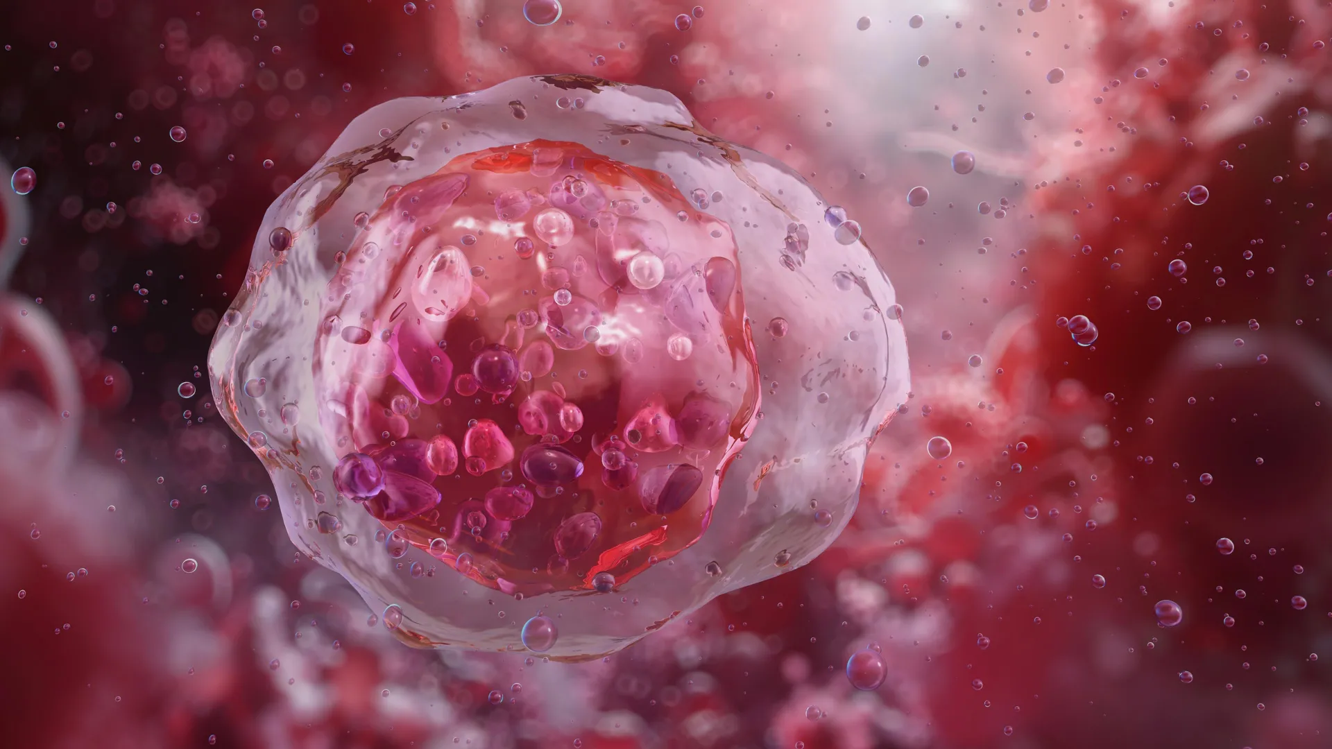
A new artificial intelligence system that examines the shape and structure of blood cells could significantly improve how diseases such as leukemia are diagnosed. Researchers say the tool can identify abnormal cells with greater accuracy and consistency than human specialists, potentially reducing missed or uncertain diagnoses.
The system, known as CytoDiffusion, relies on generative AI, the same type of technology used in image generators such as DALL-E, to analyze blood cell appearance in detail. Rather than focusing only on obvious patterns, it studies subtle variations in how cells look under a microscope.
Moving Beyond Pattern Recognition
Many existing medical AI tools are trained to sort images into predefined categories. In contrast, the team behind CytoDiffusion demonstrated that their approach can recognize the full range of normal blood cell appearances and reliably flag rare or unusual cells that may signal disease. The work was led by researchers from the University of Cambridge, University College London, and Queen Mary University of London, and the findings were published in Nature Machine Intelligence.
Identifying small differences in blood cell size, shape, and structure is central to diagnosing many blood disorders. However, learning to do this well can take years of experience, and even highly trained doctors may disagree when reviewing complex cases.
“We’ve all got many different types of blood cells that have different properties and different roles within our body,” said Simon Deltadahl from Cambridge’s Department of Applied Mathematics and Theoretical Physics, the study’s first author. “White blood cells specialize in fighting infection, for example. But knowing what an unusual or diseased blood cell looks like under a microscope is an important part of diagnosing many diseases.”
Handling the Scale of Blood Analysis
A standard blood smear can contain thousands of individual cells, far more than a person can realistically examine one by one. “Humans can’t look at all the cells in a smear — it’s just not possible,” Deltadahl said. “Our model can automate that process, triage the routine cases, and highlight anything unusual for human review.”
This challenge is familiar to clinicians. “The clinical challenge I faced as a junior hematology doctor was that after a day of work, I would face a lot of blood films to analyze,” said co-senior author Dr. Suthesh Sivapalaratnam from Queen Mary University of London. “As I was analyzing them in the late hours, I became convinced AI would do a better job than me.”
Training on an Unprecedented Dataset
To build CytoDiffusion, the researchers trained it on more than half a million blood smear images collected at Addenbrooke’s Hospital in Cambridge. The dataset, described as the largest of its kind, includes common blood cell types, rare examples, and features that often confuse automated systems.
Instead of simply learning how to separate cells into fixed categories, the AI models the entire range of how blood cells can appear. This makes it more resilient to differences between hospitals, microscopes, and staining techniques, while also improving its ability to detect rare or abnormal cells.
Detecting Leukemia With Greater Confidence
When tested, CytoDiffusion identified abnormal cells associated with leukemia with much higher sensitivity than existing systems. It also performed as well as or better than current leading models, even when trained with far fewer examples, and was able to quantify how confident it was in its own predictions.
“When we tested its accuracy, the system was slightly better than humans,” said Deltadahl. “But where it really stood out was in knowing when it was uncertain. Our model would never say it was certain and then be wrong, but that is something that humans sometimes do.”
Co-senior author Professor Michael Roberts from Cambridge’s Department of Applied Mathematics and Theoretical Physics said the system was evaluated against real-world challenges faced by medical AI. “We evaluated our method against many of the challenges seen in real-world AI, such as never-before-seen images, images captured by different machines and the degree of uncertainty in the labels,” he said. “This framework gives a multi-faceted view of model performance which we believe will be beneficial to researchers.”
When AI Images Fool Human Experts
The team also found that CytoDiffusion can generate synthetic images of blood cells that look indistinguishable from real ones. In a ‘Turing test’ involving ten experienced hematologists, the specialists were no better than random chance at telling real images apart from those created by the AI.
“That really surprised me,” Deltadahl said. “These are people who stare at blood cells all day, and even they couldn’t tell.”
Opening Data to the Global Research Community
As part of the project, the researchers are releasing what they describe as the world’s largest publicly available collection of peripheral blood smear images, totaling more than half a million samples.
“By making this resource open, we hope to empower researchers worldwide to build and test new AI models, democratize access to high-quality medical data, and ultimately contribute to better patient care,” Deltadahl said.
Supporting, Not Replacing, Clinicians
Despite the strong results, the researchers emphasize that CytoDiffusion is not intended to replace trained doctors. Instead, it is designed to assist clinicians by quickly flagging concerning cases and automatically processing routine samples.
“The true value of healthcare AI lies not in approximating human expertise at lower cost, but in enabling greater diagnostic, prognostic, and prescriptive power than either experts or simple statistical models can achieve,” said co-senior author Professor Parashkev Nachev from UCL. “Our work suggests that generative AI will be central to this mission, transforming not only the fidelity of clinical support systems but their insight into the limits of their own knowledge. This ‘metacognitive’ awareness — knowing what one does not know — is critical to clinical decision-making, and here we show machines may be better at it than we are.”
The team notes that additional research is needed to increase the system’s speed and to validate its performance across more diverse patient populations to ensure accuracy and fairness.
The research received support from the Trinity Challenge, Wellcome, the British Heart Foundation, Cambridge University Hospitals NHS Trust, Barts Health NHS Trust, the NIHR Cambridge Biomedical Research Centre, NIHR UCLH Biomedical Research Centre, and NHS Blood and Transplant. The work was carried out by the Imaging working group within the BloodCounts! consortium, which aims to improve blood diagnostics worldwide using AI. Simon Deltadahl is a Member of Lucy Cavendish College, Cambridge.
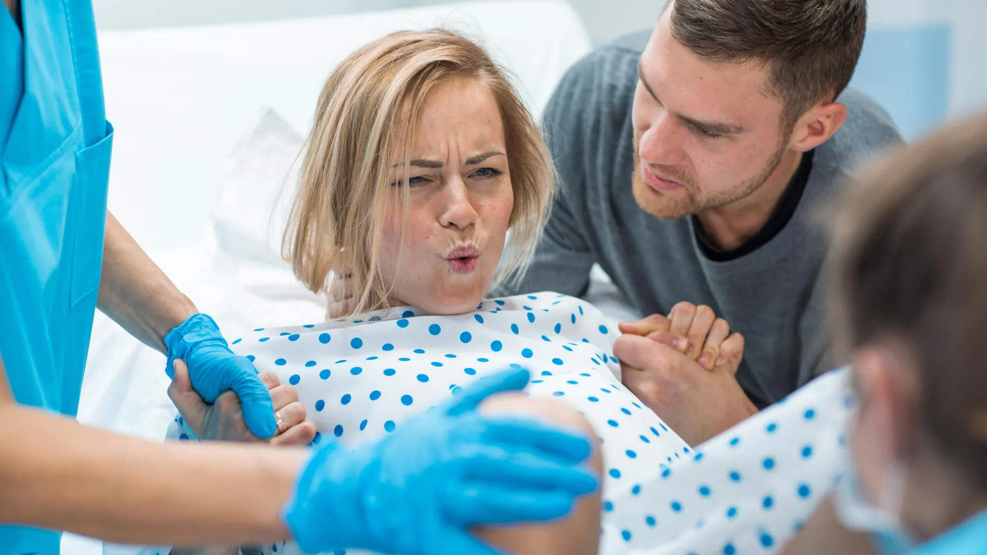
Successful childbirth depends on the uterus producing steady, well-organized contractions that move the baby safely through delivery. Hormones such as progesterone and oxytocin play a major role in controlling this process. For years, however, researchers have also suspected that physical forces involved in pregnancy and birth, including stretching and pressure, contribute in important ways.
New research from Scripps Research, published in Science, now shows how the uterus detects and responds to these physical forces at the molecular level. The findings shed light on why labor sometimes slows or begins too early and could guide future efforts to improve treatments for pregnancy and delivery complications.
Pressure and Stretch as Biological Signals
“As the fetus grows, the uterus expands dramatically, and those physical forces reach their peak during delivery,” says senior author Ardem Patapoutian, a Howard Hughes Medical Institute Investigator and the Presidential Endowed Chair in Neurobiology at Scripps Research. “Our study shows that the body relies on special pressure sensors to interpret these cues and translate them into coordinated muscle activity.”
Patapoutian shared the 2021 Nobel Prize in Physiology or Medicine for identifying the cellular sensors that allow organisms to detect touch and pressure. These sensors are ion channels built from proteins known as PIEZO1 and PIEZO2, which enable cells to respond to mechanical force.
Two Sensors With Different Roles in Childbirth
In the new study, researchers found that PIEZO1 and PIEZO2 perform separate but complementary tasks during labor. PIEZO1 operates primarily within the smooth muscle of the uterus, where it detects rising pressure as contractions strengthen. PIEZO2, in contrast, is located in sensory nerves in the cervix and vagina. It becomes activated as the baby stretches these tissues, triggering a neural reflex that boosts uterine contractions.
Together, these sensors convert stretch and pressure into electrical and chemical signals that help synchronize contractions. If one pathway is disrupted, the other can partially compensate, helping labor continue.
What Happens When Force Sensors Are Removed
To test how essential these sensors are, the team used mouse models in which PIEZO1 and PIEZO2 were selectively removed from either uterine muscle or surrounding sensory nerves. Tiny pressure sensors measured contraction strength and timing during natural labor.
Mice lacking both PIEZO proteins showed weaker uterine pressure and delayed births, indicating that muscle-based sensing and nerve-based sensing normally work together. When both systems were lost, labor was significantly impaired.
Wiring the Uterus for Strong Contractions
Further investigation revealed that PIEZO activity helps regulate levels of connexin 43, a protein that forms gap junctions. These microscopic channels connect neighboring smooth muscle cells so they contract together rather than independently. When PIEZO signaling was reduced, connexin 43 levels dropped and contractions became less coordinated.
“Connexin 43 is the wiring that allows all the muscle cells to act together,” says first author Yunxiao Zhang, a postdoctoral research associate in Patapoutian’s lab. “When that connection weakens, contractions lose strength.”
Evidence From Human Tissue
Samples of human uterine tissue showed patterns of PIEZO1 and PIEZO2 expression similar to those seen in mice. This suggests that a comparable force-sensing system likely operates in people. The findings may help explain labor problems marked by weak or irregular contractions that prolong delivery.
The results also align with clinical observations that fully blocking sensory nerves can lengthen labor.
“In clinical practice, epidurals are given in carefully controlled doses because blocking sensory nerves completely can make labor much longer,” notes Zhang. “Our data mirror that phenomenon; when we removed the sensory PIEZO2 pathway, contractions weakened, suggesting that some nerve feedback promotes labor.”
Potential Implications for Labor Care
The study opens the door to more targeted approaches to managing labor and pain. If researchers can develop safe ways to adjust PIEZO activity, it may become possible to either slow or strengthen contractions when needed. For those at risk of preterm labor, a PIEZO1 blocker, if developed, could work alongside current medications that relax uterine muscle by limiting calcium entry into cells. On the other hand, activating PIEZO channels might help restore contractions in stalled labor.
Although these applications remain far off, the underlying biology is becoming clearer.
How Hormones and Force Work Together
The research team is now examining how mechanical sensing interacts with hormonal control during pregnancy. Earlier studies show that progesterone, the hormone that keeps the uterus relaxed, can suppress connexin 43 expression even when PIEZO channels are active. This helps prevent contractions from starting too soon. As progesterone levels fall near the end of pregnancy, PIEZO-driven calcium signals may help set labor in motion.
“PIEZO channels and hormonal cues are two sides of the same system,” points out Zhang. “Hormones set the stage, and force sensors help determine when and how strongly the uterus contracts.”
Mapping the Nerve Pathways of Labor
Future studies will focus on the sensory nerve networks involved in childbirth, since not all nerves around the uterus contain PIEZO2. Some may respond to different signals and act as backup systems. Distinguishing nerves that promote contractions from those that transmit pain could eventually lead to more precise pain relief methods that do not slow labor.
For now, the findings highlight that the body’s ability to sense physical force extends beyond touch and balance. It also plays a central role in one of biology’s most critical processes.
“Childbirth is a process where coordination and timing are everything,” says Patapoutian. “We’re now starting to understand how the uterus acts as both a muscle and a metronome to ensure that labor follows the body’s own rhythm.”
In addition to Patapoutian and Zhang, authors of the study “PIEZO channels link mechanical forces to uterine contractions in parturition,” include Sejal A. Kini, Sassan A. Mishkanian, Oleg Yarishkin, Renhao Luo, Saba Heydari Seradj, Verina H. Leung, Yu Wang, M. Rocío Servín-Vences, William T. Keenan, Utku Sonmez, Manuel Sanchez-Alavez, Yuejia Liu, Xin Jin, Li Ye and Michael Petrascheck of Scripps Research; Darren J. Lipomi of the University of California San Diego; and Antonina I. Frolova and Sarah K. England of WashU Medicine.
This work was supported by the Abide-Vividion Foundations; the Baxter Foundation; the BRAIN Initiative; the Chan Zuckerberg Initiative; the Dana Foundation; the Dorris Scholar Award; the George E. Hewitt Foundation for Medical Research postdoctoral fellowship; the Howard Hughes Medical Institute Investigators; the Merck Fellow of the Damon Runyon Cancer Research Foundation (DRG-2405-20); the National Institutes of Health (NIH Director’s New Innovator Award DP2DK128800, and grants R35 NS105067, R01 AT012051 and R01 AG067331); the National Science Foundation (grant CMMI-2135428); the WashU Reproductive Specimen Processing and Banking Biorepository (ReProBank); and the Whitehall Foundation.

The Sun completes one full rotation about every 28 days. Because of this slow spin, observers on Earth can only see any given active region on the Sun’s surface for about two weeks. Once that region rotates away from our line of sight, it disappears from view for another two weeks before returning.
“Fortunately, the Solar Orbiter mission, launched by the European Space Agency (ESA) in 2020, has broadened our perspective,” says Ioannis Kontogiannis, solar physicist at ETH Zurich and the Istituto ricerche solari Aldo e Cele Daccò (IRSOL) in Locarno.
Unlike Earth-based observatories, Solar Orbiter follows a wide orbit that circles the Sun once every six months. This path allows the spacecraft to observe areas of the Sun that are normally hidden from Earth, including its far side.
A Rare View of an Exceptionally Active Solar Region
Between April and July 2024, Solar Orbiter captured detailed observations of one of the most intense solar regions seen in the past two decades. In May 2024, this region, known as NOAA 13664, rotated into view from Earth and immediately made its presence known.
It went on to trigger the strongest geomagnetic storms to hit Earth since 2003. “This region caused the spectacular aurora borealis that was visible as far south as Switzerland,” says Louise Harra, professor at ETH Zurich and director of the Davos Physical Meteorological Observatory.
Combining Data From Two Spacecraft
To better understand how extreme solar regions form and evolve, Harra and Kontogiannis assembled an international research team. The scientists combined observations from two different spacecraft to create a much more complete picture of NOAA 13664.
Solar Orbiter provided data from the far side of the Sun, while NASA’s Solar Dynamics Observatory supplied continuous observations from the Earth-Sun line, where it monitors the side of the Sun facing Earth.
By merging these datasets, researchers were able to follow NOAA 13664 almost without interruption for 94 days.
A Record-Breaking Solar Observation
“This is the longest continuous series of images ever created for a single active region: it’s a milestone in solar physics,” says Kontogiannis.
The team observed NOAA 13664 from its initial emergence on 16 April 2024, when it first appeared on the far side of the Sun, through its full evolution and eventual decay after July 18, 2024. This extended timeline allowed scientists to capture changes that would normally go unseen.
How Magnetic Fields Drive Solar Storms
Active regions on the Sun are dominated by powerful and complex magnetic fields. These regions form when highly magnetized plasma rises from the Sun’s interior and breaks through its surface. When magnetic fields become tangled and unstable, they can release energy in dramatic ways.
Such eruptions produce intense bursts of electromagnetic radiation called solar flares. They can also hurl massive amounts of plasma and high-energy particles into space, creating solar storms that travel across the solar system.
Real-World Impacts on Modern Technology
While solar storms are famous for producing auroras, their effects extend far beyond colorful skies. Severe space weather can disrupt power grids, interfere with communication systems, and increase radiation exposure for aircraft crews. Satellites are also vulnerable.
One recent example occurred in February 2022, when 38 of 49 Starlink satellites belonging to US space company SpaceX were lost just two days after launch due to heightened solar activity.
Disruptions Closer to Home
“Even signals on railway lines can be affected and switch from red to green or vice versa,” says Harra. “That’s really scary.”
NOAA 13664 caused additional disruptions in May 2024. “Modern digital agriculture was particularly affected,” says the scientist. “Signals from satellites, drones and sensors were disrupted, causing farmers to lose working days and leading to crop failures with considerable economic losses.”
“It’s a good reminder that the sun is the only star that influences our activities,” adds Kontogiannis. “We live with this star, so it’s really important we observe it and try to understand how it works and how it affects our environment.”
Watching a Solar Region Across Multiple Rotations
For the first time, researchers were able to follow a single superactive solar region through three full solar rotations. This allowed them to observe how its magnetic structure evolved step by step, becoming increasingly complex over time.
Eventually, the magnetic fields formed a tightly intertwined structure. This buildup culminated in the most powerful solar flare of the past twenty years, which erupted on the far side of the Sun on May 20, 2024.
Improving Space Weather Forecasts
Scientists hope these observations will lead to better predictions of solar storms and their potential effects on Earth. More accurate space weather forecasts could help protect satellites, power systems, and other sensitive technologies.
“When we see a region on the sun with an extremely complex magnetic field, we can assume that there is a large amount of energy there that will have to be released as solar storms,” explains Harra.
For now, predicting the exact timing and strength of eruptions remains difficult. Researchers cannot yet determine whether a region will produce one major event or several smaller ones, or precisely when those eruptions will occur.
“We’re not there yet. But we’re currently developing a new space probe at ESA called Vigil which will be dedicated exclusively to improving our understanding of space weather,” says the scientist. The mission is planned for launch in 2031.
Party leader Ed Davey calls for £1.5bn to be spent on ending a “deadly corridor crisis” in the NHS.
Health editor Hugh Pym revamped his diet after a test suggested his gut health appeared to look five years older than he was
More children in England are in drug and alcohol treatment, but families say many cannot get help.
Health editor Hugh Pym revamped his diet after a test suggested his gut health appeared to look five years older than he was

Your skin care routine deserves way more TLC than a mere wash-and-moisturize combo, especially as you age or as you face different health challenges. That said, an area that too many people tend to forget is the chin and jawline. Thankfully, we’ve discovered a V-line lifting collagen face mask that Amazon reviewers love not only for how it defines and moisturizes the chin area, but also for how comfortable it feels to sleep in. Not only is this bad boy just under $25, but shoppers also admire the mask’s softness, non-odor, cooling abilities and, of course, the way it provides a “magical facelift” with just a few uses.
Advertisement
“Color me surprised,” one reviewer shares. “I can’t believe this worked. I have lost 100lbs after having gastric sleeve and my neck has noticeably been sagging and looking rough. I have only done two treatments so far, but even after one, the changes were super noticeable! I’m definitely going to add these masks to my nighttime skin routine.”
Designed with Japanese and power-packed hydrogel, this mask is a true treasure for folks determined to sculpt their jawlines to perfection. Unlike other V-line face masks, these masks provide a precise lift and a stretchable fit, designed to accommodate all face shapes.
Advertisement
This beloved jaw-tightening mask also features a mix of natural ingredients — collagen, aloe vera, and hyaluronic acid — that work to deeply moisturize and refresh skin, while also boosting skin radiance. Granted, while skincare goals vary, everyone appreciates a product that can provide the basics while snatching your jawline to perfection.
“These masks really work! I can tell a huge difference in my jawline and neck,” says Cashlady. “These are much more comfortable on my ears because they stretch without breaking like some of the other brands. My face feels moisturized, soft, and smooth after each use, and I can see my jawline and neck tightening up after each use! I have tried several brands, and this is by far the best.”
Available in various packs, these masks work like a charm to depuff, contour, and revitalize your jawline, while also featuring ultra-soft ear loops that ensure a non-slip construction for seamless wear.
Advertisement
If you’re ready to stop spending money on firming creams, painful masks, or trying to define your jawline with targeted exercises, these Stylia masks may be the missing piece to your nighttime routine.
“I came across an ad for these. I figured it was just another scam and I was going to waste my money. I am pleasantly surprised by these. I feel like these are very high-quality and fit nicely. They stretich nicely, the silicone on the inside sticks very well and the outer side is a soft, felt like material. I’ve been wearing these with an additional chin strap at night for a few hours while watching TV and doing things around the house. After I remove this, I do my normal skin care routine and I have been wearing a chin strap at bedtime. I really didn’t think I would see any results. This morning I woke up and I honestly couldn’t believe my neck skin is tighter. I also started a few facial exercises. So I believe these along with the strap at bedtime and some facial exercises are making an impact on my skin. I absolutely recommend these. I think the key is to treat before it becomes a big problem. I wish they had these 30 years ago. I would’ve started treatment when I was 20 as a preventative.” — Dobie Girl
“I love this product. I had some facial injections in my chin, lip, and jaw area, and had significant bruising. I put this on right when I go to bed, and I leave it on all night. I also store it in the refrigerator. It really helped with the bruising, the overall texture of my skin, and especially improved the hydration of my skin. I also think it helps improve the jawline. I wake up, looking so refreshed. I have worn one every night for the past week and will probably go to once or twice a week, or when I need a pick-me-up. I will continue to re-purchase.” — DRA
“I was skeptical at first. However, using it according to the directions, it actually lifted and tightened my chin. I started noticing the difference after two weeks, so please be patient. People have noticed without me saying anything. I’m about to be 58 years old, and it looks like I took about 10 years off my face. It’s very comfortable; sometimes I forget I’ve put it on. It is definitely not sticky and fits your chin perfectly. It does say to stretch it before you put it on, but I recommend stretching it as you’re going along this way; you don’t overstretch it, so it fits perfectly around the ear without missing any part behind the jaw. They have no odor. I highly recommend these because it actually works. I am very pleased with the results.” — peppercorn
“This product is really good for lifting and smoothing out wrinkles on the neck. It doesn’t just work on the double chin; it also helps with the wrinkles on the neck while lifting up the chin. I really like these chin and neck lifts.” — Josephine Harris
Advertisement
The Real Deal: We use deal trackers and commerce experience to sift through “fake” hike-and-drop deals and other deceptive sales tactics. Products will usually be rated at least 4 stars with a minimum 15% discount. (And when there’s an exception, we’ll tell you why.)

Using a specially designed analysis tool, scientists at Brown University have identified a brain-based biomarker that may help predict whether mild cognitive impairment will progress into Alzheimer’s disease. The approach focuses on measuring electrical activity produced by neurons, offering a new way to spot early signs of the disease directly in the brain.
“We’ve detected a pattern in electrical signals of brain activity that predicts which patients are most likely to develop the disease within two and a half years,” said Stephanie Jones, a professor of neuroscience affiliated with Brown’s Carney Institute for Brain Science who co-led the research. “Being able to noninvasively observe a new early marker of Alzheimer’s disease progression in the brain for the first time is a very exciting step.”
The results were published in the journal Imaging Neuroscience.
Tracking Brain Activity in People With Mild Cognitive Impairment
In collaboration with researchers at the Complutense University of Madrid in Spain, the team studied brain activity recordings from 85 people diagnosed with mild cognitive impairment. The researchers followed these participants for several years to see how their conditions changed over time.
Brain activity was recorded using magnetoencephalography, or MEG — a noninvasive method that captures electrical signals from the brain. During the recordings, participants were resting quietly with their eyes closed.
A New Way to See Neuronal Signals
Traditional approaches to analyzing MEG data often rely on averaging signals, which can blur important details about how individual neurons behave. To overcome this limitation, Jones and her colleagues at Brown developed a computational method known as the Spectral Events Toolbox.
This tool breaks brain activity down into distinct events, revealing when signals occur, how frequently they appear, how long they last, and how strong they are. The Spectral Events Toolbox has gained wide adoption and has been cited in more than 300 academic studies.
Memory-Related Brain Signals Reveal Key Differences
Using this tool, the researchers focused on brain activity in the beta frequency band, which has been linked to memory processes and is especially relevant in Alzheimer’s research, according to Jones. They compared beta activity patterns in people with mild cognitive impairment who later developed Alzheimer’s disease with those who did not.
Clear differences emerged. Participants who went on to develop Alzheimer’s within two and a half years showed noticeable changes in their beta activity compared with those whose condition remained stable.
“Two and a half years prior to their Alzheimer’s disease diagnosis, patients were producing beta events at a lower rate, shorter in duration and at a weaker power,” said Danylyna Shpakivska, the Madrid-based first author of the study. “To our knowledge, this is the first time scientists have looked at beta events in relation to Alzheimer’s disease.”
Why Brain-Based Biomarkers Matter
Current biomarkers found in spinal fluid or blood can detect beta amyloid plaques and tau tangles, proteins that accumulate in the brain and are believed to drive Alzheimer’s symptoms. However, these markers do not directly show how brain cells respond to this damage.
A biomarker based on brain activity itself offers a more direct look at how neurons are functioning under this stress, said David Zhou, a postdoctoral researcher in Jones’ lab at Brown who will lead the next stage of the research.
Toward Earlier Diagnosis and Better Treatments
Jones believes the Spectral Events Toolbox could eventually help clinicians identify Alzheimer’s disease earlier, before significant cognitive decline occurs.
“The signal we’ve discovered can aid early detection,” Jones said. “Once our finding is replicated, clinicians could use our toolkit for early diagnosis and also to check whether their interventions are working.”
The team is now moving into a new phase of the project, supported by a Zimmerman Innovation Award in Brain Science from the Carney Institute.
“Now that we’ve uncovered beta event features that predict Alzheimer’s disease progression, our next step is to study the mechanisms of generation using computational neural modeling tools,” Jones said. “If we can recreate what’s going wrong in the brain to generate that signal, then we can work with our collaborators to test therapeutics that might be able to correct the problem.”
The research was funded by the National Institutes of Health, including the Brain Research through Advancing Innovative Neurotechnologies (BRAIN) Initiative, along with support from funding agencies in Spain.
