What scientists found inside Titan was not what anyone expected

A new examination of spacecraft data collected more than ten years ago suggests that Saturn’s largest moon, Titan, probably does not contain a massive ocean beneath its frozen surface, as scientists once believed. Instead, moving downward through Titan’s icy shell would likely reveal additional layers of ice that gradually transition into slushy pathways and isolated pockets of liquid water closer to the moon’s rocky interior.

Earlier interpretations of data from NASA’s Cassini mission to Saturn led scientists to propose a deep ocean of liquid water hidden beneath Titan’s ice. When researchers tested that idea using computer models, however, the results did not align with the physical characteristics seen in the data. A closer reanalysis produced new — slushier — conclusions. These results may prompt scientists to revisit assumptions about other icy worlds and refine how they search for life on Titan.

“Instead of an open ocean like we have here on Earth, we’re probably looking at something more like Arctic sea ice or aquifers, which has implications for what type of life we might find, but also the availability of nutrients, energy and so on,” said Baptiste Journaux, a University of Washington assistant professor of Earth and space sciences.

The study, published Dec. 17 in Nature, was led by NASA, with contributions from Journaux and Ula Jones, a UW graduate student of Earth and space sciences in his lab.

Cassini’s Legacy and Titan’s Unusual Surface

The Cassini mission began in 1997 and continued for nearly two decades, gathering extensive information about Saturn and its 274 moons. Titan — shrouded by a hazy atmosphere — stands out as the only place besides Earth where liquid is known to exist on the surface. With temperatures near -297 degrees Fahrenheit, that liquid is methane, not water. Methane forms lakes on Titan and even falls from the sky as rain.

As Titan travels around Saturn in an elongated orbit, scientists noticed that the moon stretches and compresses depending on its position relative to the planet. In 2008, researchers argued that this pronounced flexing could only occur if a large ocean existed beneath Titan’s crust.

“The degree of deformation depends on Titan’s interior structure. A deep ocean would permit the crust to flex more under Saturn’s gravitational pull, but if Titan were entirely frozen, it wouldn’t deform as much,” Journaux said. “The deformation we detected during the initial analysis of the Cassini mission data could have been compatible with a global ocean, but now we know that isn’t the full story.”

A Subtle Time Lag Reveals a Slushy Interior

The new research adds an important factor that earlier studies did not fully consider: timing. Titan’s changes in shape lag roughly 15 hours behind the strongest pull from Saturn’s gravity. Moving a thick, sticky material requires more energy than shifting a free flowing liquid, similar to how stirring honey takes more effort than stirring water. By measuring this delay, scientists could estimate how much energy Titan absorbs as it deforms, offering insight into how thick or viscous its interior must be.

The amount of energy lost, or dissipated, inside Titan turned out to be far greater than expected if a global liquid ocean were present.

“Nobody was expecting very strong energy dissipation inside Titan. That was the smoking gun indicating that Titan’s interior is different from what was inferred from previous analyses,” said Flavio Petricca, a postdoctoral fellow at NASA’s Jet Propulsion Laboratory and lead author of the study.

Based on these findings, the researchers propose an interior made up largely of slush, with significantly less liquid water than previously assumed. This slushy material is thick enough to explain the delayed response to Saturn’s gravity, while still containing enough water to allow Titan to change shape.

Radio Signals and Extreme Physics Support the Model

Petricca reached these conclusions by analyzing the frequencies of radio waves transmitted from the Cassini spacecraft during close fly-bys of Titan. Journaux helped interpret the results using thermodynamics. His work focuses on how water and minerals behave under intense pressure, knowledge that is critical for understanding whether other planetary environments might support life.

“The watery layer on Titan is so thick, the pressure is so immense, that the physics of water changes. Water and ice behave in a different way than sea water here on Earth,” Journaux said.

At his planetary cryo-mineral physics laboratory at UW, researchers have spent years developing methods to recreate the extreme conditions found on other worlds. Using this work, Journaux provided Petricca and his colleagues with data describing how water and ice are expected to behave deep inside Titan.

“We could help them determine what gravitational signal they should expect to see based on the experiments made here at UW,” Journaux said. “It was very rewarding.”

What Slush Could Mean for Life on Titan

“The discovery of a slushy layer on Titan also has exciting implications for the search for life beyond our solar system,” Jones said. “It expands the range of environments we might consider habitable.”

While the idea of a vast ocean once fueled optimism about life on Titan, the researchers suggest the updated picture may actually improve the odds. Their analysis indicates that Titan’s freshwater pockets could reach temperatures as high as 68 degrees Fahrenheit. In these smaller volumes of water, nutrients would be more concentrated than in a large ocean, potentially making it easier for simple life forms to survive.

Although scientists do not expect to find fish swimming through Titan’s slushy channels, any life discovered there might resemble organisms found in Earth’s polar regions.

Journaux is also part of NASA’s upcoming Dragonfly mission to Titan, which is scheduled to launch in 2028. The findings from this study will help inform that mission, and Journaux hopes future data will provide both evidence of life and a definitive answer about the presence of an ocean beneath Titan’s ice.

Co-authors include Steven D. Vance, Marzia Parisi, Dustin Buccino, Gael Cascioli, Julie Castillo-Rogez, Mark Panning and Jonathan I. Lunine from NASA; Brynna G. Downey at Southwest Research Institute; Francis Nimmo and Gabriel Tobie from the University of Nantes; Andrea Magnanini from the University of Bologna; Amirhossein Bagheri from the California Institute of Technology and Antonio Genova from Sapienza University of Rome.

This research was funded by NASA, the Swiss National Science Foundation and the Italian Space Agency.


Share Button

Deaths of despair were rising long before opioids

A new study suggests that falling participation in organized religion among middle-aged white Americans with lower levels of education may have contributed to the rise in so-called “deaths of despair.” These deaths include fatalities linked to drug overdoses, suicide, and alcoholic liver disease.

The researchers found a clear pattern at the state level. States that experienced the sharpest drops in church attendance between 1985 and 2000 also saw the largest increases in deaths from these causes during the same period.

A Trend That Began Before the Opioid Crisis

Deaths of despair are often associated with the arrival of OxyContin and other powerful opioids in the late 1990s. However, the new analysis indicates that the upward trend started earlier, at the same time religious participation began to decline.

“What we see in this study is the beginning of the story, before opioids became a major issue, and it shows rises in deaths of despair were already beginning to happen when the opioid crisis hit,” said Tamar Oostrom, co-author of the study and an assistant professor of economics at The Ohio State University.

Oostrom worked on the research with Tyler Giles of Wellsley College and Daniel Hungerman of the University of Notre Dame. The study was published online in the Journal of the European Economic Association.

Data Sources and Affected Populations

The research team combined survey data on religious involvement from the General Social Surveys with mortality records from the Centers for Disease Control and Prevention.

Their findings showed that the decline in religious participation was concentrated among white, middle-aged adults without a college degree. This was the same group that experienced the most pronounced increases in deaths of despair, Oostrom said.

The relationship between lower church attendance and higher mortality appeared consistently across genders and was seen in both rural and urban areas of the United States.

Blue Laws and Changes in Church Attendance

To strengthen their findings, the researchers examined the repeal of “blue laws,” which had previously restricted many businesses from operating on Sundays. These laws limited competition with church attendance by reducing alternative activities.

A major wave of repeals took place in 1985, when Minnesota, South Carolina, and Texas eliminated their blue laws. The researchers compared outcomes in those states with others that did not make similar changes at that time.

The analysis showed that repealing blue laws led to a 5- to 10-percentage-point drop in weekly attendance at religious services. In later years, those same states also experienced higher rates of deaths of despair.

Mortality Trends Before and After Opioids

Oostrom noted that deaths of despair among middle-aged white Americans had been steadily declining from the late 1970s through the early 1990s. That decline eventually stalled, a shift that aligns with both falling church attendance and the repeal of blue laws.

After OxyContin was introduced in 1996, mortality rates rose sharply.

“OxyContin and the opioid crisis made a bad situation worse, but the deaths of despair were already on the rise,” Oostrom said.

Why Churchgoing May Matter for Health

The study raises an important question: how could lower church attendance contribute to higher death rates?

Oostrom explained that people who stop attending religious services often lose social ties, which past research has shown play a crucial role in physical and mental health. However, the findings suggest that social connection alone does not fully explain the pattern.

The researchers did not observe similar declines in other types of social activities during the same period when church attendance was falling.

“Religion may provide some way of making sense of the world, some sense of identity in relation to others, that can’t easily be replaced by other forms of socialization,” Oostrom said.

She also emphasized that belief itself did not fade during the years studied.

“What changed is whether people identified as religious and whether they go to church. Those are the things that matter when it comes to deaths of despair,” she said.

Can Community Participation Reverse the Trend?

The findings raise the possibility that renewed involvement in religious organizations or even secular community groups might help counter rising mortality rates. However, the authors caution that existing evidence does not offer much optimism.

“To our knowledge, findings on this point have so far been pessimistic,” the researchers wrote.

Oostrom added that there is no clear sign that broader declines in community participation are reversing. She also noted that the positive effects of religious involvement on life satisfaction are difficult to reproduce through other forms of social engagement.

The growing role of social media in the 21st century may further reduce the likelihood of a meaningful reversal, she said.

“People are less religious now, and there hasn’t been a substitute that provides what religion provided to many people. And our paper suggests this could have long-term impacts on health and mortality,” Oostrom said.

Share Button

‘I thought I had flu but was told I had cancer’

Sophie Claxton, from Burnley, was 16 when she was diagnosed with acute lymphoblastic leukaemia.

Share Button

What’s really going on with flu this winter?

We’ve been told we’re facing an unprecedented superflu. Is it?

Share Button

Resident doctors in Scotland to go on strike for first time

Their union BMA Scotland has accused the government of reneging on a commitment to restore pay to 2008 levels.

Share Button

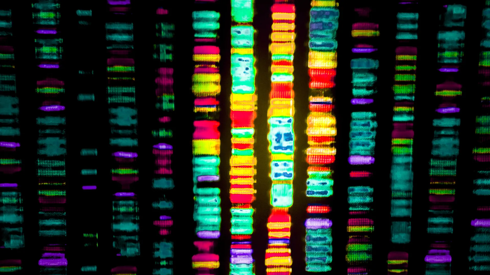
The 98% mystery: Scientists just cracked the code on “junk DNA” linked to Alzheimer’s

When people picture DNA, they often imagine a set of genes that shape our physical traits, influence behavior, and help keep our cells and organs functioning.

But genes make up only a small slice of our genetic code. Just around 2% of DNA contains our 20,000-odd genes. The other 98% has long been labelled the non-coding genome, or so-called ‘junk’ DNA. This larger portion includes many of the control switches that determine when genes turn on and how strongly they act.

Astrocytes and hidden DNA switches in the brain

Researchers from UNSW Sydney have now pinpointed DNA switches that help regulate astrocytes. Astrocytes are brain cells that support neurons, and they are known to be involved in Alzheimer’s disease.

In research published on December 18 in Nature Neuroscience, a team from UNSW’s School of Biotechnology & Biomolecular Sciences reported that they tested nearly 1000 possible switches in lab-grown human astrocytes. These switches are strings of DNA called enhancers. Enhancers can sit far from the genes they influence, sometimes separated by hundreds of thousands of DNA letters, which makes them difficult to investigate.

Testing nearly 1000 enhancers at once

To tackle that problem, the researchers combined CRISPRi with single-cell RNA sequencing. CRISPRi is a method that can switch off small stretches of DNA without cutting it. Single-cell RNA sequencing measures gene activity in individual cells. Together, the tools let the team examine the effects of nearly 1000 enhancers in a single large-scale test.

“We used CRISPRi to turn off potential enhancers in the astrocytes to see whether it changed gene expression,” says lead author Dr. Nicole Green.

“And if it did, then we knew we’d found a functional enhancer and could then figure out which gene — or genes — it controls. That’s what happened for about 150 of the potential enhancers we tested. And strikingly, a large fraction of these functional enhancers controlled genes implicated in Alzheimer’s disease.”

Cutting the list from 1000 candidates to about 150 confirmed switches greatly reduces the search area in the non-coding genome for genetic clues linked to Alzheimer’s disease.

“These findings suggest that similar studies in other brain cell types are needed to highlight the functional enhancers in the vast space of non-coding DNA”

Why “in-between” DNA matters for many diseases

Professor Irina Voineagu, who oversaw the study, says the results also provide a useful reference for interpreting other genetic research. The team’s findings create a catalogue of DNA regions that can help explain results from studies looking for disease-related genetic changes.

“When researchers look for genetic changes that explain diseases like hypertension, diabetes and also psychiatric and neurodegenerative disorders like Alzheimer’s disease — we often end up with changes not within genes so much, but in-between,” she says.

Her team directly tested those “in-between” stretches in human astrocytes and showed which enhancers truly control key brain genes.

“We’re not talking about therapies yet. But you can’t develop them unless you first understand the wiring diagram. That’s what this gives us — a deeper view into the circuitry of gene control in astrocytes.”

From gene switches to AI prediction models

Running nearly a thousand enhancer tests in the lab took painstaking effort. The researchers say this is the first time a CRISPRi enhancer screen of this size has been carried out in brain cells. Now that the groundwork has been done, the dataset can also be used to train computer models to predict which suspected enhancers are real gene switches, potentially saving years of lab work.

“This dataset can help computational biologists test how good their prediction models are at predicting enhancer function,” says Prof. Voineagu.

She adds that Google’s DeepMind team is already using the dataset to benchmark their recent deep learning model called AlphaGenome.

Potential tools for gene therapy and precision medicine

Because many enhancers are active only in specific cell types, targeting them could offer a way to fine-tune gene expression in astrocytes without changing neurons or other brain cells.

“While this is not close to being used in the clinic yet — and much work remains before these findings could lead to treatments — there is a clear precedent,” Prof. Voineagu says.

“The first gene editing drug approved for a blood disease — sickle cell anemia — targets a cell-type specific enhancer.”

Dr. Green says enhancer research could become an important part of precision medicine.

“This is something we want to look at more deeply: finding out which enhancers we can use to turn genes on or off in a single brain cell type, and in a very controlled way,” she says.

Share Button

An 11-year-old needed two new organs and doctors made history

Children’s Hospital Colorado (Children’s Colorado) has completed its first-ever dual heart and liver transplant, marking a major milestone for the hospital. The complex procedure involved dozens of specialists working across 25 multidisciplinary care teams. Nationwide, only 38 pediatric patients have previously received both a heart and liver transplant.

“Performing Children’s Colorado’s first-ever heart and liver dual organ transplant is an amazing accomplishment for our Pediatric Transplant Program,” said Dr. Megan Adams, surgical director of the Pediatric Liver Transplant and Kidney Transplant Programs. “Thanks to years of dedication and a team committed to being the trusted leaders in pediatric transplant across our seven-state region, we’re grateful to provide this level of care to even more kids who need complex organ transplants to treat life-threatening illnesses and help them live healthy and happy lives.”

Years of Preparation Lead to a Life-Saving Moment

Care teams at Children’s Colorado had spent years preparing for the possibility of a dual heart and liver transplant. Close coordination among specialists in surgery, cardiology, hepatology, and other fields, along with strong backing from hospital leadership, ensured the team was ready when 11-year-old Gracie Greenlaw and her family needed help.

Gracie was born with hypoplastic left heart syndrome (HLHS), a condition in which her heart developed with only one functioning pumping chamber. Before turning three, she underwent three major surgeries, the Norwood, the Glenn and the Fontan, to allow her heart to circulate blood effectively. Although many children with HLHS now survive into adulthood, the condition and its treatments can lead to serious long-term complications, including liver damage and liver failure.

Managing the Long-Term Effects of Congenital Heart Disease

To address these ongoing challenges, Children’s Colorado established the Fontan Multidisciplinary Clinic in 2016 as part of its Single Ventricle Program. The clinic focuses on caring for patients with HLHS and other single ventricle conditions, such as tricuspid atresia and unbalanced common atrioventricular canal, by providing coordinated, whole-patient care.

Through this program, Gracie received continuous monitoring and treatment for both her heart and liver. Her care team included experts like cardiologist Dr. Kathleen Simpson and hepatologist Dr. Dania Brigham, who worked together to manage her condition until a transplant became the best option.

“The Fontan is a lifesaving surgery, but the longer someone lives after the procedure, there is an increased chance of developing comorbidities,” Simpson said. “Our care team worked to keep her healthy and living a typical day-to-day life as long as possible before we determined a dual organ transplant would give her the best long-term quality of life.”

Preparing for a Complex Dual Organ Transplant

For years, Gracie lived with plastic bronchitis, a condition that causes thick, protein-like material to build up in the airways. Over the past year, her symptoms worsened, and signs of liver failure began to appear. Her medical team concluded that moving forward with a dual transplant was necessary, and she was placed on the transplant waitlist in April.

In preparation, dozens of specialists met regularly to plan for the surgery. They carefully accounted for the challenges of transplanting two organs at once, including differences in blood volume needs and electrolyte management during the operation.

A Carefully Orchestrated 16-Hour Surgery

Less than a month after joining the waitlist, compatible donor organs became available, made possible by another family’s decision to donate. Because the heart can only remain viable for a short time, the surgical team began with the heart transplant. Dr. Matthew Stone, surgical director of the Pediatric Heart Transplant Program, and congenital heart surgeon Dr. Emily Downs led the nine-hour procedure.

While the heart surgery was underway, the donor liver was maintained on a TransMedics Organ Care System — a specialized device designed to replicate normal liver function. This technology preserved the liver and allowed the heart surgeons the time they needed to complete their work. Dr. Adams and transplant surgeon Dr. Kendra Conzen then performed the liver transplant, which took an additional seven hours. Throughout the process, close coordination with anesthesiology teams was essential to protect Gracie’s health.

Recovery and a Return to Everyday Life

The surgery was successful. Gracie left the cardiac progress care unit just over a month later. Seven months after the transplant, she continues to attend monthly follow-up visits, but she has returned to school and is back home with her dogs.

Like other pediatric heart transplant recipients, Gracie will need another heart transplant later in life. Her transplanted liver, however, is expected to last for the rest of her lifetime.

“This procedure showcases the expertise, talent and level of care Children’s Colorado provides to our patients, including those with complex medical needs,” said Dr. Duncan Wilcox, Surgeon in Chief. “As the top-ranked pediatric hospital in Colorado and the Rocky Mountain region, we are proud of our leading-edge transplant care and look forward to supporting more patients who need dual organ transplants in the future.”

Share Button

Young people will feel burden of UK’s ageing society, report suggests

The House of Lords said raising the state pension age and increasing immigration would not be a solution.

Share Button

Neurons aren’t supposed to regrow but these ones brought back vision

For decades, neuroscientists have taught that neurons do not regenerate once they are damaged or destroyed. This belief has shaped how brain injuries are understood and treated. Yet people often regain at least some lost abilities after trauma, raising an important question: if neurons do not grow back, how does recovery happen?

A new JNeurosci paper offers insight into this puzzle. Athanasios Alexandris and colleagues at Johns Hopkins University used mice to study what happens inside the visual system after traumatic brain injury. The visual system includes cells in the eye that send information to the brain, allowing animals and humans to see. Damage to this system can disrupt communication between the eye and the brain, leading to vision problems.

Surviving Cells Rebuild Eye to Brain Connections

After injury, the researchers closely tracked the connections between cells in the eye and neurons in the brain. Instead of finding widespread regrowth of new cells, they observed something different. The cells that survived the injury began to adapt.

These surviving cells grew extra branches, which allowed them to connect with more neurons in the brain than before. This process, known as sprouting, helped compensate for cells that were lost due to injury. Over time, the number of connections between the eye and the brain returned to levels similar to those seen before the injury occurred.

Importantly, these rebuilt connections were not just structural. Measurements of brain activity showed that the new pathways were working properly and could transmit signals effectively. In practical terms, this means the visual system was able to function again despite the damage.

Sex Differences in Visual System Recovery

The study also revealed a significant difference between male and female mice. While male mice showed strong recovery through this compensatory sprouting process, female mice experienced slower or incomplete repair. The eye to brain connections in females did not always fully return to preinjury levels.

According to the authors, these findings point to a recovery mechanism that operates differently depending on sex. As Alexandris explains, “We didn’t expect to see sex differences, but this aligns with clinical observations in humans. Women experience more lingering symptoms from concussion or brain injury than men. Understanding the mechanism behind the branch sprouting we observed — and what delays or prevents this mechanism in females — could eventually point toward strategies to promote recovery from traumatic or other forms of neural injury.”

The research team plans to continue investigating why this repair process differs between females and males. By uncovering the biological factors that influence neural recovery, they hope to identify new ways to improve healing after brain injuries, including concussions and other forms of trauma.

Share Button

Has flu peaked? What the figures tell us

NHS remains on high alert over flu, health bosses say, but there are signs infections are levelling off.

Share Button