Astronomers discover dying stars eating their planets

A new study from astronomers at UCL (University College London) and the University of Warwick suggests that aging stars may be destroying the giant planets orbiting closest to them.

When stars like the Sun exhaust their hydrogen fuel, they begin to cool and expand, transforming into red giants. For our Sun, this dramatic phase is expected to occur in roughly five billion years.

The research, published in the Monthly Notices of the Royal Astronomical Society, analyzed nearly half a million stars that had recently entered this “post-main sequence” stage of evolution.

Searching for Planets Around Evolving Stars

The team identified 130 planets and potential planet candidates (i.e., that still need to be confirmed) orbiting closely around these aging stars, including 33 new candidates never detected before.

They found that such planets were far less common around stars that had expanded and cooled enough to qualify as red giants (i.e., those further along in their post-main sequence evolution). This pattern suggests that many of these planets may already have been destroyed.

Evidence of Planetary Destruction

Lead author Dr. Edward Bryant (Mullard Space Science Laboratory at UCL and the University of Warwick) explained: “This is strong evidence that as stars evolve off their main sequence they can quickly cause planets to spiral into them and be destroyed. This has been the subject of debate and theory for some time but now we can see the impact of this directly and measure it at the level of a large population of stars.

“We expected to see this effect but we were still surprised by just how efficient these stars seem to be at engulfing their close planets.”

According to Dr. Bryant, the destruction occurs through a powerful gravitational struggle known as tidal interaction. As a star grows and expands, these forces intensify. “Just like the Moon pulls on Earth’s oceans to create tides, the planet pulls on the star,” he said. “These interactions slow the planet down and cause its orbit to shrink, making it spiral inwards until it either breaks apart or falls into the star.”

What It Means for the Solar System

Co-author Dr. Vincent Van Eylen (Mullard Space Science Laboratory at UCL) added perspective: “In a few billion years, our own Sun will enlarge and become a red giant. When this happens, will the solar system planets survive? We are finding that in some cases planets do not.

“Earth is certainly safer than the giant planets in our study, which are much closer to their star. But we only looked at the earliest part of the post-main sequence phase, the first one or two million years of it — the stars have a lot more evolution to go.

“Unlike the missing giant planets in our study, Earth itself might survive the Sun’s red giant phase. But life on Earth probably would not.”

To carry out their research, the team used data from NASA’s Transiting Exoplanet Survey Satellite (TESS). They employed an algorithm to identify small, repeated dips in starlight caused by planets passing in front of their stars. Their focus was on giant planets with short orbits (i.e., taking no more than 12 days to circle their star).

Starting with more than 15,000 possible signals, the researchers used rigorous checks to eliminate false positives, ultimately narrowing the list to 130 confirmed or candidate planets. Of these, 48 were already known, 49 were known candidates awaiting confirmation, and 33 were completely new discoveries.

Fewer Planets Around Older Stars

The researchers found that stars further along in their evolution were significantly less likely to host nearby giant planets. The overall occurrence rate was just 0.28%, with younger post-main sequence stars showing a higher rate (0.35%) comparable to main sequence stars. The most evolved stars — those classified as red giants — showed a sharp drop to 0.11%. (For this analysis, the smallest 12 of the 130 identified planets were excluded.)

Using TESS data, astronomers can estimate a planet’s size (radius). To confirm whether these objects are true planets or low-mass stars or brown dwarfs (“failed stars” that never ignited nuclear fusion), their mass must be determined.

This is done by measuring the tiny shifts in the motion of the host star caused by a planet’s gravitational pull. These “stellar wobbles” allow scientists to infer the planet’s mass.

Dr. Bryant added: “Once we have these planets’ masses, that will help us understand exactly what is causing these planets to spiral in and be destroyed.”

The research was supported by the UK Science and Technology Facilities Council (STFC).

Share Button

Scientists find hidden brain source that fuels dementia

Scientists at Weill Cornell Medicine have identified a surprising culprit that may contribute to dementia: free radicals generated in a particular region of brain support cells known as astrocytes. The study, published Nov. 4 in Nature Metabolism, found that blocking this specific site reduced inflammation and protected neurons. The results point toward a promising new strategy for treating neurodegenerative diseases such as frontotemporal dementia and Alzheimer’s disease.

“I’m really excited about the translational potential of this work,” said Dr. Anna Orr, the Nan and Stephen Swid Associate Professor of Frontotemporal Dementia Research in the Feil Family Brain and Mind Research Institute and member of the Appel Alzheimer’s Disease Research Institute at Weill Cornell, who co-led the study. “We can now target specific mechanisms and go after the exact sites that are relevant for disease.”

How Mitochondria and Free Radicals Affect the Brain

The research focused on mitochondria, the cell’s energy-producing structures that convert food into usable energy. In the process, mitochondria release reactive oxygen species (ROS) — molecules commonly known as free radicals. At normal levels, ROS help regulate essential cell functions, but excessive or poorly timed production can damage cells.

“Decades of research implicate mitochondrial ROS in neurodegenerative diseases,” said Dr. Adam Orr, an assistant professor of research in neuroscience in the Feil Family Brain and Mind Research Institute at Weill Cornell, who co-led the work.

Because of this connection, scientists have long tested antioxidants as a potential way to neutralize ROS and slow neurodegeneration. However, these clinical trials have largely failed. “That lack of success might be related to the inability of antioxidants to block ROS at their source and do so selectively without altering cell metabolism,” Dr. Adam Orr explained.

A New Way to Stop Harmful Free Radicals

As a postdoctoral researcher, Dr. Orr developed a drug discovery platform designed to find molecules that specifically suppress ROS at individual mitochondrial sites while leaving normal functions intact. Through this approach, the team identified a group of compounds called S3QELs (“sequels”), which showed potential to block harmful ROS activity.

The researchers focused on Complex III, a mitochondrial site known for producing ROS that can leak into the rest of the cell, potentially causing damage. To their surprise, the excess ROS did not originate from neurons, but from astrocytes — non-neuronal cells that provide structural and metabolic support to neurons.

“When we added S3QELs, we found significant neuronal protection but only in the presence of astrocytes,” said Daniel Barnett, a graduate student in the Orr lab and the study’s lead author. “This suggested that ROS coming from Complex III caused at least some of the neuronal pathology.”

Further experiments showed that when astrocytes were exposed to disease-related factors such as inflammatory molecules or proteins linked to dementia (including amyloid-beta), their mitochondrial ROS production increased dramatically. Treatment with S3QELs suppressed much of this rise, while blocking other ROS sources did not have the same effect.

Barnett discovered that ROS oxidized certain immune and metabolic proteins involved in neurological disease, altering the activity of thousands of genes tied to inflammation and dementia.

“The precision of these mechanisms had not been previously appreciated, especially not in brain cells,” said Dr. Anna Orr. “This suggests a very nuanced process in which specific triggers induce ROS from specific mitochondrial sites to affect specific targets.”

Promising Results in Animal Models

When the team administered the S3QEL compound to mice engineered to model frontotemporal dementia, they observed reduced astrocyte activation, lower levels of inflammatory gene expression, and a decrease in a tau modification linked to dementia. Remarkably, these effects appeared even when treatment began after symptoms had already started.

Extended treatment improved lifespan, was well tolerated, and produced no significant side effects. Dr. Anna Orr attributes this to the compound’s highly targeted action.

The team plans to continue developing the S3QEL compounds in collaboration with medicinal chemist Dr. Subhash Sinha, professor of research in neuroscience in the Brain and Mind Research Institute and member of the Appel Alzheimer’s Disease Research Institute at Weill Cornell.

They also intend to investigate how disease-associated genes influence ROS production and whether certain genetic variants that raise or lower dementia risk might do so by altering mitochondrial ROS activity.

Changing How Scientists Think About Free Radicals

“The study has really changed our thinking about free radicals and opened up many new avenues of investigation,” said Dr. Adam Orr. The potential of these findings to open new research approaches to inflammation and neurodegeneration is highlighted in the journal.

Share Button

How caffeine can help you manage headaches and other tips

If you’re getting a pattern of milder, recurring headaches here are four things you can do to help manage it.

Share Button

German nurse gets life in jail after murdering 10 to reduce workload

The palliative care nurse was convicted of the murder of 10 patients, and the attempted murder of 27 others.

Share Button

ADHD services shutting door to new NHS patients as demand soars, BBC finds

A BBC investigation finds that a host of areas in England are closing waiting lists and others are rationing care.

Share Button

BMA rejects fresh offer to end doctor strikes

An offer was made to avert the resident doctors’ strike but the union said it did not go far enough.

Share Button

LED mask ads banned over acne and rosacea claims

Adverts for cosmetic devices not registered with the medicines regulator must not make medical claims.

Share Button

A 480-million-year-old parasite still infects oysters today

A surprising new study has revealed that a parasite still troubling modern oysters first began infecting shell-dwelling sea creatures hundreds of millions of years before the dinosaurs vanished.

Researchers reporting in iScience used high-resolution 3D imaging to examine 480-million-year-old fossil shells from Morocco, a site famous for its exceptionally preserved marine life. The scans uncovered a pattern of unusual markings etched both on the surfaces of the shells and inside them.

“The marks weren’t random scratches,” explained Karma Nanglu, a paleobiologist at UC Riverside and lead author of the study. “We saw seven or eight of these perfect question mark shapes on each shell fossil. That’s a pattern.”

Javier Ortega-Hernandez, a Harvard evolutionary biologist and co-author, recalled the team’s initial confusion. “It took us a while to figure out the mystery behind these peculiar-looking traces. It was as if they were taunting us with their question mark-like shape,” he said. “But as often happens, we came across the answer while deep in obscure literature before our eureka moment.”

Ancient Worms Behind the Mystery Marks

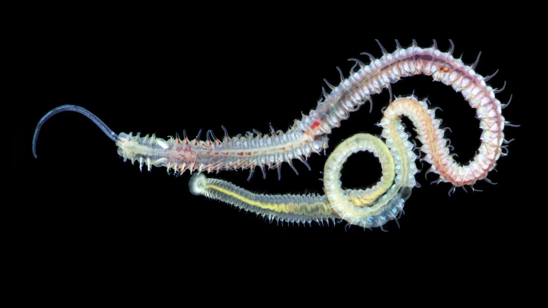
After comparing the markings with modern examples, the scientists concluded that they were made by a soft-bodied marine bristle worm belonging to a group known as spionids. These worms, still common today, bore into the shells of mussels and oysters but typically don’t kill their hosts outright.

“They parasitize the shells of bivalves like oysters, not the flesh of the animals themselves,” said Nanglu. “But damaging their shells may increase oyster death rates.”

The fossils studied came from early relatives of modern clams that lived during the Ordovician Period, a time of rapid ecological expansion when marine life became increasingly mobile, predatory, and parasitic. “This is a time when ocean ecosystems got more intense,” Nanglu said. “You see the rise of mobility, predation, and, clearly, parasitism.”

A Half-Billion-Year Parasitic Lineage

The researchers considered other explanations for the distinctive marks, such as self-inflicted shell growth patterns or traces from unrelated organisms. However, the evidence most strongly matched spionid activity.

“There’s one image in particular, from a study of modern worms, that shows exactly the same shape inside a shell,” Nanglu said. “That was the smoking gun.”

The finding offered more than just an identification — it provided a rare evolutionary insight. “This group of worms hasn’t changed its lifestyle in nearly half a billion years,” said Nanglu. “We tend to think of evolution as constant change, but here’s an example of a behavior that worked so well, it stayed the same through multiple mass extinction events.”

Peering Inside Fossils with High-Tech Scans

To reveal the interior structures of the shells, the team used a technique similar to medical CT imaging, called micro-CT scanning. This high-resolution method allowed them to visualize internal burrows and hidden shells embedded within the rock layers, which were stacked like a layered cake.

“We never would’ve seen this without the scanner,” Nanglu said.

The Parasite’s Ancient Life Cycle

The worm’s life cycle helped confirm its identity. The researchers determined that it likely began as a larva that attached itself to a shell, dissolved a small spot to secure its position, and then tunneled deeper as it grew — creating the recognizable question mark shape.

No other known species leaves this precise trace. “”If it’s not a spionid, then it’s something we’ve never seen before,” Nanglu said. “But it would have to have evolved the same behavior, in the same place, in the same way.”

A Survivor Across Deep Time

Remarkably, the same burrowing behavior continues in today’s oceans. Although spionid worms do not consume their hosts directly, the damage they inflict on oyster shells still increases mortality rates in modern fisheries.

“This parasite didn’t just survive the cutthroat Ordovician period, it thrived,” Nanglu said. “It’s still interfering with the oysters we want to eat, just as it did hundreds of millions of years ago.”

Fossil Time Capsules of Ancient Life

The Moroccan fossil site where these discoveries were made is celebrated for capturing behavior frozen in time. Other fossils there have preserved scenes of animals interacting, such as creatures feeding on the remains of squid-like ancestors.

“You’re lucky to get any record of an animal from that long ago,” Nanglu said. “But to see evidence of two animals interacting? That’s gold.”

Share Button

A shapeshifting protein explains rabies’ deadly power

  • Viruses are masters of efficiency, able to take over our cells and control vital processes using only a handful of genes.
  • For years, scientists have wondered how something so small could do so much.
  • Researchers have now uncovered the answer — a discovery that could reshape our understanding of how viruses work and lead to new ways to fight them.

Breakthrough Reveals How Viruses Outsmart Human Cells

A team of Australian scientists has uncovered how certain viruses manage to seize control of human cells, a finding that could lead to the next generation of antivirals and vaccines.

The research, led by Monash University and the University of Melbourne and published in Nature Communications, explains how the rabies virus can manipulate a wide range of cellular activities despite producing only a few proteins.

Scientists believe this same mechanism could also be at work in other deadly pathogens, including Nipah and Ebola viruses. If so, the discovery could pave the way for new treatments that block these viral strategies.

How Viruses Do So Much With So Little

Co-senior author Associate Professor Greg Moseley, head of the Monash Biomedicine Discovery Institute’s (BDI) Viral Pathogenesis Laboratory, described the remarkable efficiency of viruses.

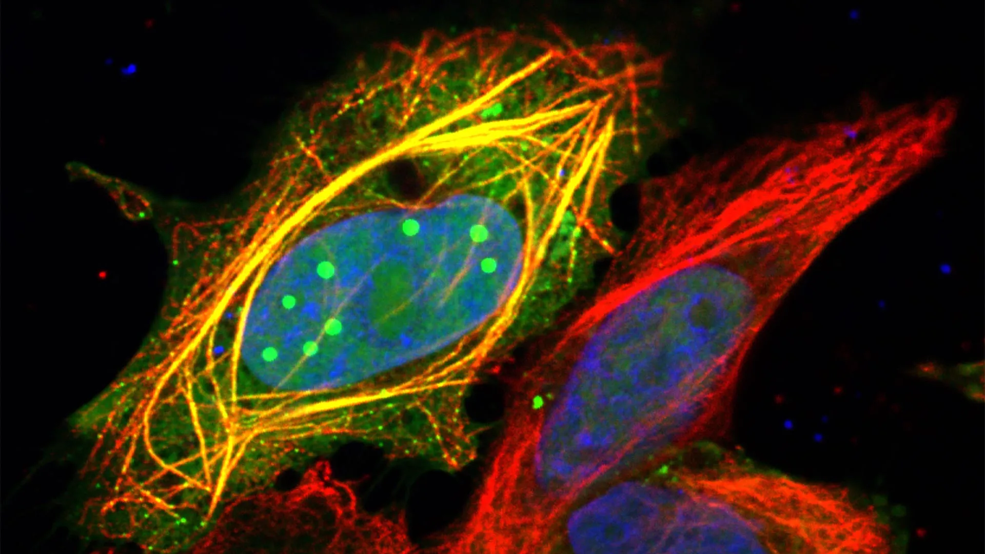
“Viruses such as rabies can be incredibly lethal because they take control of many aspects of life inside the cells they infect,” Associate Professor Moseley said. “They hijack the machinery that makes proteins, disrupt the ‘postal service’ that sends messages between different parts of the cell, and disable the defenses that normally protect us from infection.”

He explained that scientists have long puzzled over how viruses with such limited genetic material could be so powerful. “Rabies virus, for example, has the genetic material to make only five proteins, compared with about 20,000 in a human cell,” he said.

The Key: A Shape-Shifting Viral Protein

Co-first author Dr. Stephen Rawlinson, a research fellow in the Moseley Lab, said the team’s work offers a long-sought answer.

“Our study provides an answer,” he said. “We discovered that one of rabies virus’s key proteins, called P protein, gains a remarkable range of functions through its ability to change shape and to bind to RNA.”

“RNA is the same molecule used in new-generation RNA vaccines, but it plays essential roles inside our cells, carrying genetic messages, coordinating immune responses, and helping make the building blocks of life.”

Taking Over the Cell’s Inner World

Co-senior author Professor Paul Gooley, who leads the University of Melbourne’s Gooley Laboratory, said the viral P protein’s ability to interact with RNA allows it to shift between different physical ‘phases’ within a cell.

“This allows it to infiltrate many of the cell’s liquid-like compartments, take control of vital processes, and turn the cell into a highly efficient virus factory,” Professor Gooley said.

Although this research focused on rabies, he noted that similar tactics may be used by other deadly viruses, including Nipah and Ebola. “Understanding this new mechanism opens exciting possibilities for developing antivirals or vaccines that block this remarkable adaptability,” he added.

Rethinking How Viral Proteins Work

Dr. Rawlinson said the findings challenge how scientists have traditionally viewed multifunctional viral proteins. “Until now, these proteins were often viewed like trains made up of several carriages, with each carriage (or module) responsible for a specific task,” he said.

“According to this view, shorter versions of a protein should simply lose functions as carriages are removed. However, this simple model could not explain why some shorter viral proteins actually gain new abilities. We found that multifunctionality can also arise from the way the ‘carriages’ interact and fold together to create different overall shapes, as well as forming new abilities such as binding to RNA.”

A New Perspective on Viral Adaptability

Associate Professor Moseley said that the ability of the P protein to bind RNA allows it to move between different physical ‘phases’ inside the cell.

“In doing so, it can access and manipulate many of the cell’s own liquid-like compartments that control key processes, such as immune defense and protein production,” he said. “By revealing this new mechanism, our study provides a fresh way of thinking about how viruses use their limited genetic material to create proteins that are flexible, adaptable, and able to take control of complex cellular systems.”

This study involved Monash University, the University of Melbourne, the Australian Nuclear Science and Technology Organisation (Australian Synchrotron), Peter Doherty Institute for Infection and Immunity, Commonwealth Scientific and Industrial Research Organisation (CSIRO), the Australian Centre for Disease Preparedness (ACDP), and Deakin University.

Share Button

Frozen for 6 million years, Antarctic ice rewrites Earth’s climate story

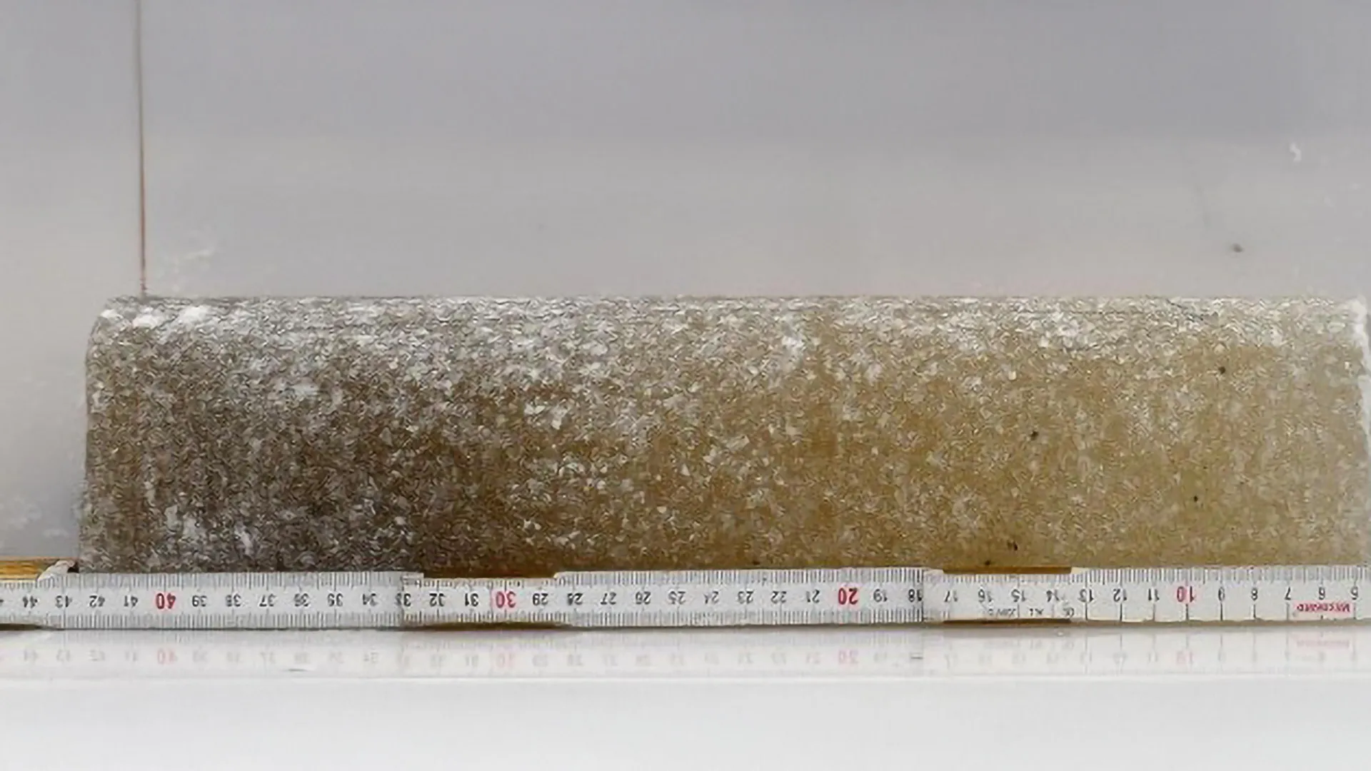
In a breakthrough for climate science, a team of U.S. researchers has identified the oldest directly dated ice and air ever discovered on Earth. The samples were found in the Allan Hills region of East Antarctica, an area long known to preserve ancient ice near the surface.

According to a study published on October 28 in the Proceedings of the National Academy of Sciences, the ice is about 6 million years old. Tiny air bubbles sealed within the ice provide a rare and direct glimpse into Earth’s ancient atmosphere, offering scientists an unmatched record of the planet’s past climate.

A Glimpse Into a Warmer Ancient World

The oldest sample collected from the Allan Hills dates back 6 million years, a period when geological records indicate Earth was significantly warmer and sea levels were much higher than they are today.

The research effort was led by Sarah Shackleton of the Woods Hole Oceanographic Institution and John Higgins of Princeton University. Both are part of the National Science Foundation-funded Center for Oldest Ice Exploration (COLDEX), a multi-institution collaboration of 15 U.S. research partners coordinated by Oregon State University.

“Ice cores are like time machines that let scientists take a look at what our planet was like in the past,” explained Shackleton, who has worked on several Antarctic drilling expeditions. “The Allan Hills cores help us travel much further back than we imagined possible.”

Ed Brook, COLDEX Director and paleoclimatologist at Oregon State University, described the find as the center’s most important discovery so far. The NSF Science and Technology Center, established in 2021, was created to explore the Antarctic ice sheet, the largest reservoir of frozen water on Earth.

“We knew the ice was old in this region,” Brook said. “Initially, we had hoped to find ice up to 3 million years old, or maybe a little older, but this discovery has far exceeded our expectations.”

A Global Race to Reach Deeper Into Time

COLDEX is among several international teams striving to extend the ice core record beyond its previous 800,000-year limit. A European project recently announced a continuous deep core reaching 1.2 million years in East Antarctica, but the Allan Hills samples push the timeline back several million years further, though in discrete fragments rather than a single continuous core.

The COLDEX researchers work at a remote field site in the Allan Hills, spending months drilling through one to two hundred meters of ice near the edge of the Antarctic ice sheet. In this region, rugged terrain and ice flow patterns act together to preserve extremely old ice closer to the surface. By contrast, obtaining continuous cores from the Antarctic interior typically requires drilling to depths of more than 2,000 meters.

“We’re still working out the exact conditions that allow such ancient ice to survive so close to the surface,” said Shackleton. “Along with the topography, it’s likely a mix of strong winds and bitter cold. The wind blows away fresh snow, and the cold slows the ice to almost a standstill. That makes Allan Hills one of the best places in the world to find shallow old ice, and one of the toughest places to spend a field season.”

The age of the Allan Hills samples was determined directly from the ice itself using precise measurements of an isotope of the noble gas argon. This approach allows scientists to date the ice without relying on surrounding sediments or other indirect evidence.

Although the record is not continuous, the samples are far older than any previously studied ice. As Higgins noted, “Tthe team has built up a library of what we call ‘climate snapshots’ roughly six times older than any previously reported ice core data, complementing the more detailed younger data from cores in the interior of Antarctica.”

Reconstructing 6 Million Years of Cooling

Analysis of oxygen isotopes in the ice revealed that the region cooled by about 12 degrees Celsius (roughly 22 degrees Fahrenheit) over the past 6 million years. This is the first direct evidence quantifying how much Antarctica’s climate has cooled since that ancient warm period.

Future research will focus on reconstructing concentrations of greenhouse gases and ocean heat levels preserved within the ice bubbles — key clues to understanding the natural forces driving long-term climate change.

A new COLDEX expedition is scheduled to return to the Allan Hills in the coming months for additional drilling. The team hopes to recover even older samples and produce a more detailed record of Earth’s ancient atmosphere.

“Given the spectacularly old ice we have discovered at Allan Hills, we also have designed a comprehensive longer-term new study of this region to try to extend the records even further in time, which we hope to conduct between 2026 and 2031,” Brook said.

Collaboration and Support

Contributors to the study include Julia Marks Peterson, Christo Buizert, and Jenna Epifanio of Oregon State; Valens Hishamunda, Austin Carter, and Michael Bender of Princeton; Lindsey Davidge, Eric Steig, and Andrew Schauer of the University of Washington; Sarah Aarons, Jacob Morgan, and Jeff Severinghaus of Scripps Institution of Oceanography at the University of California, San Diego; Andrei V. Kurbatov and Douglas Introne of the University of Maine; Yuzhen Yan of Tongji University; and Peter Neff of the University of Minnesota.

COLDEX is supported by the NSF Office of Polar Programs; the NSF Office of Integrative Activities’ Science and Technology Center Program; and Oregon State University. Field operations in Antarctica are carried out with support from the U.S. Antarctic Program and NSF funding, with drilling assistance from the NSF U.S. Ice Drilling Program and sample storage managed by the NSF Ice Core Facility in Denver, Colorado.

Share Button