Siobhan Gorman was 16 weeks pregnant and home alone when she gave birth to baby Archie.


Siobhan Gorman was 16 weeks pregnant and home alone when she gave birth to baby Archie.
The aim is to help people return to work quicker and reduce the length of time they need fit notes – better known as sick notes.
The study looked at vaccines deployed during outbreaks of five deadly diseases.
The Labour peer says the strikes could damage public trust in doctors.

In the last year, there has been a surge in proteins developed by AI that will eventually be used in the treatment of everything from snakebites to cancer. What would normally take decades for a scientist to create — a custom-made protein for a particular disease — can now be done in seconds.
For the first time, Australian scientists have used Artificial Intelligence (AI) to generate a ready-to-use biological protein, in this case, one that can kill antibiotic resistant bacteria like E. coli.
This study, published in Nature Communications, provides a new way to combat the growing crisis caused by antibiotic resistant super bugs. By using AI in this way, Australian science has now joined countries like the US and China having developed AI platforms capable of rapidly generating thousands of ready-to-use proteins, paving the way for faster, more affordable drug development and diagnostics that could transform biomedical research and patient care.
The Nature Communications paper is co-led by Dr. Rhys Grinter and Associate Professor Gavin Knott, a Snow Medical Fellow, who lead the new AI Protein Design Program with nodes at the University of Melbourne Bio21 Institute and Monash Biomedicine Discovery Institute.
According to Dr. Grinter and A/Prof. Knott, the AI Protein Design Platform used in this work is the first in Australia that models the work done by David Baker (who won the Nobel Prize in Chemistry last year) developing an end-to-end approach that could create a wide range of proteins. “These proteins are now being developed as pharmaceuticals, vaccines, nanomaterials and tiny sensors, with many other applications yet to be tested” Associate Professor Knott said.
For this study, the AI Protein Design Platform used AI-driven protein design tools that are freely available for scientists everywhere. “It’s important to democratize protein design so that the whole world has the ability to leverage these tools,” said Daniel Fox, the PhD student who performed most of the experimental work for the study. “Using these tools and those we are developing in-house, we can engineer proteins to bind a specific target site or ligand, as inhibitors, agonists or antagonists, or engineered enzymes with improved activity and stability.”
According to Dr Grinter, currently proteins used in the treatment of diseases like cancer or infections are derived from nature and repurposed through rational design or in vitro evolution and selection. “These new methods in deep learning enable efficient de novo design of proteins with specific characteristics and functions, lowering the cost and accelerating the development of novel protein binders and engineered enzymes,” he said.
Since the work of David Baker, new tools and software are being developed, such as Bindcraft and Chai which have been incorporated into an AI Protein Design Platform co-led by Dr. Grinter and A/Prof. Knott..
Professor John Carroll, Director of the Monash Biomedicine Discovery Institute, said the new AI Protein Design Program ‘brings Australia “right up to speed in this exciting new modality for designing novel therapeutics and research tools. It is testament to the entrepreneurial spirit of two fabulous young scientists who have worked night and day to build this capability from scratch.”
“The Program, based at Monash University and the University of Melbourne, is run by a team of talented structural biologists and computer scientists who understand the design process from end-to-end. This in-depth knowledge of protein structure and machine learning makes us a highly agile program capable of regularly onboarding cutting edge tools in AI-protein design,” Associate Professor Knott said.

Pain isn’t just a physical sensation — it also carries emotional weight. That distress, anguish, and anxiety can turn a fleeting injury into long-term suffering.
Researchers at the Salk Institute have now identified a brain circuit that gives physical pain its emotional tone, revealing a new potential target for treating chronic and affective pain conditions such as fibromyalgia, migraine, and post-traumatic stress disorder (PTSD).
Published on July 9, 2025, in Proceedings of the National Academy of Sciences, the study identifies a group of neurons in a central brain area called the thalamus that appears to mediate the emotional or affective side of pain in mice. This new pathway challenges the textbook understanding of how pain is processed in the brain and body.
“For decades, the prevailing view was that the brain processes sensory and emotional aspects of pain through separate pathways,” says senior author Sung Han, associate professor and holder of the Pioneer Fund Developmental Chair at Salk. “But there’s been debate about whether the sensory pain pathway might also contribute to the emotional side of pain. Our study provides strong evidence that a branch of the sensory pain pathway directly mediates the affective experience of pain.”
The physical sensation of pain is what allows you to immediately detect it, assess its intensity, and identify its source. The affective part of pain is what makes it so unpleasant. This emotional discomfort motivates you to take action and helps you learn to associate negative feelings with the situation so you can avoid it in the future.
This is a critical distinction. Most people start to perceive pain at the same stimulus intensities, meaning we all process the sensory side of pain fairly similarly. In comparison, our ability to tolerate pain varies greatly. How much we suffer or feel threatened by pain is determined by our affective processing, and if that becomes too sensitive or lasts too long, it can result in a pain disorder. This makes it important to understand which parts of the brain control these different dimensions of pain.
Sensory pain was thought to be mediated by the spinothalamic tract, a pathway that sends pain signals from the spinal cord to the thalamus, which then relays them to sensory processing areas across the brain.
Affective pain was generally thought to be mediated by a second pathway called the spinoparabrachial tract, which sends pain information from the spinal cord into the brainstem.
However, previous studies using older research methods have suggested the circuitry of pain may be more complex. This long-standing debate inspired Han and his team to revisit the question with modern research tools.
Using advanced techniques to manipulate the activity of specific brain cells, the researchers discovered a new spinothalamic pathway in mice. In this circuit, pain signals are sent from the spinal cord into a different part of the thalamus, which has connections to the amygdala, the brain’s emotional processing center. This particular group of neurons in the thalamus can be identified by their expression of CGRP (calcitonin gene-related peptide), a neuropeptide originally discovered in Professor Ronald Evans’ lab at Salk.
When the researchers “turned off” (genetically silenced) these CGRP neurons, the mice still reacted to mild pain stimuli, such as heat or pressure, indicating their sensory processing was intact. However, they didn’t seem to associate lasting negative feelings with these situations, failing to show any learned fear or avoidance behaviors in future trials. On the other hand, when these same neurons were “turned on” (optogenetically activated), the mice showed clear signs of distress and learned to avoid that area, even when no pain stimuli had been used.
“Pain processing is not just about nerves detecting pain; it’s about the brain deciding how much that pain matters,” says first author Sukjae Kang, a senior research associate in Han’s lab. “Understanding the biology behind these two distinct processes will help us find treatments for the kinds of pain that don’t respond to traditional drugs.”
Many chronic pain conditions — such as fibromyalgia and migraine — involve long, intense, unpleasant experiences of pain, often without a clear physical source or injury. Some patients also report extreme sensitivity to ordinary stimuli like light, sound, or touch, which others would not perceive as painful.
Han says overactivation of the CGRP spinothalamic pathway may contribute to these conditions by making the brain misinterpret or overreact to sensory inputs. In fact, transcriptomic analysis of the CGRP neurons showed that they express many of the genes associated with migraine and other pain disorders.
Notably, several CGRP blockers are already being used to treat migraines. This study may help explain why these medications work and could inspire new nonaddictive treatments for affective pain disorders.
Han also sees potential relevance for psychiatric conditions that involve heightened threat perception, such as PTSD. Growing evidence from his lab suggests that the CGRP affective pain pathway acts as part of the brain’s broader alarm system, detecting and responding to not only pain but a wide range of unpleasant sensations. Quieting this pathway with CGRP blockers could offer a new approach to easing fear, avoidance, and hypervigilance in trauma-related disorders.
Importantly, the relationship between the CGRP pathway and the psychological pain associated with social experiences like grief, loneliness, and heartbreak remains unclear and requires further study.
“Our discovery of the CGRP affective pain pathway gives us a molecular and circuit-level explanation for the difference between detecting physical pain and suffering from it,” says Han. “We’re excited to continue exploring this pathway and enabling future therapies that can reduce this suffering.”
Other authors include Shijia Liu, Jong-Hyun Kim, Dong-Il Kim, Tae Gyu Oh, Jiahang Peng, Mao Ye, Kuo-Fen Lee, Ronald M. Evans, and Martyn Goulding of Salk.
The work was supported by the National Institutes of Mental Health (BRAINS grant 1R01MH116203) and the Simons Foundation (Bridge to Independence award SFARI #388708).
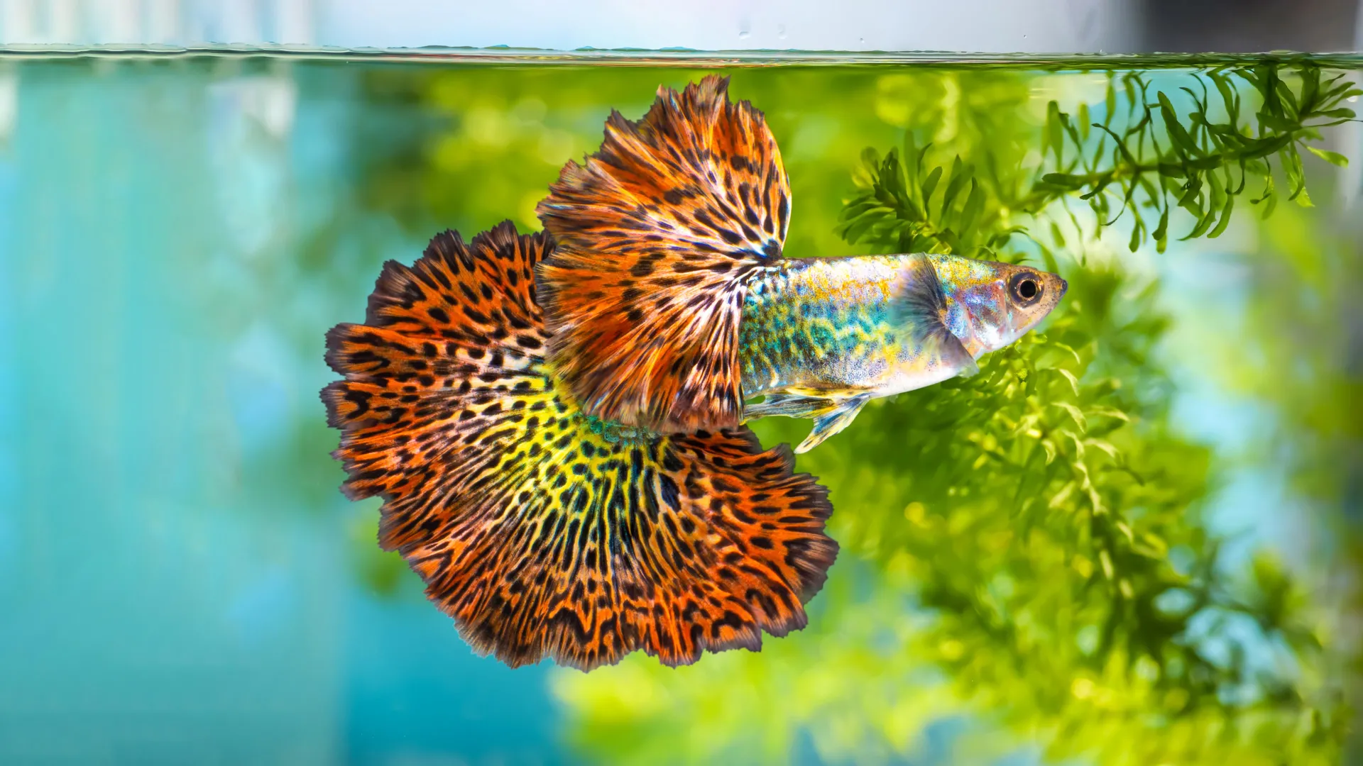
It turns out color isn’t just fashionable for guppies: According to a new UBC study, the more orange a male, the more virile it is.
The research published in Nature Ecology & Evolution shines light on an enduring evolutionary mystery: why male guppies have such vibrant and varied colors and patterns.
Virile me up
Zoologists Drs. Wouter van der Bijl and Judith Mank used deep learning, genetic studies and bred three generations of increasingly orange guppies to investigate. They found the more colorful males were up to two times more sexually active, performing for females at a greater rate and for longer periods of time, and attempting to sneakily copulate more often.
Orange you glad to see me, baby?
It’s known that female guppies prefer orange, and unusual, patterns in their male partners, but the team found that the color diversity of guppies comes from the same cells that are responsible for forming the brain, suggesting a genetic link between how guppies look and how they behave.
“Previously, people thought perhaps males realized that if they were more orange, they were more sexy. With the genetic link, it may be that they’re healthier and fitter,” said Dr. Mank.
Let’s recombine sometime
The researchers found the guppy color genes, and the locations they appeared in, were tied to multiple chromosomes, creating a vast architecture of genetic possibilities. Seven orange and eight black color types were identified overall, allowing for a potential 32,768 unique pattern combinations.
“Genetic variation is the raw material that evolution uses to produce resilient, adapted animals and plants, including for things like climate change or disease,” said van der Bijl. “We often look at extreme examples to understand where genetic variation comes from and how it’s maintained.”
Marc Lamah, dropped by a private provider, continues to operate on NHS patients in Brighton.

What can lemurs tell us about inflammation and aging, aka “inflammaging” in humans? That’s the question Elaine Guevara, a biological anthropologist who studies the evolution of life history and aging in primates, set out to understand.
In newly published research on age-related inflammation in ring-tailed and sifaka lemurs, Guevara discovered that perhaps we should rethink the inevitability of inflammaging in humans.
Although similar in many ways, ring-tailed and sifaka lemurs show differences in life pacing and lifespan, making useful comparisons. Because lemurs and humans are primates and share a common ancestor that lived millions of years ago, they offer valuable insights into human evolution.
Her findings, she said, were “surprising.”
“Contrary to our predictions, neither species showed age-related change in either marker of oxidative stress. Neither lemur species exhibited age-related change in inflammation; if anything, contrary to our prediction, ring-tailed lemurs showed marginal declines in inflammation with age,” Guevara said.
This finding, consistent with a few recent studies of other non-human primates, suggests that lemurs avoid the phenomenon of “inflammaging” widely observed in humans.
The study shows inflammaging is not a universal feature of primates, pointing to some differences that might suggest it turns out it’s not even a universal feature of humans, according to Christine Drea, a professor of evolutionary anthropology who was one of the researchers working with Guevara.
What is Inflammaging?
As we grow older, low-grade chronic inflammation sets in, which in turn can cause health problems such as heart disease, strokes, diabetes, cancer and osteoarthritis.
Why inflammaging increases with age in humans, what causes it and how it can be prevented are answers to questions that can unlock critical information to help humans live longer and healthier lives.
Collecting Data from Lemurs
Drea said the team first had to find a way to measure oxidative stress, which can be found in blood, urine and saliva. They settled on urine.
“Our role at the beginning was planning, designing, brainstorming, comparing and getting these samples,” said Drea, who has worked with the Duke Lemur Center since 1999. The Lemur Center does not allow research that will harm the animals.
The next step says Guevara is to conduct similar research with lemurs in the wild.
“There are a lot of good reasons to think that aging can be quite different in captivity and in the wild, and that in itself, is informative to evaluating the degree to which human inflammation is intrinsic versus environmental,” she said.
In the meantime, Guevara says this study serves as the first step in unraveling the question of why humans are suffering from inflammatory-related and age-related conditions and finding ways to treat them.
With a rapidly aging global population, “these insights are essential for mitigating disability and improving quality of life in later years,” she said.
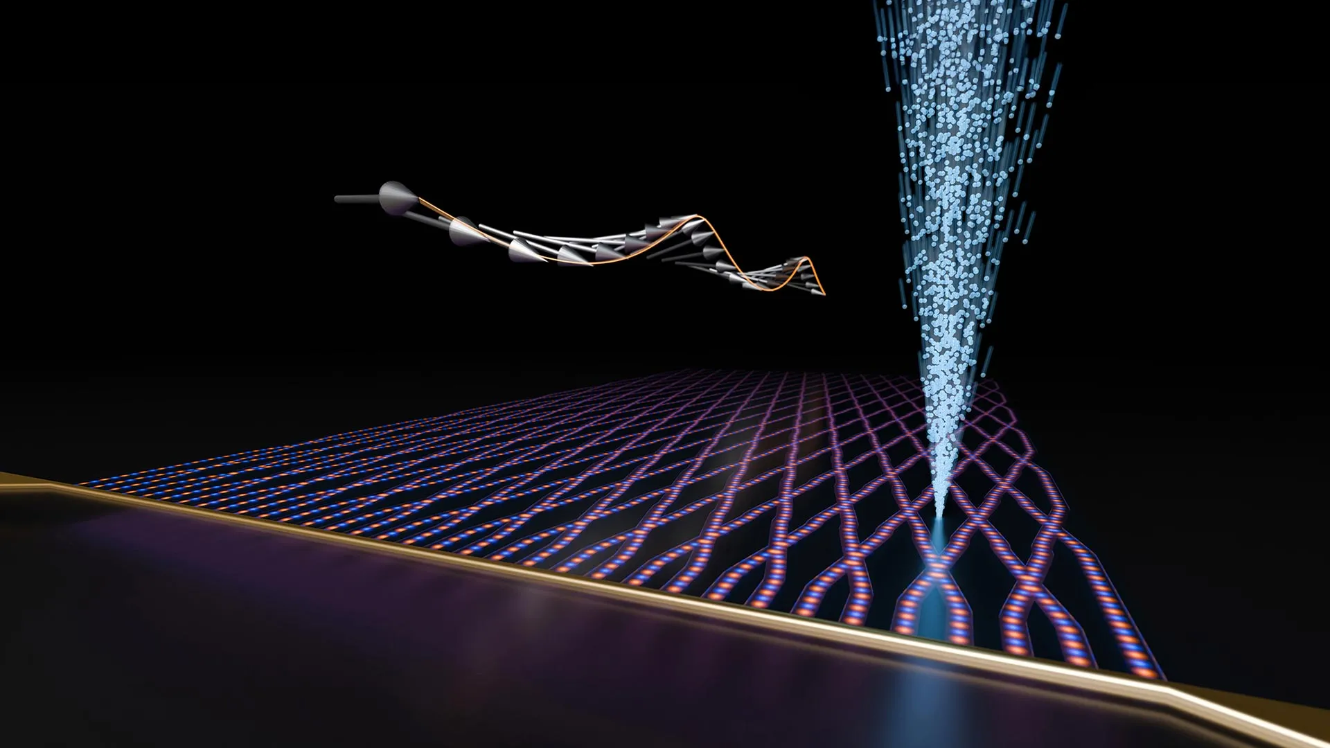
The rapid rise in AI applications has placed increasingly heavy demands on our energy infrastructure. All the more reason to find energy-saving solutions for AI hardware. One promising idea is the use of so-called spin waves to process information. A team from the Universities of Münster and Heidelberg (Germany) led by physicist Prof. Rudolf Bratschitsch (Münster) has now developed a new way to produce waveguides in which the spin waves can propagate particularly far. They have thus created the largest spin waveguide network to date. Furthermore, the group succeeded in specifically controlling the properties of the spin wave transmitted in the waveguide. For example, they were able to precisely alter the wavelength and reflection of the spin wave at a certain interface. The study was published in the scientific journal Nature Materials.
The electron spin is a quantum mechanical quantity that is also described as the intrinsic angular momentum. The alignment of many spins in a material determines its magnetic properties. If an alternating current is applied to a magnetic material with an antenna, thereby generating a changing magnetic field, the spins in the material can generate a spin wave.
Spin waves have already been used to create individual components, such as logic gates that process binary input signals into binary output signals, or multiplexers that select one of various input signals. Up until now, however, the components were not connected to form a larger circuit. “The fact that larger networks such as those used in electronics have not yet been realised, is partly due to the strong attenuation of the spin waves in the waveguides that connect the individual switching elements – especially if they are narrower than a micrometre and therefore on the nanoscale,” explains Rudolf Bratschitsch.
The group used the material with the lowest attenuation currently known: yttrium iron garnet (YIG)., The researchers inscribed individual spin-wave waveguides into a 110 nanometre thin film of this magnetic material using a silicon ion beam and produced a large network with 198 nodes. The new method allows complex structures of high quality to be produced flexibly and reproducibly.
The German Research Foundation (DFG) funded the project as part of the Collaborative Research Centre 1459 “Intelligent Matter.”
