When rainforests died, the planet caught fire: New clues from Earth’s greatest extinction

The collapse of tropical forests during Earth’s most catastrophic extinction event was the primary cause of the prolonged global warming which followed, according to new research.

The Permian-Triassic Mass Extinction – sometimes referred to as the “Great Dying,” happened around 252 million years ago, leading to the massive loss of marine species and significant declines in terrestrial plants and animals.

The event has been attributed to intense global warming triggered by a period of volcanic activity in Siberia, known as the Siberian Traps, but scientists have been unable to pinpoint why super-greenhouse conditions persisted for around five million years afterwards.

Now a team of international researchers led by the University of Leeds and the China University of Geosciences in Wuhan has gathered new data which supports the theory that the demise of tropical forests, and their slow recovery, limited carbon sequestration – a process where carbon dioxide is removed from the atmosphere and held in plants, soils or minerals.

During extensive field studies, the team used a new type of analysis of fossil records as well as clues about past climate conditions found in certain rock formations to reconstruct maps of changes in plant productivity during the Permian-Triassic Mass Extinction.

Their results, which are published on July 2 in Nature Communications,show that vegetation loss during the event led to greatly reduced levels of carbon sequestration resulting in a prolonged period where there were high levels of CO2.

The paper’s lead author, Dr Zhen Xu, from the School of Earth and Environment, University of Leeds, said: “The causes of such extreme warming during this event have been long discussed, as the level of warming is far beyond any other event.

“Critically, this is the only high temperature event in Earth’s history in which the tropical forest biosphere collapses, which drove our initial hypothesis. Now, after years of fieldwork, analysis and simulations, we finally have the data which supports it.”

The researchers believe their results reinforce the idea that thresholds, or ‘tipping points’ exist in Earth’s climate-carbon system which, when reached, means that warming can be amplified.

China is home to the most complete geological record of the Permian-Triassic mass Extinction and this work leverages an incredible archive of fossil data that has been gathered over decades by three generations of Chinese geologists.

The lead author Dr Zhen Xu is the youngest of these and is continuing the work begun by Professor Hongfu Yin and Professor Jianxin Yu, who are also authors of the study. Since 2016, Zhen and her colleagues have travelled throughout China from subtropical forests to deserts, including visiting areas accessible only by boat or on horseback.

Zhen came to the University of Leeds in 2020 to work with Professor Benjamin Mills on simulating the extinction event and assessing the climate impacts of the loss of tropical vegetation which is shown by the fossil record. Their results confirm that the change in carbon sequestration suggested by the fossils is consistent with the amount of warming that occurred afterwards.

Professor Mills added: “There is a warning here about the importance of Earth’s present day tropical forests. If rapid warming causes them to collapse in a similar manner, then we should not expect our climate to cool to preindustrial levels even if we stop emitting CO2.

“Indeed, warming could continue to accelerate in this case even if we reach zero human emissions. We will have fundamentally changed the carbon cycle in a way that can take geological timescales to recover, which has happened in Earth’s past.”

Reflecting on the study’s broader mission, Professor Hongfu Yin and Professor Jianxin Yu of the China University of Geosciences, underscored the urgency of blending tradition with innovation: “Paleontology needs to embrace new techniques — from numerical modelling to interdisciplinary collaboration — to decode the past and safeguard the future,” explained Professor Yin.

Professor Yu added: “Let’s make sure our work transcends academia: it is a responsibility to all life on Earth, today and beyond. Earth’s story is still being written, and we all have a role in shaping its next chapter.”

This research is primarily funded by the UK Research and Innovation (UKRI) and the National Natural Science Foundation of China (NSFC), with additional funding for collaborators provided by UKRI, ETH+, and the Australian Research Council. The work was conducted in collaboration with the following institutions:

  • School of Earth and Environment, University of Leeds, Leeds, LS2 9JT, UK
  • State Key Laboratory of Geomicrobiology and Environmental Changes, School of Earth Sciences, China University of Geosciences, Wuhan, 430074, P.R. China
  • School of Physics, Chemistry and Earth Science, University of Adelaide, Adelaide, SA 5005, Australia
  • Birmingham Institute of Forest Research, University of Birmingham, Edgbaston, Birmingham, B15 2TT, UK
  • Department of Biosystems Science and Engineering, ETH Zürich, Basel, 4056, Switzerland
  • Computational Evolution Group, Swiss Institute of Bioinformatics, Lausanne, 1015, Switzerland
  • State Key Laboratory of Geological Processes and Mineral Resources, China University of Geosciences, Wuhan, 430074, P.R. China
  • Department of Biology, Howard University, Washington DC, USA
  • Géosciences Environnement Toulouse, CNRS-Université de Toulouse III, Toulouse, France
  • CEREGE, Aix Marseille Université, CNRS, IRD, INRA, Coll France, Aix-en-Provence, France
Share Button

Sweet-smelling molecule halts therapy-resistant pancreatic cancer

Cancer cells have the capacity to multiply rapidly. The aggressive cancer cells undergo conversion from their tightly connected epithelial state into a mesenchymal state, which lacks contact restrictions and spreads easily to other parts of the body. Such epithelial-to-mesenchymal plasticity also makes the cancer cells resistant to elimination by anticancer therapies.

The search is ongoing for newer anticancer agents that can overcome this acquired resistance to therapy and destroy the ‘rogue’ cancer cells. A group of researchers led by Dr. Hideyuki Saya, Director of the Oncology Innovation Center, Fujita Health University, Japan, has uncovered the mechanism of the anticancer activity of benzaldehyde, a compound responsible for the aroma of almonds, apricots, and figs.

Giving insights into their motivation for this study, Dr. Saya explains, “In the 1980s, researchers demonstrated the anticancer activity of benzaldehyde and its derivatives. The first author of our study, Dr. Jun Saito, is the daughter of one of the researchers involved in those early studies, and she was driven by a strong desire to uncover the mechanism behind benzaldehyde’s anticancer effects.” This study, published online in the British Journal of Cancer on May 02, 2025, shows the impact of benzaldehyde on key signaling protein interactions within the cancer cells and the resulting cytotoxicity.

Early studies reported the ability of benzaldehyde to inhibit the progressive development of mouse embryonic cells, indicating its potential in preventing rapid cell proliferation. Here, the anticancer effects of benzaldehyde were studied by using a mouse model grafted to have a growing pancreatic cancer.

In cell culture studies, benzaldehyde inhibited the growth of cancer cells resistant to radiation therapy and also those resistant to treatment with osimertinib, an agent blocking tyrosine kinases in growth factor signaling. Benzaldehyde synergized with radiation to eliminate previously radiation-resistant cancer cells.

The study findings revealed that benzaldehyde exerted its anticancer effects by preventing interactions of the signaling protein 14-3-3ζ with the Ser28-phosphorylated form of histone H3 (H3S28ph). This interaction, key to cancer cell survival, was also responsible for treatment resistance and the expression of genes related to epithelial-mesenchymal plasticity.

Here, benzaldehyde prevented 14-3-3ζ-dependent phosphorylation of the serine28 amino acid of histone H3. Consequently, benzaldehyde treatment reduced the expression of genes responsible for treatment resistance. Treatment of mice with a benzaldehyde derivative inhibited the growth of pancreatic tumors and suppressed the epithelial-to-mesenchymal plasticity, thus preventing the spread of cancer to distant organs like the lungs.

By blocking an interaction key to cancer cell survival, benzaldehyde overcomes therapy resistance and prevents metastasis. Sharing the implications of their findings, Dr. Saya concludes, “The 14-3-3ζ protein has long been considered a target for cancer therapy, but its direct inhibition is not feasible due to its important functions in normal cells. Our results suggest that inhibition of the interaction between 14-3-3ζ and its client proteins by benzaldehyde has the potential to overcome the problem.”

The present study shows benzaldehyde is effective against cancer cells that have acquired resistance to radiation and tyrosine kinase inhibitors commonly used in cancer treatment. In the long term, this study suggests its potential as a combinatorial anticancer agent, alongside molecular-targeted therapies.

Dr. Jun Saito is a researcher in the laboratory of Dr. Hideyuki Saya, Oncology Innovation Center, Fujita Health University. She obtained her Ph.D. from the Nihon University Graduate School of Medicine. Continuing the legacy of one of her parents, who pioneered the breakthrough research on the anticancer activity of benzaldehyde in the 1980s, Dr. Saito has uncovered the underlying mechanism of benzaldehyde’s anticancer effects. Her expertise includes oncology, pathophysiology, immunology, applied physics, and chemistry.

This work was supported by Grants-in-Aid for Scientific Research from the Ministry of Education, Culture, Sports, Science, and Technology of Japan (KAKENHI 19K22568).

Share Button

Women poisoned by fake Botox get apology from beautician

As the BBC names a beautician who gave illegal jabs, two victims share their story.

Share Button

Digital imaging cuts waiting times for skin cancer

A digital imaging service has reduced waiting times for patients with suspected skin cancers.

Share Button

New plan will fundamentally rewire NHS, says PM

But nurses, doctors and health experts say that more funding and extra staff are needed to make it a reality.

Share Button

Covid care home policy was ‘least worst decision’ – Hancock

Former health secretary Matt Hancock denied claims the government’s attempt to throw a protective ring around care homes in early 2020 was empty rhetoric.

Share Button

Catherine talks candidly of ‘life-changing’ cancer treatment

Catherine says “you put on a sort of brave face” during treatment but afterwards it can also feel “really difficult”.

Share Button

A midlife MRI that spots rapid aging and signals disease long before symptoms

Any high school reunion is a sharp reminder that some people age more gracefully than others. Some enter their older years still physically spry and mentally sharp. Others start feeling frail or forgetful much earlier in life than expected.

“The way we age as we get older is quite distinct from how many times we’ve traveled around the sun,” said Ahmad Hariri, professor of psychology and neuroscience at Duke University.

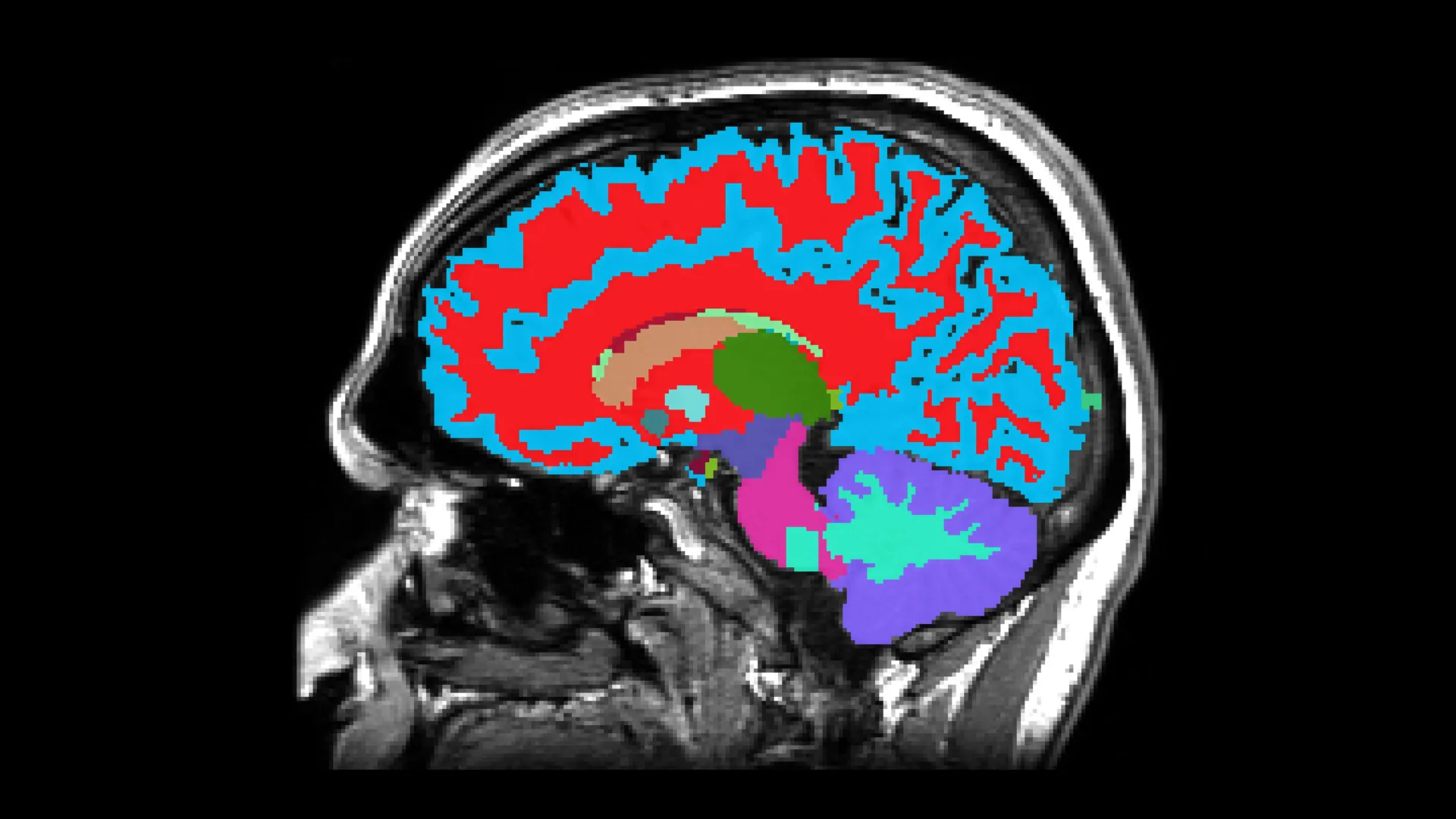
Now, scientists at Duke, Harvard and the University of Otago in New Zealand have developed a freely available tool that can tell how fast someone is aging, and while they’re still reasonably healthy — by looking at a snapshot of their brain.

From a single MRI brain scan, the tool can estimate your risk in midlife for chronic diseases that typically emerge decades later. That information could help motivate lifestyle and dietary changes that improve health.

In older people, the tool can predict whether someone will develop dementia or other age-related diseases years before symptoms appear, when they might have a better shot at slowing the course of disease.

“What’s really cool about this is that we’ve captured how fast people are aging using data collected in midlife,” Hariri said. “And it’s helping us predict diagnosis of dementia among people who are much older.”

The results were published July 1 in the journal Nature Aging.

Finding ways to slow age-related decline is key to helping people live healthier, longer lives. But first “we need to figure out how we can monitor aging in an accurate way,” Hariri said.

Several algorithms have been developed to measure how well a person is aging. But most of these “aging clocks” rely on data collected from people of different ages at a single point in time, rather than following the same individuals as they grow older, Hariri said.

“Things that look like faster aging may simply be because of differences in exposure” to things such as leaded gasoline or cigarette smoke that are specific to their generation, Hariri said.

The challenge, he added, is to come up with a measure of how fast the process is unfolding that isn’t confounded by environmental or historical factors unrelated to aging.

To do that, the researchers drew on data gathered from some 1,037 people who have been studied since birth as part of the Dunedin Study, named after the New Zealand city where they were born between 1972 and 1973.

Every few years, Dunedin Study researchers looked for changes in the participants’ blood pressure, body mass index, glucose and cholesterol levels, lung and kidney function and other measures — even gum recession and tooth decay.

They used the overall pattern of change across these health markers over nearly 20 years to generate a score for how fast each person was aging.

The new tool, named DunedinPACNI, was trained to estimate this rate of aging score using only information from a single brain MRI scan that was collected from 860 Dunedin Study participants when they were 45 years old.

Next the researchers used it to analyze brain scans in other datasets from people in the U.K., the U.S., Canada and Latin America.

Faster aging and higher dementia risk

Across data sets, they found that people who were aging faster by this measure performed worse on cognitive tests and showed faster shrinkage in the hippocampus, a brain region crucial for memory.

More soberingly, they were also more likely to experience cognitive decline in later years.

In one analysis, the researchers examined brain scans from 624 individuals ranging in age from 52 to 89 from a North American study of risk for Alzheimer’s disease.

Those who the tool deemed to be aging the fastest when they joined the study were 60% more likely to develop dementia in the years that followed. They also started to have memory and thinking problems sooner than those who were aging slower.

When the team first saw the results, “our jaws just dropped to the floor,” Hariri said.

Links between body and brain

The researchers also found that people whose DunedinPACNI scores indicated they were aging faster were more likely to suffer declining health overall, not just in their brain function.

People with faster aging scores were more frail and more likely to experience age-related health problems such as heart attacks, lung disease or strokes.

The fastest agers were 18% more likely to be diagnosed with a chronic disease within the next several years compared with people with average aging rates.

Even more alarming, they were also 40% more likely to die within that timeframe than those who were aging more slowly, the researchers found.

“The link between aging of the brain and body are pretty compelling,” Hariri said.

The correlations between aging speed and dementia were just as strong in other demographic and socioeconomic groups than the ones the model was trained on, including a sample of people from Latin America, as well as United Kingdom participants who were low-income or non-White.

“It seems to be capturing something that is reflected in all brains,” Hariri said.

The work is important because people worldwide are living longer. In the coming decades, the number of people over age 65 is expected to double, reaching nearly one fourth of the world’s population by 2050.

“But because we live longer lives, more people are unfortunately going to experience chronic age-related diseases, including dementia,” Hariri said.

Dementia’s economic burden is already huge. Research suggests that the global cost of Alzheimer’s care, for example, will grow from $1.33 trillion in 2020 to $9.12 trillion in 2050 — comparable or greater than the costs of diseases like lung disease or diabetes that affect more people.

Effective treatments for Alzheimer’s have proven elusive. Most approved drugs can help manage symptoms but fail to stop or reverse the disease.

One possible explanation for why drugs haven’t worked so far is they were started too late, when the Alzheimer’s proteins that build up in and around nerve cells have already done too much damage.

“Drugs can’t resurrect a dying brain,” Hariri said.

But in the future, the new tool could make it possible to identify people who may be on the way to Alzheimer’s sooner, and evaluate interventions to stop it — before brain damage becomes extensive, and without waiting decades for follow-up.

In addition to predicting our risk of dementia over time, the new clock will also help scientists better understand why people with certain risk factors, such as poor sleep or mental health conditions, age differently, said first author Ethan Whitman, who is working toward a Ph.D. in clinical psychology with Hariri and study co-authors Terrie Moffitt and Avshalom Caspi, also professors of psychology and neuroscience at Duke.

More research is needed to advance DunedinPACNI from a research tool to something that has practical applications in healthcare, Whitman added.

But in the meantime, the team hopes the tool will help researchers with access to brain MRI data measure aging rates in ways that aging clocks based on other biomarkers, such as blood tests, can’t.

“We really think of it as hopefully being a key new tool in forecasting and predicting risk for diseases, especially Alzheimer’s and related dementias, and also perhaps gaining a better foothold on progression of disease,” Hariri said.

The authors have filed a patent application for the work. This research was supported by the U.S. National Institute on Aging (R01AG049789, R01AG032282, R01AG073207), the UK Medical Research Council (MR/X021149/1), and the New Zealand Health Research Council (Program Grant 16-604).

Share Button

The pandemic pet boom was real. The happiness boost wasn’t

A new study challenges the belief in a universal “pet effect” on human well-being. Using data collected during COVID-19 lockdowns, researchers found no significant change in respondents’ well-being when they acquired or lost a pet in their household. The findings suggest that, even during a time of extreme isolation, human-animal bonds may not be as emotionally transformative as we like to believe.

Humans and their pets, a match made in heaven? Does adopting a new dog make you happier and less lonely? It is now commonplace to associate pet ownership with health and happiness for the human and the animal. Still, science has had a hard time pinning down the ‘pet effect’ — a hypothesised boost in life quality for those who surround themselves with cats, dogs, or other companion animals. Only a few years ago, circumstances presented us with a severe test of the importance of human-animal bonds — a global pandemic, COVID-19, which confined people to their homes, cutting them off from face-to-face contact in both work and personal life.

Researchers at ELTE Eötvös Loránd University have examined how pet acquisition and loss were experienced during the pandemic and the short- and long-term effects of acquiring a pet on the participants. The study was published in Scientific Reports. “Through a collaboration with a psychologist team led by Zsolt Demetrovics and Róbert Urbán, we had access to a unique data set,” explains Eniko Kubinyi, head of the MTA-ELTE ‘Momentum’ Companion Animals Research Group. “During the 2020 COVID-19 lockdowns, almost three thousand people across Hungary participated three times in data collection, several months apart. We noticed that 65 people acquired a pet and 75 lost one during the study, and decided to investigate how their well-being changed over time.”

The researchers found little support for the romanticised view we hold of pet owners and their emotional well-being. A short-lived boost in cheerfulness appeared after acquiring a dog, however, in a long run, dog owners’ calmness, life-satisfaction, cheerfulness, and activity had gone down. Most surprisingly, the researchers found that losing a pet did not leave a mark on the well-being of their former owners.

Ádám Miklósi, who initiated the data collection on companion animals, emphasises, “We rarely have access to data that documents spontaneous pet acquisition from people unbiased in their attitude toward pet ownership. Usually, pet lovers are identified and studied when the decision to adopt an animal is already settled. It appears that, at least during stressful periods, the average person, who may not be the primary caregiver but simply shares a household with the pet, is not significantly affected by the pet’s loss, nor is their well-being a strong predictor of the decision to acquire one.”

“What surprised me most,’ adds Judit Mokos, data scientist and one of the paper’s first authors, ‘was that a new pet in the household had no effect on the respondents’ loneliness. Dog adoption is often promoted as a solution for elderly and/or lonely people. Shelters and pet food companies promote adoption as a means of alleviating loneliness. However, our research suggests that dogs do not provide a real solution to loneliness; rather, they make the new owners more anxious.”

Kubinyi concludes, “Based on the data, most people, living together with a companion animal, do not seem to experience any long-term ‘pet effect’, nor do they bond strongly with their animal. It is possible that the dynamics of the pandemic have led many to make impulsive choices against their long-term interest, or that only certain groups — like devoted animal lovers or older adults living alone — truly benefit from pets in stressful times.”

It appears that during the COVID-19 pandemic, the emotional bonds people formed with animals often fell short of expectations.

Share Button

Tiny gut “sponge” bacteria found to flush out toxic PFAS “forever chemicals”

Scientists have discovered that certain species of microbe found in the human gut can absorb PFAS — the toxic and long-lasting ‘forever chemicals.’ They say boosting these species in our gut microbiome could help protect us from the harmful effects of PFAS.

PFAS have been linked with a range of health issues including decreased fertility, developmental delays in children, and a higher risk of certain cancers and cardiovascular diseases.

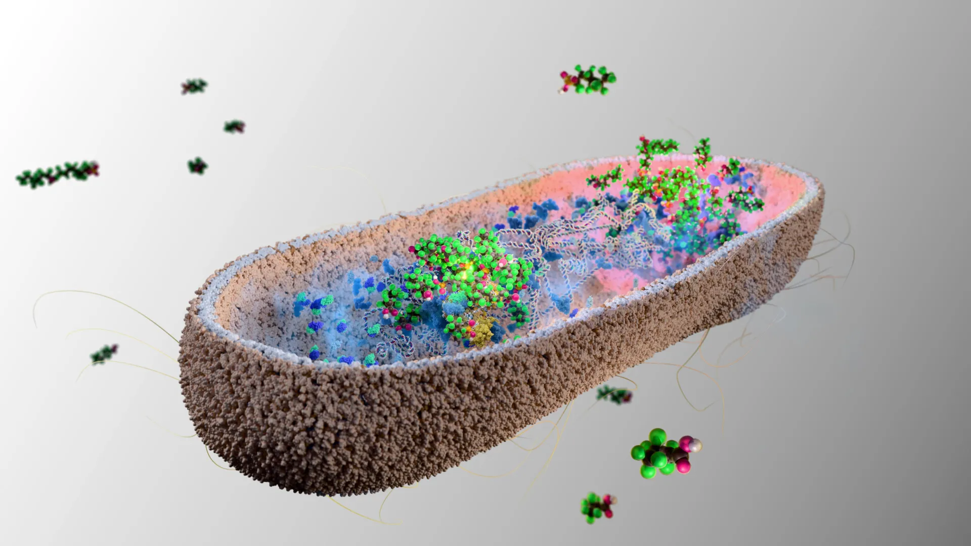
Scientists at the University of Cambridge have identified a family of bacterial species, found naturally in the human gut, that absorb various PFAS molecules from their surroundings. When nine of these bacterial species were introduced into the guts of mice to ‘humanise’ the mouse microbiome, the bacteria rapidly accumulated PFAS eaten by the mice — which were then excreted in faeces.

The researchers also found that as the mice were exposed to increasing levels of PFAS, the microbes worked harder, consistently removing the same percentage of the toxic chemicals. Within minutes of exposure, the bacterial species tested soaked up between 25% and 74% of the PFAS.

The results are the first evidence that our gut microbiome could play a helpful role in removing toxic PFAS chemicals from our body — although this has not yet been directly tested in humans.

The researchers plan to use their discovery to create probiotic dietary supplements that boost the levels of these helpful microbes in our gut, to protect against the toxic effects of PFAS.

The results are published today in the journal Nature Microbiology.

PFAS (Perfluoroalkyl and Polyfluoroalkyl Substances) can’t be avoided in our modern world. These man-made chemicals are in many everyday items including waterproof clothing, non-stick pans, lipsticks and food packaging, used for their resistance to heat, water, oil and grease. But because they take thousands of years to break down, they are accumulating in large quantities in the environment – and in our bodies.

Dr Kiran Patil, in the University of Cambridge’s MRC Toxicology Unit and senior author of the report, said: “Given the scale of the problem of PFAS ‘forever chemicals’, particularly their effects on human health, it’s concerning that so little is being done about removing these from our bodies.”

“We found that certain species of human gut bacteria have a remarkably high capacity to soak up PFAS from their environment at a range of concentrations, and store these in clumps inside their cells. Due to aggregation of PFAS in these clumps, the bacteria themselves seem protected from the toxic effects.”

Dr Indra Roux, a researcher at the University of Cambridge’s MRC Toxicology Unit and a co-author of the study said: “The reality is that PFAS are already in the environment and in our bodies, and we need to try and mitigate their impact on our health now. We haven’t found a way to destroy PFAS, but our findings open the possibility of developing ways to get them out of our bodies where they do the most harm.”

There is increasing concern about the environmental and health impacts of PFAS, and in April 2025 the UK launched a parliamentary inquiry into their risks and regulation.

There are over 4,700 PFAS chemicals in widespread use. Some get cleared out of the body in our urine in a matter of days, but others with a longer molecular structure can hang around in the body for years.

Dr Anna Lindell, a researcher at the University of Cambridge’s MRC Toxicology Unit and first author of the study said: “We’re all being exposed to PFAS through our water and food – these chemicals are so widespread that they’re in all of us.

“PFAS were once considered safe, but it’s now clear that they’re not. It’s taken a long time for PFAS to become noticed because at low levels they’re not acutely toxic. But they’re like a slow poison.”

Lindell and Patil have co-founded a startup, Cambiotics, with serial entrepreneur Peter Holme Jensen to develop probiotics that remove PFAS from the body, and they are investigating various ways of turbo-charging the microbes’ performance. Cambiotics is supported by Cambridge Enterprise, the innovation arm of the University of Cambridge, which helps researchers translate their work into globally-leading economic and social impact.

While we wait for new probiotics to become available, the researchers say the best things we can do to help protect ourselves against PFAS are to avoid PFAS-coated cooking pans, and use a good water filter.

Share Button