Sitting for prolonged periods is associated with health complications – but you can counteract the risks of a sedentary life.


Sitting for prolonged periods is associated with health complications – but you can counteract the risks of a sedentary life.
Testing the vagina microbiome is increasing in popularity with companies offering at-home tests
Prices change quickly on Prime Day, so keep checking back to see the latest updates. Make sure to sign up for an Amazon Prime account to get the deals.
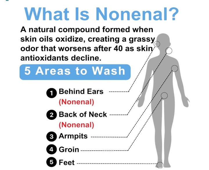
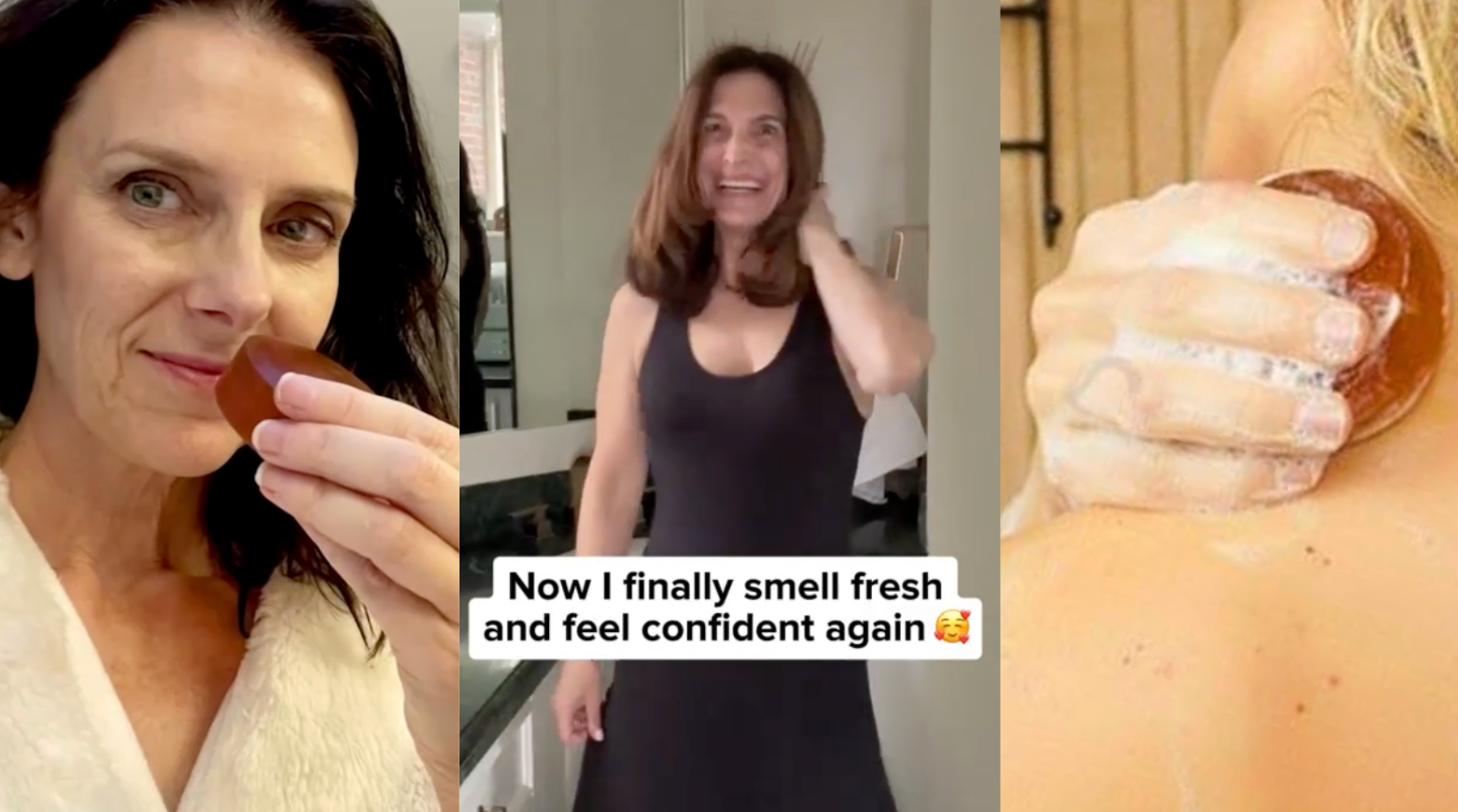
In previous HuffPost coverage, experts have shared that our body odor can actually change with aging, thanks to our skin naturally producing different compounds over time. The smell is distinct from regular BO, and while it isn’t a poor hygiene indicator, it generally doesn’t come off with just ordinary soap and water. It may, however, be addressed with this special bar of Japanese persimmon soap from the brand Mirai Clinical. It was runaway hit with HuffPost readers from the first time we covered it. For Amazon Prime Day, you can nab it for a whopping 38% off (aka, the lowest price we’ve seen it at, ever).
Advertisement
Advertisement
Typically beginning after age 40 and caused by antioxidant defenses declining in the skin, nonenal odors can have a mild to moderate organic or musty smell that varies from person to person, we’re told by dermatologists. And you might not even be able to notice it on yourself due to something called olfactory adaptation, where you become accustomed to the scent since its development is so gradual. It’s also not necessarily unpleasant to all humans, since smell is so subjective. But, if it is a concern for you, this is a possible solution.
“Persimmon soap, which contains tannins, has a plausible mechanism and a long history of use, though large randomized trials are lacking,” dermatologist Dr. Naana Boakye, founder of Bergen Dermatology, previously suggested.
And while Boakye did not mention a specific brand, the Mirai bar soap appears to be the most popular and widely reviewed option. (The founder even appeared on “Shark Tank” last year.) This formula has a dense concentration of persimmon-derived tannins, along with green tea extract, an ingredient that’s rich in antioxidants for continual defense throughout the day.
Advertisement
One Amazon customer put it to the test behind their ears, another part of the body where this smell can commonly develop.
Advertisement
“The results were pretty remarkable,” they claimed in their review. “I washed behind my ears on day one and then didn’t do it again for three days, constantly smelling that area each day. The smell stayed gone for two days, and by about the third day, it was starting to become slightly noticeable. That made me a believer.”
Others claim the soap worked on perimenopausal-related odors and just body odors in general that had previously been resistant to other soap formulas.
Advertisement
“Magical!! This stuff really works! No more perimenopause weird odor. I was going crazy! Bought a bunch more because it’s an essential now.” — An
“The perfect soap for seniors. This is a fantastic product. I had been changing soaps, shampoos, laundry soaps and sheets, and the unpleasant smell remained. Within days of using Mirai clinical persimmon soap I smell clean and fresh again. I also use the deodorant. I like the round shape of the bar and it makes a nice lather. I recommend this to everyone.” — Kindle customer
“Now get ready because I am going to get slightly gross for a minute. I rubbed my finger behind both my ears and smelled and yeah, it wasn’t great, even though I just washed that morning. So I gave the soap a shot and in particular focused on behind the ears. The result were pretty remarkable. I washed behind my ears on day one and then didn’t do it again for 3 days. Constantly smelling that area each day. The smell stayed gone for 2 days and by about the 3rd day it was starting to become slightly noticeable. That made me a believer. So I started using it in all the places the nonenal is at. I start by washing my entire body with regular soap and then at the end of my shower I use this soap in all the places they teach you that nonenal shows up. I have to say that I feel much more confident about how I smell after using it. No one wants to go through hormonal changes but we all have to. At least now we can be more comfortable in our skin while we go through it. I know it’s very expensive for a soap. But it is worth it. And if you only use it on the parts of the body that the nonenal smell comes from you won’t have to buy it again for a couple of months.” — L.B. (This review has been edited for length. Read the full review.)
“I’m glad I decided to try this soap despite the cost. It’s a large bar that hopefully will last for months and lathers very well with a pleasant clean smell. I noticed after using the bar the sour odor that I have been struggling with is absent. I hope this soap works as well for you as it has for me.” — Patricia Kroepel
The experts consulted for this story do not necessarily endorse the products ahead unless otherwise noted.
Amazon
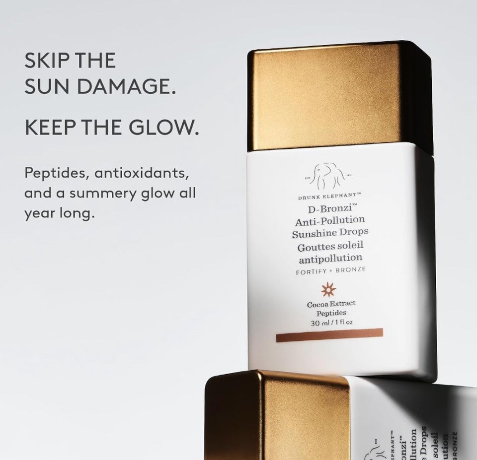
At a jaw-dropping discount of 50% off, Drunk Elephant’s D-Bronzi Sunshine Drops are for anyone who wants a glow-within-complexion with the added benefits of skin care. Popular on TikTok, these drops can be added to your foundation or used alone and are infused with peptides, antioxidants and fatty acids while delivering a bronzy wash of glossy color. HuffPost executive editor Kate Palmer calls them “potent and useful!”
Amazon
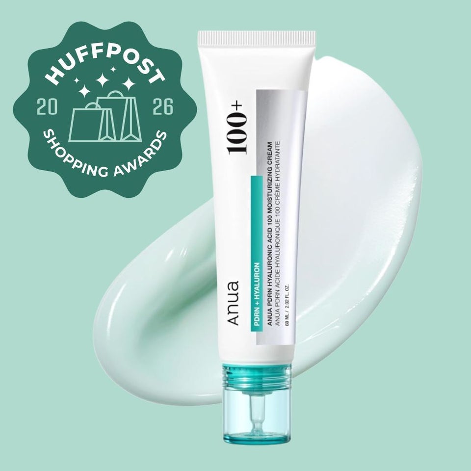
Our first impression of this Korean PDRN cream by Anua, which we awarded in 2026, was when one reviewer compared it to “a miracle cream” and said they were unable to say enough good things about this under-$25 formula.
PDRN, as it’s been previously described to us by board-certified dermatologists, is a portion of DNA that’s typically derived from salmon sperm and has been shown to accelerate repair of damaged skin and promote new tissue growth. It also boosts hydration in the skin, promotes cellular turnover and reduces hyperpigmentation.
The cream also offers other anti-aging heavy-hitters, like hyaluronic acid for plumping and hydration, niacinamide for protection, squalane for softening, glycerin for moisture, and turmeric, aloe and eggplant extras for soothing and brightening. Lightweight and non-greasy, users say it quickly soaks into your skin and plays well with other products.
One 40-year-old reviewer with combination dry and oily skin and acne says after using the cream, “I have elasticity and softness as I had in my 20s.” They also note that they’ve been getting many compliments on their skin — something they are not used to. “My face at 65 years old looks and feels about 40,” another wrote.
Amazon
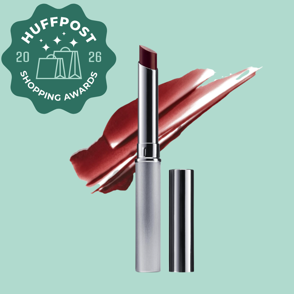
Clinique’s Black Honey Almost Lipstick has stood the test of time and remained an iconic makeup staple for many, myself included. I have always found that it’s the perfect, low-maintenance daily lip product for a little slick of color and hydration. The nostalgia factor, combined with the nourishing formula and timelessly romantic hue that looks good on everyone, has earned it legions of devotees. “My favorite part about the Almost Lipstick is how comfortable it feels on the lips,” said senior shopping writer Tessa Flores.
Part of what makes it so special is the “Almost Lipstick” formula. It’s technically an emollient-rich and ultra-moisturizing tinted balm that deposits a sheer, buildable kiss of color. It’s more hydrating than a traditional lipstick thanks to seed oils that give it a perfect sheen without the sticky feel of a gloss. There are three different “Almost Lipstick” shades, but Black Honey is the enduring star, perhaps due to the brand’s claim that it has a “chameleon-like ability to flatter all skin tones yet look different on everyone.” Apparently, Clinique sells seven tubes every minute!
While Black Honey never really went away, there has been a cultural resurgence of late thanks to social media. It’s all over TikTok, and not only do influencers love it, but so do beauty editors, and that includes the HuffPost shopping team. Black Honey’s enduring popularity and resiliency has earned it a top spot on the 2026 HuffPost Shopping Awards list of beauty must-haves.
Advertisement
Amazon
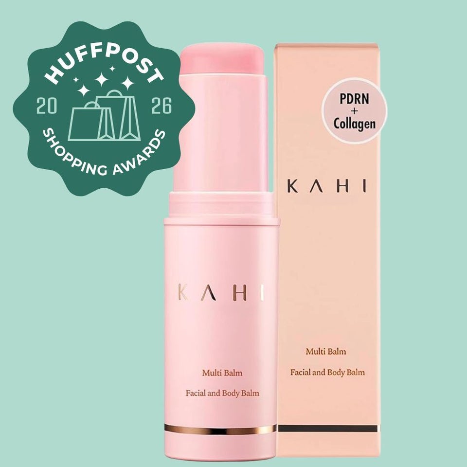
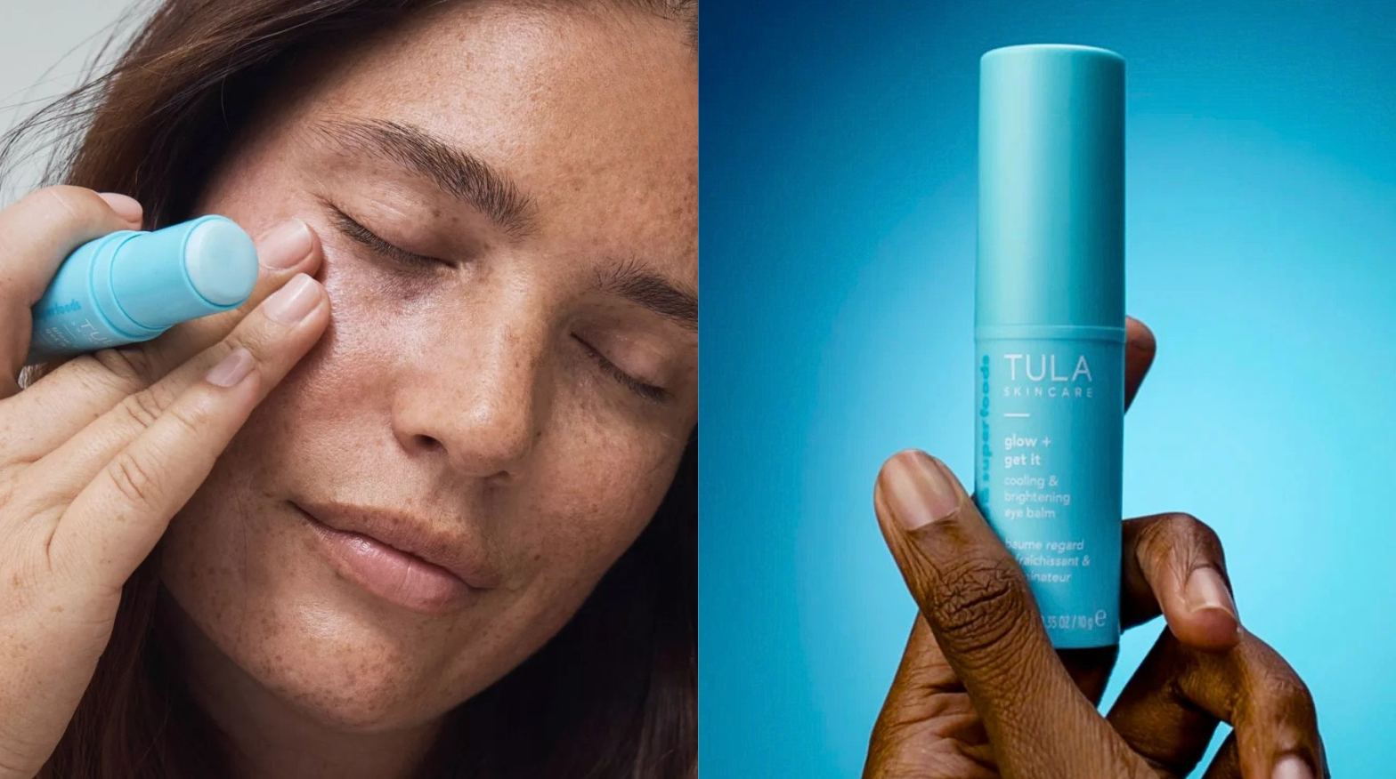
This reader-favorite multi-balm makes smoothing and hydrating the trouble areas on your face impossibly easy, earning it a 2026 HuffPost Shopping Award.
Featured in the fall, the Kahi Wrinkle Bounce balm is a lightweight and hydrating stick, enriched with fermented jeju oil and a collagen combo with salmon DNA (the PDRN portion of this formula), which works to deeply moisturize the skin and support skin renewal. Additionally, the balm helps diminish fine lines and improve skin clarity to reveal a more radiant complexion.
HuffPost readers and Kahi Balm reviewers alike have had a longstanding obsession with this treatment stick, which you can swipe under the eyes to target dehydration and crow’s feet, on the lips and forehead.
“I just turned 50. I have always taken care of my skin and looked younger than my age, but this year I started to notice more wrinkles popping up,” one Amazon shopper wrote, ” I started using this every night before bed, all around my eyes and lips. After about a week, I saw a huge difference!”
Amazon
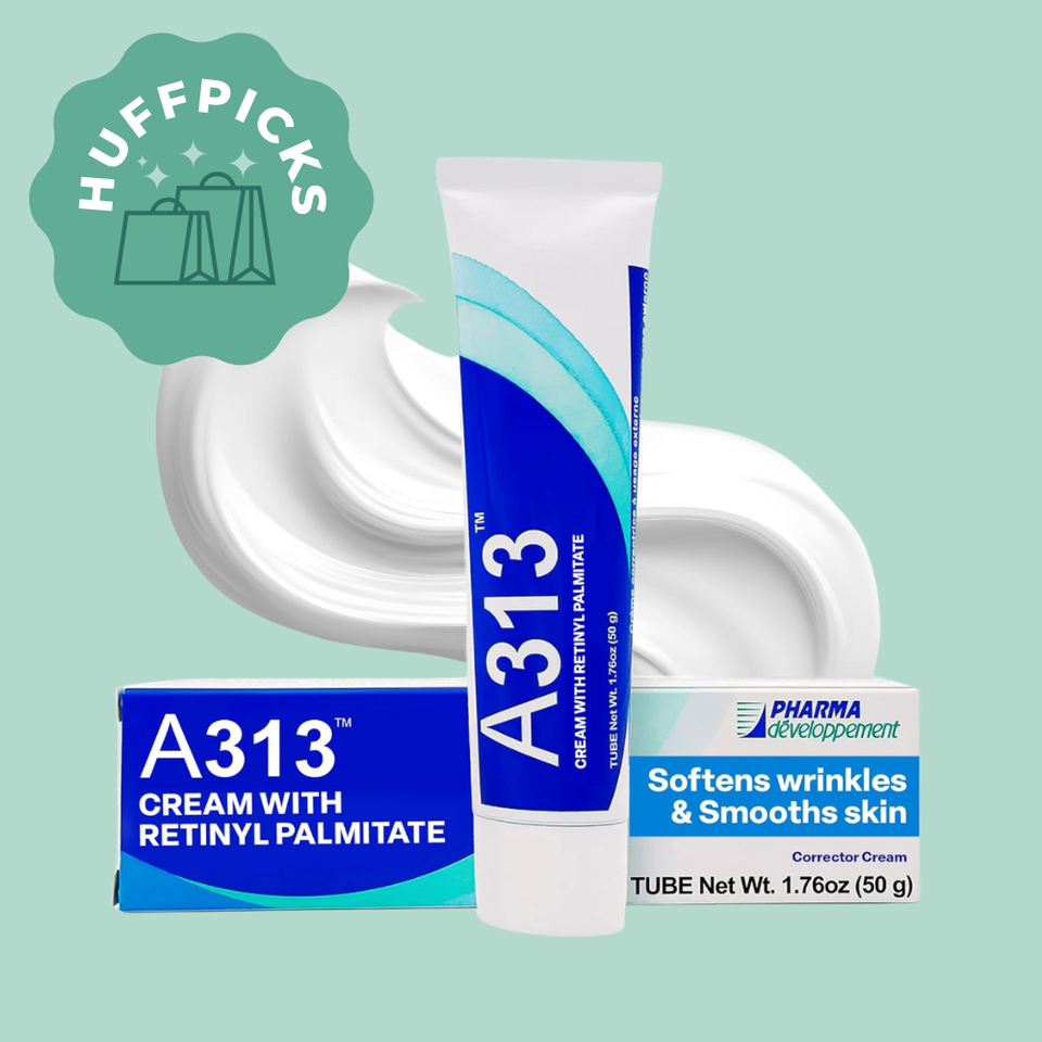
If there’s one thing that HuffPost readers and editors love, it’s French pharmacy skin care products. A313 cream is the highest-strength retinol cream you can get over the counter, with three kinds of retinol that can help stimulate collagen. Its efficacy is renowned, as is the reasonable price point, making it a unique entry in the retinol category — and a 2025 HuffPost Shopping Award winner.
Reviewers have long noted that it’s nearly as potent and effective as prescription-strength Tretinoin, without the negative side effects. In fact, some users who had been using prescription-strength Tretinoin then made the switch to this alternative, say they prefer A313 and have maintained the same results that they had with the prescription-strength formula.
In past reporting, board-certified dermatologist Dr. Rebecca Marcus explained that, “[A313] contains minimal ingredients in its vehicle, which is helpful for those who are sensitive to fragrances or other ingredients that are commonly found in OTC retinol products.”
Ultimately, what makes A313 distinctive (along with the price) is the thick occlusive texture, which is closer to a balm, and according to reviewers, can be more hydrating compared to other pricier formulations that you might find on the OTC market. The brand claims that the 2% retinol concentration can help unclog pores, minimize fine lines, reduce hyperpigmentation and even out skin texture.
Amazon
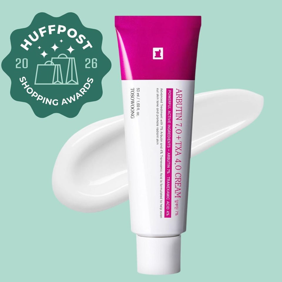
Unlike your run-of-the-mill sun spots, we know that melasma is arguably one of the most clingy and temperamental skin conditions to treat. That’s what makes this Korean drugstore cream — originally featured mid-last year and well-received by HuffPost readers — all the more impressive, and worthy of a 2026 HuffPost Shopping Award.
It contains a 7% concentration of arbutin, an antioxidant with brightening properties that works by inhibiting the over-production of melanin in the skin, which causes hyperpigmentation. It contains other well-trusted ingredients in the world of melasma care, one of which is 4% tranexamic acid, which is revered for its ability to reduce pigmentation associated with melasma, without causing any kind of irritation.
There’s also niacinamide, a workhorse antioxidant that you’re most likely already familiar with. If you’re not, just know that this all-skin-concerns ingredient can reduce redness, acne, and, most notably, brighten the complexion.
“I’ve been using it for about a third application, after washing my face and wow! I see drastic fading already!” one Amazon reviewer claims.
Advertisement
Amazon
HuffPost readers love a high-quality skin care product that can replace pricey facial treatments, like this K-beauty serum that one beauty TikToker calls “filler in a bottle.” Its prowess is attributed to an ingredient called PDRN, which is widely used in injectables overseas. It has anti-inflammatory and tissue-repairing capabilities that can improve skin firmness and leave recipients with a telltale glow. It’s usually extracted from salmon sperm cells, but Iope’s serum has a completely vegan, plant-based formula that is as unique as it is effective.
Additional ingredients like niacinamide, probiotics and caffeine round out this repairing formulation — all ingredients that are beloved by HuffPost editors and readers alike.
The combination of a buzzy new ingredient like PDRN and time-honored stalwarts like the ones we just mentioned joined forced to create the perfect beauty product storm that earned this serum a 2025 best-in-show badge. Both Flores and Uribe have covered countless serums in their many years of beauty reporting, but this one covers all their must-have bases.
Dermstore
We’ve rarely seen an audience response quite like we did when former HuffPost editor Janie Campbell introduced our readers to the Revision Skincare YouthFull lip replenisher, one of our 2025 winners. You purchased this nourishing lip product in droves, and upon closer inspection, it’s clear why. Campbell made a compelling case for this balm, calling it a “truly stellar and game-changing product.”
Formulated with several peptides, vitamins E and C, green tea and shea butter, it’s designed to target visible signs of lip aging like puckering, wrinkles, dryness and dullness.
And while it’s not typically like us to recommend such an expensive lip balm, the results speak for themselves. It has a thick, tacky texture that really grips the lips, making it a great overnight treatment. Campbell could see results upon her very first application, and other reviewers agree, including one at Dermstore who said it gives her “a plump lip filler look when left on overnight.” Say no more!
Amazon
When multiple HuffPost editors are raving about one product, you know it’s going to be pretty magical. Case in point: Londontown’s cult-fave nail concealer. It’s a game-changer whether you’re looking to take a break from gel manicures, want to touch up your polish at home between visits to the salon or simply want a quick and easy way to give your paws a refresh.
Senior reporter Lydia O’Connor first brought this nail polish to the Shopping team’s attention, and it very quickly became apparent that it’s beloved across the entire newsroom. “I am obsessed with this product,” O’Connor said in previous reporting.
Greta Geiselman, our director of office services, swears by it, saying “I love this brand! For quick manis, I can put on two coats, no top or bottom needed and it dries quickly. Buildable coverage and fun colors.”
Aiken proclaimed, “I love this stuff!…I have really weird nails (the whites are jagged because I use my hands too much and have ‘nailbed trauma’) and I’m too rugged to paint my nails because it instantly comes off, but I love the nail concealer because it looks natural but just, better. And it doesn’t show when it starts to chip off.”
Their glowing reviews, combined with our audience’s fervor for this nail concealer, has earned it a coveted 2025 HuffPost Shopping Award designation. The concealer comes in four different colors that look great on a variety of skin tones: bubble, pink, milky and quartz. Each formula contains optical brighteners to give nails a subtle gleam and hide imperfections and discoloration, along with plant extracts and a nourishing complex.
Advertisement
Amazon
Lancôme’s Lash Idôle, a 2026 winner, has a lightweight, long-lasting formula that’s infused with white tea extract for conditioning your lashes as it coats them. It helps fan your lashes out and give them a whole lot of fluttery oomph without smudging. “Goes on smoothly,” user me q. wrote. “I am 68 years old and my lashes look like lashes of a 20-year-old. I won’t be using anything else.”
The inner curve has smaller bristles for building volume, and the exterior of the curve has longer ones for helping lengthen and separate your lashes. Reviewers can’t get enough. “It really does lift and separate the lashes, giving a wide-eyed look without clumps,” A’Ville Triathlon said. “The brush grabs every lash, even the tiny ones and builds nice volume without making them feel heavy or sticky.”
Amazon
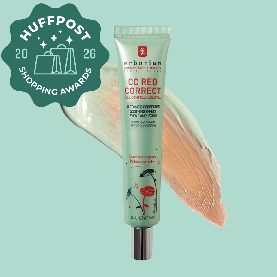
Whether you’re struggling with a condition like rosacea or are simply prone to irritation, there’s one item that doubles as both skin care and makeup with legions of devoted fans (both online and in the HuffPost newsroom) that is formulated to correct even the ruddiest complexion, and it is worth every penny.
According to many, the Erborian CC Red Correct color-correcting cream is one of the best K-beauty products out there. It’s designed to help neutralize redness and calm skin in both the short and long term, with more visible benefits the longer you use it. It also has SPF 25 sun protection. It’s formulated with centella asiatica, also known as tiger grass, which has been used for thousands of years for its soothing and calming properties. The inclusion of glycerin can hydrate and strengthen the skin’s natural moisture barrier, so your skin stays soft and supple even while the active ingredients work hard to nourish the skin.
The critical mass of positive testimony from our colleagues here at HuffPost made it a no-brainer when it came to including it in 2026’s HuffPost Shopping Awards. O’Connor called it “a miracle product.” She came across it while searching for something to counter her skin’s redness and blotchiness, saying, “It’s very your-skin-but-better and just sort of blurs everything while cutting down on red undertones.” She uses it by itself on her “no makeup days” but finds that it’s great under foundation and concealer as well.
HuffPost senior reporter Carly Ledbetter said, “As someone with rosacea, it is the only ‘green’ product that makes my skin look normal.” Senior reporter Jessica Schulberg chimed in, saying, “I’ve influenced every single one of my friends to try it, it’s literally the only makeup I wear now. It’s magical.”
The finish is lighter than expected from such a transformative product, so even those with dry skin or who are acne-prone can enjoy it. It is also worth noting that this product is best suited for those with lighter skin tones who are prone to redness. Those with more melanated skin may find that it leaves a white cast.
Amazon
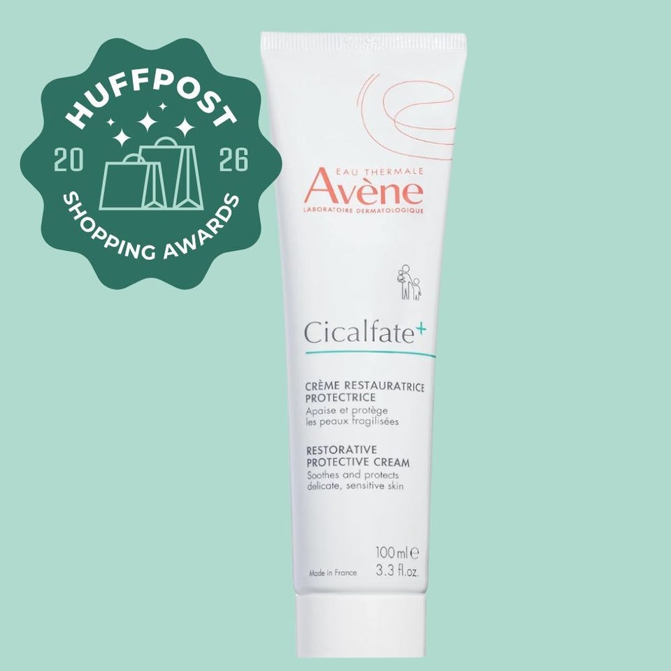
In the fall of 2025, senior shopping writer, Tessa Flores, wrote about how the Avène Cicalfate+ restorative protective cream saved her skin during an eczema breakout when she didn’t have access to the prescription steroid cream that she usually depends on in these situations. “After it managed to instantly soothe and soften the unbearably itchy and burning patches on my fingers and hands, I quickly learned why this French treatment lotion has such a hearty reputation for helping those with the most troubled and sensitive skin,” she said.
It would appear that HuffPost readers also hoped that the Avène cream would be their skin savior, because, during the colder winter months, our shopping trackers marked it a consistent top seller. Ultimately, we named it a winner among skin care products in 2026.
Functioning as a shielding and isolating layer to compromised skin, this richly textured cream has 45 uses and works by creating the optimal condition for damaged skin to recover by employing protein-rich probiotics and a copper-zinc-sulfate complex.
Advertisement
Amazon
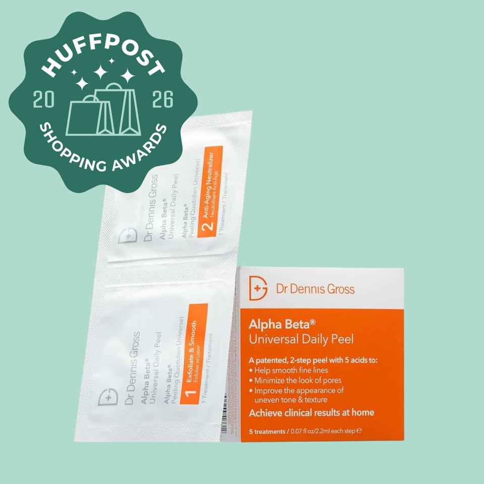
HuffPost contributor Carolin Lehmann previously described her experience with the Dr. Dennis Gross Alpha Beta daily peel pads, reporting that, since she started using them, people keep underestimating her age.
“I simply wipe down your face with these exfoliating pads, go to sleep, and wake up glowing like never before. These wipes are seemingly “magic,” helping my pores look smaller, minor acne clear up, fine lines iron out, and my skin [feel] baby smooth,” Lehmann said of the 2026 winner.
Also previously suggested to HuffPost by board-certified dermatologists, the Dr. Dennis Gross daily peel pads can offer the benefit of a chemical peel, but with a gentler approach than what you might find in a dermatologist’s office. Each treatment contains two pads: one to exfoliate and smooth your skin, and one that provides anti-aging benefits. The pads are pre-dosed with five acids, including glycolic, salicylic and lactic, that each work to reduce dullness and uneven texture, pores and hyperpigmentation. They were even compared (and preferred) to an in-office chemical peel by Amazon customers.
Amazon
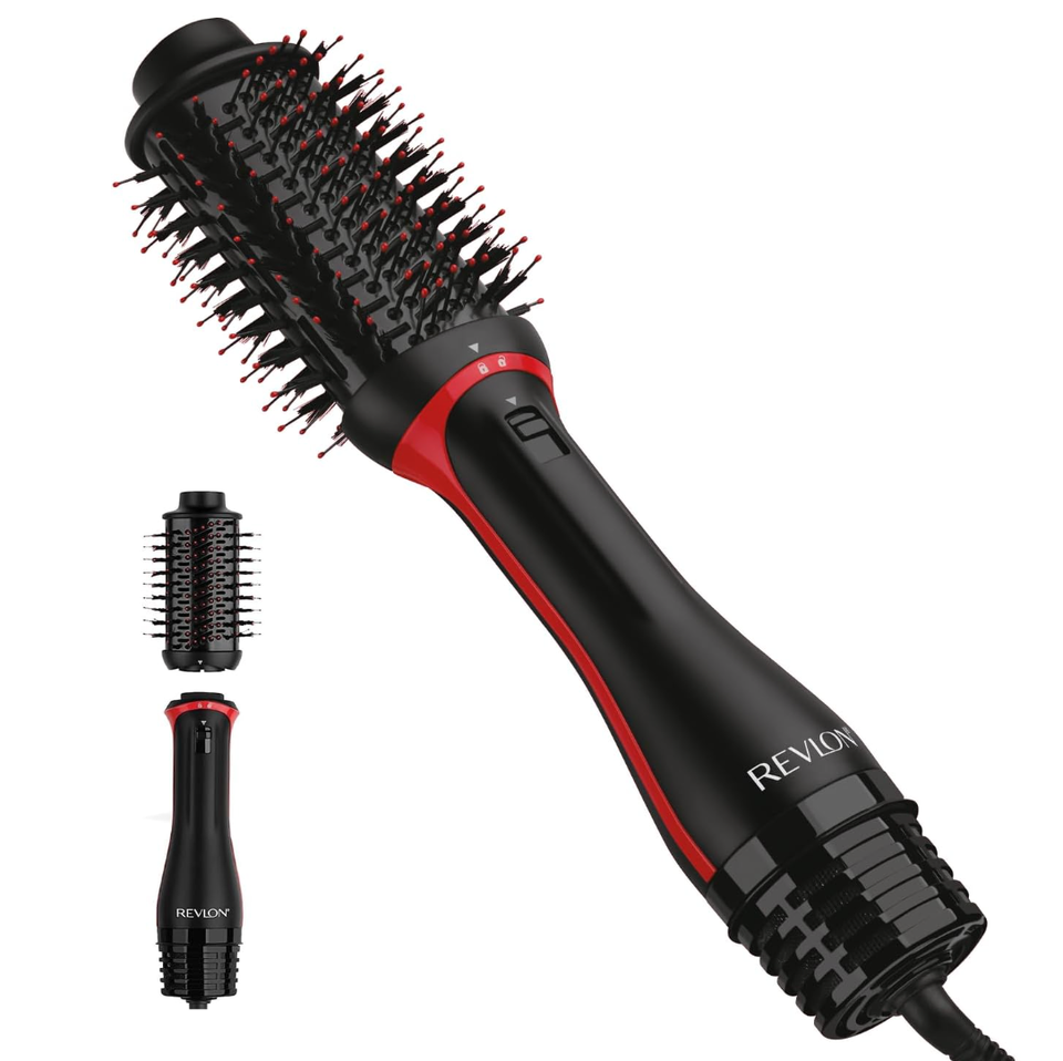
HuffPost shoppers still can’t get enough of this cult-fave styler and dryer from Revlon — a surefire way to get glossy, sleek hair. It’s shaped like a regular round brush with detangling bristles, and comes with three heat settings so you can style, dry and smooth your hair all in one go. It’s a must for anyone who loves that freshly blown-out feeling. Once you add this to your hair arsenal, you may not even need any other styling tools cluttering up your bathroom.
Amazon
Advertisement

Amazon
Washing your face daily can feel like an arduous task, made more so when you factor in the labor or having to launder washcloths. As if that weren’t annoying enough on its own, dermatologist Joshua Zeichner previously told HuffPost, “Items like washcloths can be a breeding ground for microorganisms if they are left out wet and reused.”
The search for a sanitary solution led former HuffPost Shopping writer Haley Zovickian to the cult-favorite Clean Skin Club towels. Plant-based and disposable, they’re designed to help keep skin germ-free post-wash, so you can apply products with a fresh face, clean conscience and without added microorganisms.
Endorsed by thousands of happy reviewers, these soft, plush and surprisingly large towels are as good as it gets. Some users even noted that they helped to reduce skin irritation. It’s no wonder they’ve become a staple in myriad HuffPost readers’ skin care routines, making them worthy of high praise.
Amazon
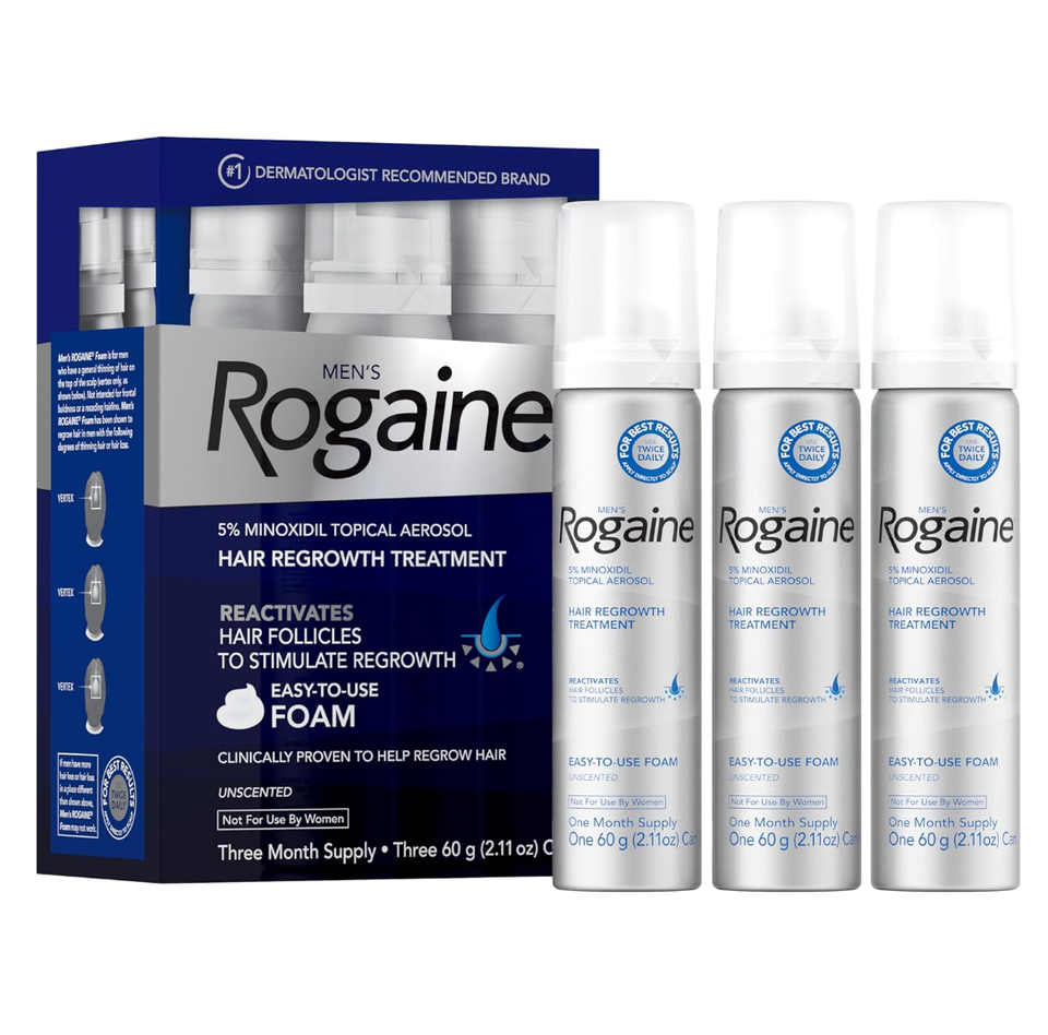
Rogaine is probably the best-known product that features minoxidil as the active ingredient for hair growth. This three-month supply features a 5% minoxidil along with a variety of botanical extracts and humectants, which can draw moisture into the hair.
The once-a-day treatment is also in the form of a foam, an important requirement according to experts we’ve previously spoken with, so the product won’t drip down to other areas of the face, thus risking unwanted hair growth.
Amazon
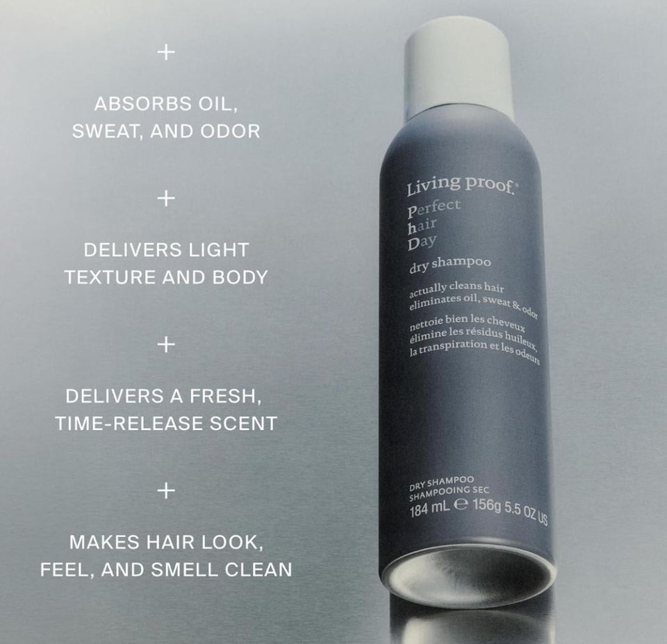
Both Flores and fellow senior shopping writer, Lourdes Avila Uribe, are longtime fans of this dry shampoo by Living Proof because it’s as close as you can get to freshly washed hair. It’s definitely on the higher end of the price spectrum for a product like this one, but it’s well worth it for the results. It doesn’t leave my scalp or hair feeling gritty with excess buildup; it’s like starting over with a clean slate without the hassle of drying and styling your hair again. But what has amazed us the most over the years of use is that it so effectively soaks up oil and dirt without leaving behind an unappealing chalky residue. Flores added that “despite the fact that I routinely spray on quite a bit of dry shampoo for multiple days in a row, it is never visible on my dark hair.”
It is formulated with a mix of starch and mineral blend powders that are known to quickly absorb excess oil and sweat, while an odor neutralizer gives hair a surprisingly fresh scent that makes it smell freshly washed. But it isn’t just absorbing oil; it leaves hair looking and feeling fresh and clean while also adding volume and movement.
Advertisement
Amazon
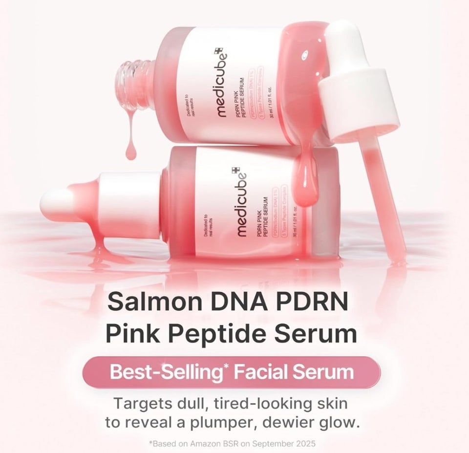
Medicube is one of Flores’ and Uribe’s favorite brands for reliable K-beauty products, and this salmon PDRN serum is one of its top-sellers (it’s even been previously suggested to us by dermatologists). In addition to salmon sperm-derived PDRN, it’s enriched with a complex containing five different types of peptides. According to dermatologists that we previously spoke to, peptides are amino acid proteins known to boost collagen production, improve skin texture and promote wound healing.
Amazon
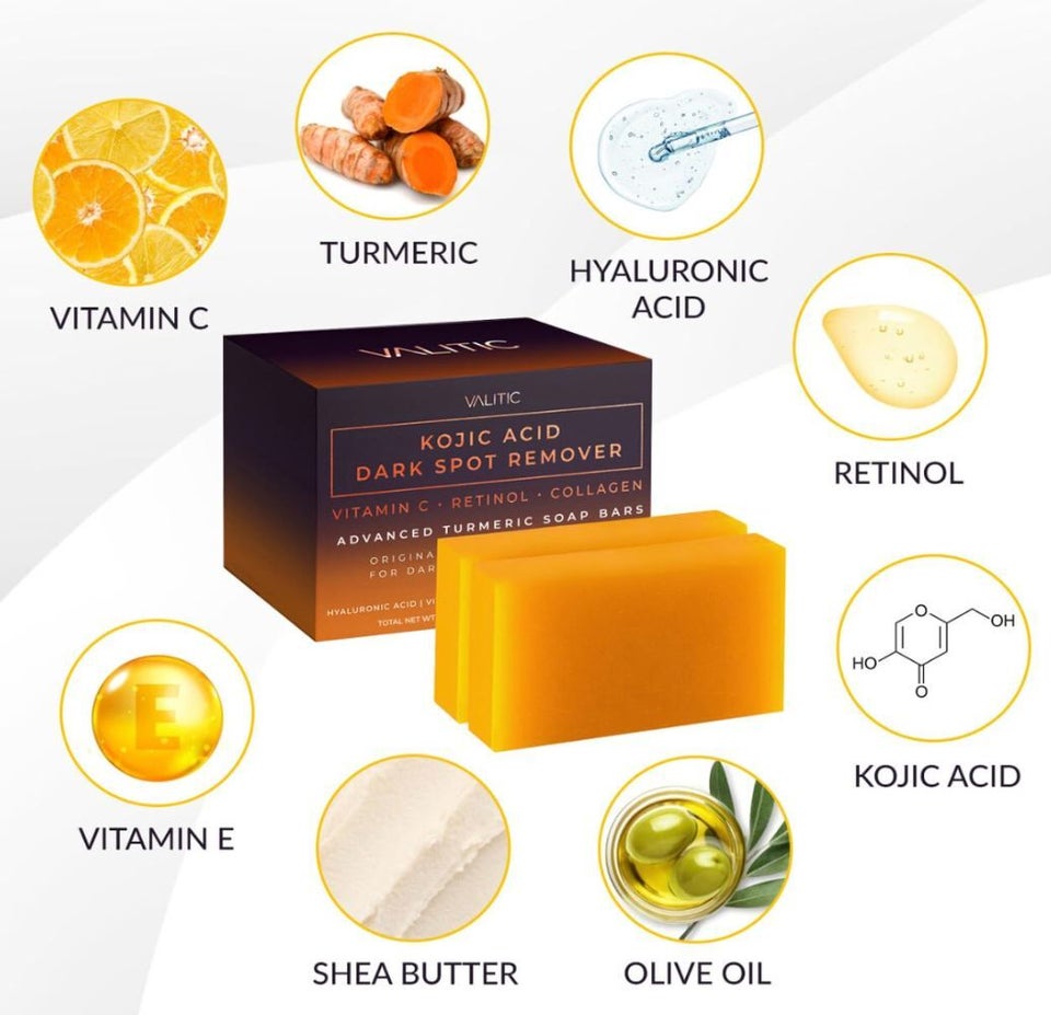
Flores said, “I have used this kojic acid bar soap as the first in-line product in my shower routine for about two years now, particularly on my arms for my KP and my underarms for any hyperpigmentation — and it really works.”
These bar soaps harness the power of active ingredients like vitamin C and retinol combined with the nourishing benefits of hyaluronic acid, vitamin E, shea butter and olive oil to potentially help even out skin tone, lighten dark spots, smooth skin and reduce blemishes. This two-pack has myriad glowing reviews attesting to its prowess, and could be a good tool in your skin health arsenal.
Amazon
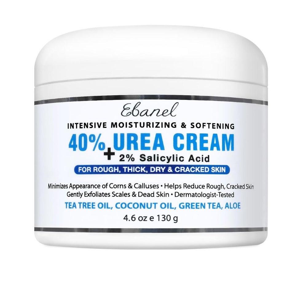
Experts previously put us on the skin-smoothing benefits of urea, especially when it comes to softening rough and cracked callused heels, psoriasis, keratosis pilaris and more. The Ebanel intensive moisturizing and softening cream, which, at 40%, contains the highest concentration of urea out of many market options out there. Protective, intensely moisturizing and softening, this cream also features 2% salicylic acid to help exfoliate away patches of rough and flaky skin, as well as targeting things like calluses, corns and cracked skin. The shea butter-based formula is enriched with trusted hydrators like hyaluronic acid, coconut and jojoba oils, and antioxidants like green tea and vitamin E. It’s a combination that can be useful for feet and elbows and even promote healthier-looking nails.
Advertisement
Amazon
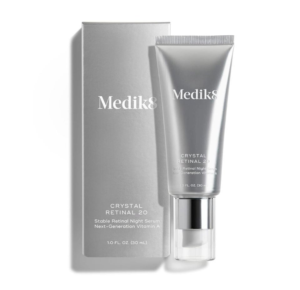
If you’ve ever wondered how to get your hands on some of the strongest retinoids, without a perscription, this is a much-talked-about option, which actually comes in six strengths for everyone from the retinol beginner to the seasoned expert. The Medik8 Crystal Retinal (in the mid-strength option) is also a favorite of Uribe’s, who called it an effective yet gentle addition to her repertoire.
“It leaves my skin looking and feeling great. Not only does it address typical signs of aging like wrinkles and texture quickly and efficiently, but it’s also helped to round out my anti-hyperpigmentation routine without stripping my skin of moisture or being overly harsh,” Uribe said. “The formula is balanced with hyaluronic acid and glycerin to help keep skin supple, soft, smooth and hydrated.”
Amazon
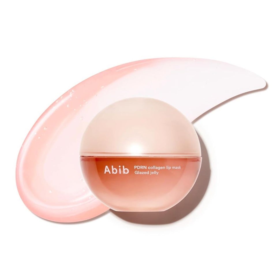
Flores swears by this Korean lip mask with a distinctive jelly texture and a heavy dose of PDRN to minimize the appearance of lip lines, make pouts look more plump and, above all, stave off her chronic dryness — especially around the corners of the mouth where regular retinol use can make it go flaky. “Not quite a balm and not quite a gloss, but somewhere in a delightful jammy middle, the Abib treatment feels cushiony and velvet-like on the lips and not at all sticky. Because of this, it’s much more comfortable to wear as an overnight lip mask and I love waking up with noticeably softer lips,” Flores said.
Many users are also that this thick and nourishing lip gel “was the best product” they had ever used and worked miraculously on wrinkled, aging lips.
Amazon
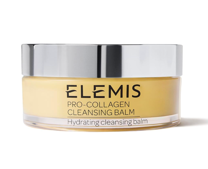
Uribe said that the first time she tried the famous Elemis cleansing balm, she couldn’t believe how soft, supple and clean her skin felt. “I had always assumed that someone with acne-prone, oily skin like me should stay away from oil cleansing, but I couldn’t have been more wrong,” she said.
According to Uribe, It has a buttery soft consistency that starts off like a rich, thick oil and slowly melts into the skin, dissolving makeup, grime and impurities. It’s formulated with fatty acids that can help to improve skin elasticity and moisture retention, and the brand says its starfish and elderberry oils help soothe irritation and smooth out the complexion.
Our colleague Kristen Aiken is also a huge fan of this cleansing balm. She told Uribe that “this item is way pricier than what I’m usually willing to spend on cleanser (which is usually from the drugstore and under $10), but I’ve found it’s one of the only products that can effectively cleanse my skin AND nourish it without making me break out. I have perpetually dry skin no matter how much water I drink, and this saves me from drying out (especially in the winter months). Bonus: If you use it as a second round of double-cleansing (i.e. after you’ve already removed the day’s initial layer of gunk), you can use your favorite facial massage tool over top it, to help your tool slide smoothly along your face and de-puff.”
Advertisement
Amazon
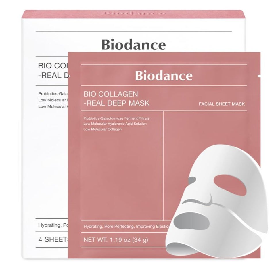
It’s possible you’ve seen some version of these overnight hydrogel masks on TikTok that start off opaque and become transparent as all of the infused goodness absorbs into skin, leaving behind a complexion that’s plump, hydrated and glass-like. These Biodance masks contain a collagen-peptide that’s of a low molecular weight to penetrate deeper into the skin. It also has brightening niacinamide, moisture-trapping hyaluronic acid and galactomyces to improve skin tone and texture.
Amazon
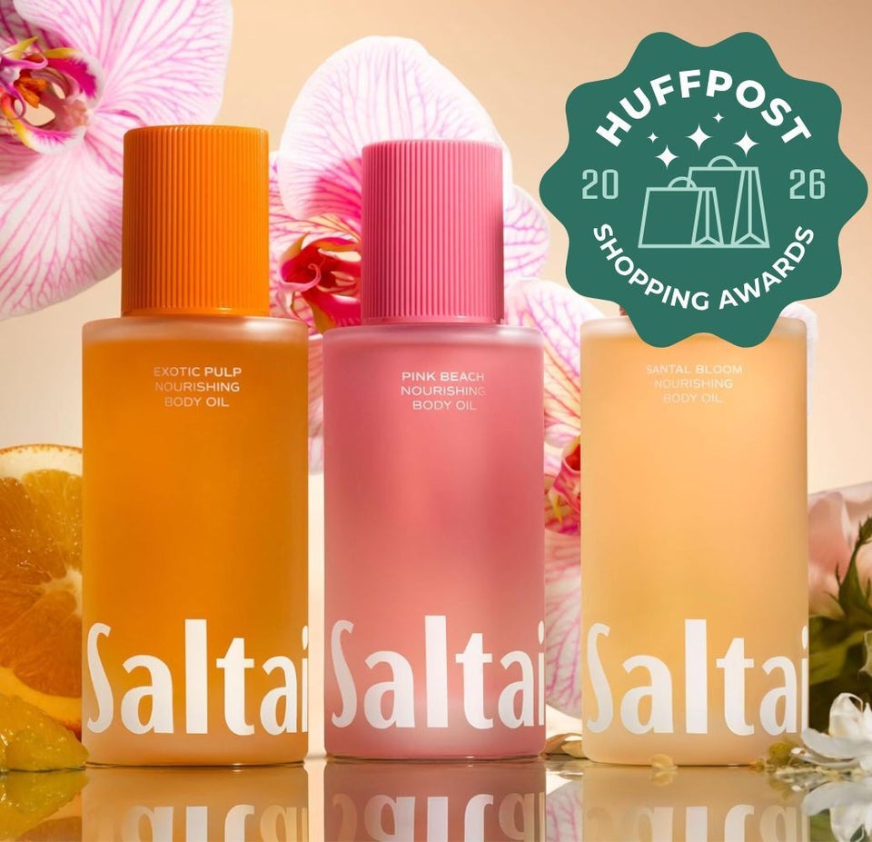
Flores has had chronically dry and troubled skin since childhood, but my years’ worth of experience in parched-skin troubles have finally led her to curate a body care routine that has actually made her skin the healthiest it’s ever been. This past winter, she even shared this effective ritual with readers and one product seemed to lead the pack in consumer interest.
“My beloved Saltair nourishing body oil is one of the best formulas of its kind that I’ve tried (including better than the uber-popular Osea Undaria algae body oil), and I’m pleased to know that it’s finally getting the flowers it deserves,” Flores wrote.
“As a person who is opposed to certain textures, I’m in love with the fact that this formula doesn’t sit greasy or stick atop the skin. It actually soaks in quickly and thoroughly to the point that I’m able to throw on my clothes right after and not leave a stain. Deceptively luxurious and available in five mild scents, it’s enriched with three different oils that are nourishing and rich in antioxidants like squalane and moringa. Since incorporating this glossy goodness into my daily post-shower routine, it has noticeably helped trap in hydration, improve texture and succeeded in making my skin feel bouncy and soft, well into the following day. I’ve also noticed that my tattoos appear more vibrant and renewed.”
Amazon
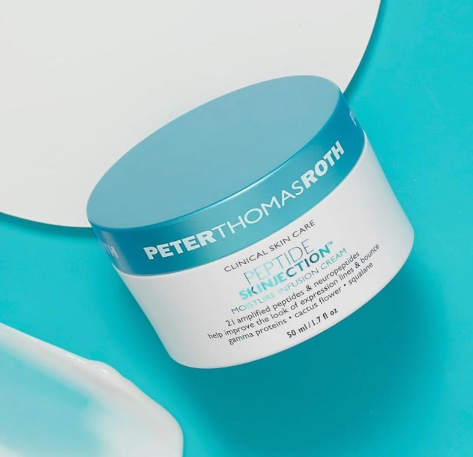
Flores said that she will always opt for a moisturizer with peptides for their proven skin-plumping power and the way they always make her skin feel juicy and hydrated. Peter Thomas Roth created an amped up moisturizer that contains both regular peptides and neuropeptides, which may reduce the appearance of expression lines, similarly to how neurotoxins like Botox work, but on a much lesser scale. “It also contains some of my favorite hydrators like squalane and hyaluronic acid,” Flores said.
Advertisement
Amazon
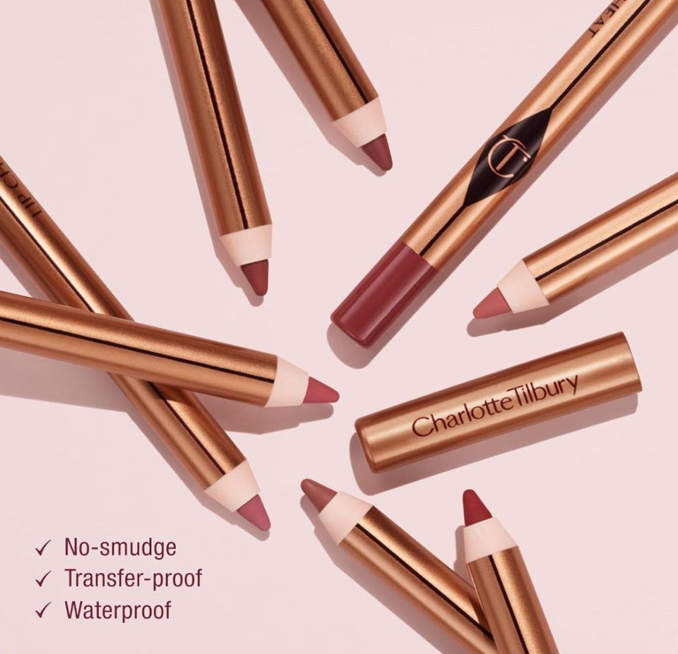
Charlotte Tilbury’s lip liner in the shade Pillow Talk — a famous rosy hue that comes in several depth options to suit everyone — is a makeup product that needs no introduction. “I always have one of these in my bag to slightly overline my lips without looking completely unnatural,” Flores said. The creamy formula is also really hydrating, infused with jojoba oil, hyaluronic acid and ceresin to help prevent smudging and transfer.
Amazon
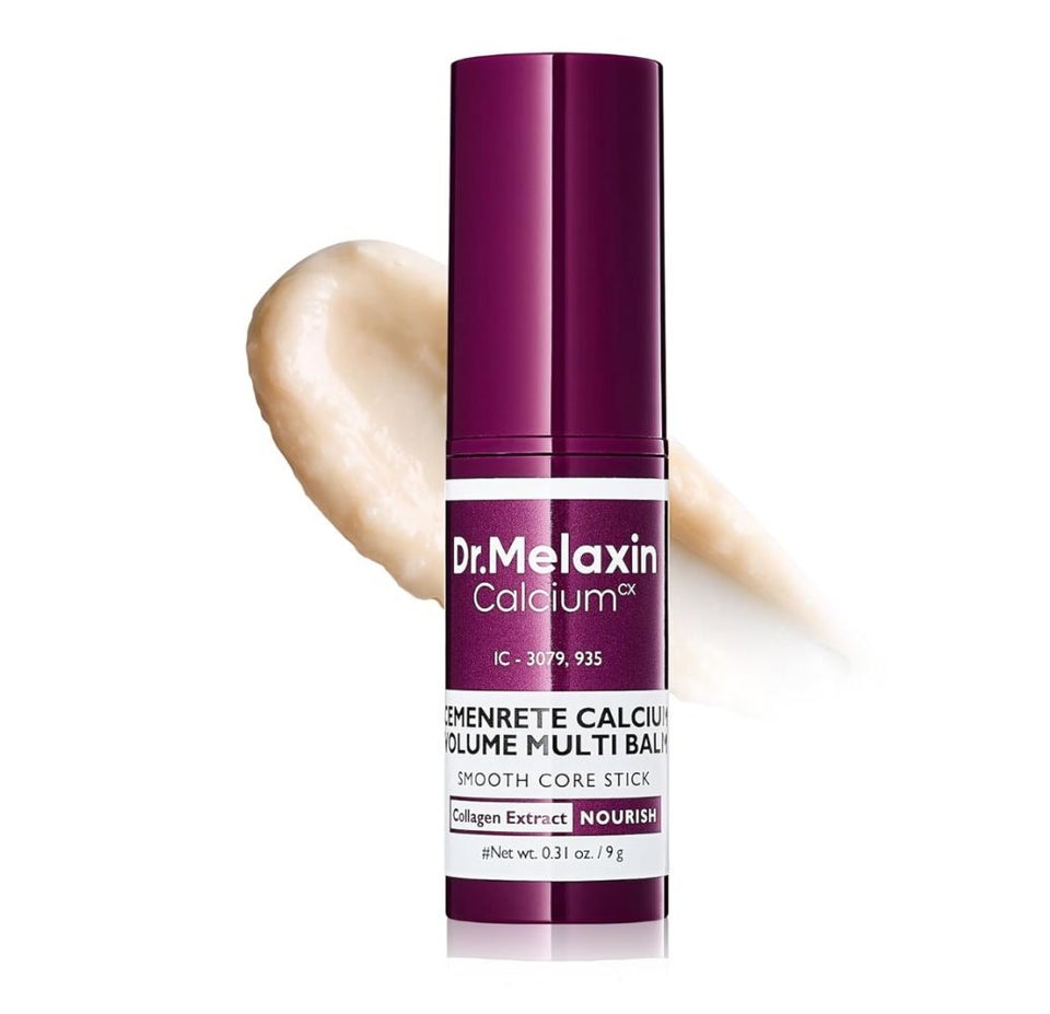
In a previous story, HuffPost shopping writer Griffin Wynne reported that topical calcium can strengthen the skin barrier and increase cell turnover, making it a useful ingredient for anti-aging skin care. That’s where this Dr. Melaxin calcium multi-balm stick comes in: This non-sticky balm contains calcium, vitamin D, collagen, elastin and adenosine to smooth, hydrate and firm the skin, plus reduce the appearance of wrinkles.
The stick packaging makes it so easy to apply this balm wherever needed on the face and neck without making a mess. Best of all, reviewers in their 50s and 60s love it for its help with “turkey neck” and fine lines.
Amazon
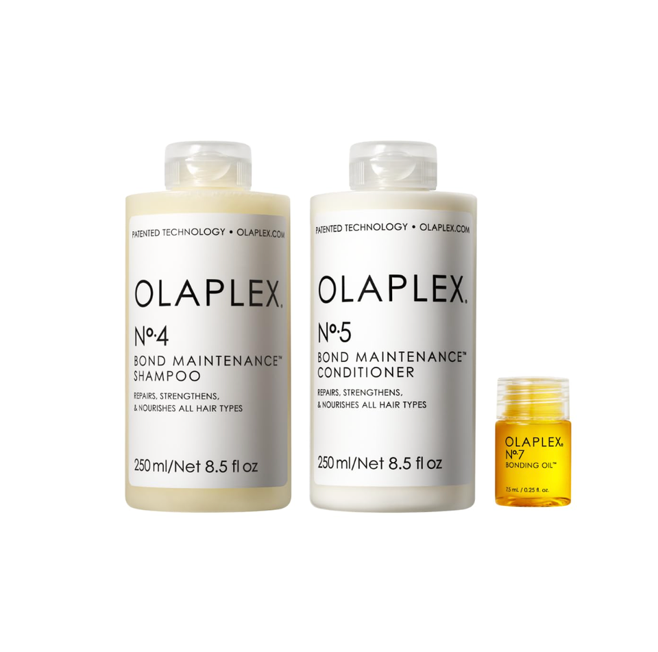
Olaplex is a must for people with bleached or color-treated hair. It’s designed to reduce breakage while conditioning and strengthening your hair, with reviewers claiming that it leaves their hair soft, smooth and ultra-shiny. This set of three includes some of the brand’s most popular products: No. 4 Bond Maintenance shampoo, No. 5 Bond Maintenance conditioner and No. 7 Bonding Oil. It is formulated for all hair types, and those with curly or damaged hair can also get a lot out of it.
Although Flores said that she’s been using the bonding oil pre-heat styling for many years now, she just started using the Bond Maintenance shampoo and conditioner about a year ago, and, “almost with the first use, I felt my hair was softer and less frizzy after blow drying — it’s a popular choice of many for a reason,” she said.
Advertisement
Amazon
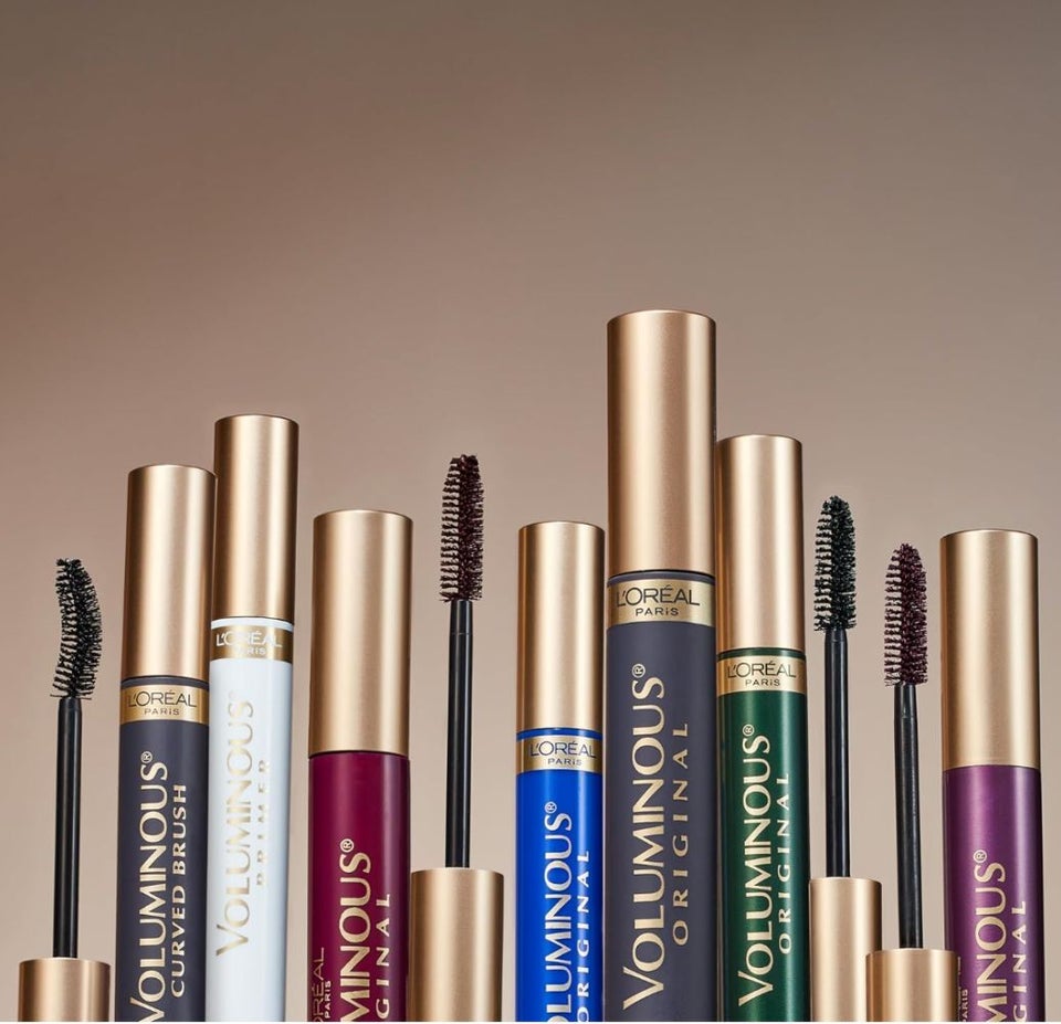
According to Flores, this was the tube of mascara that solidified the potential of drugstore formulas. The L’Oreal Voluminous mascara line was previously recommended to us by professional makeup artists as a good budget option for anyone looking to experiment with mascara shades that go beyond just black or brown.
“There’s something about the product consistency of this mascara and the fluffy barrel brush that makes my lashes look longer and noticeably volumized, without looking weighed down or spidery. It also never flakes or smudges, even when the tube is a little older. I’ve even opted to try a few other colors, including the deep violet shade that I like so much, I find myself grabbing for it more than my go-to black. You’ll want to opt for this if you’re after a set of lashes that look fanned, wispy and not overly unnatural,” Flores said.
Amazon
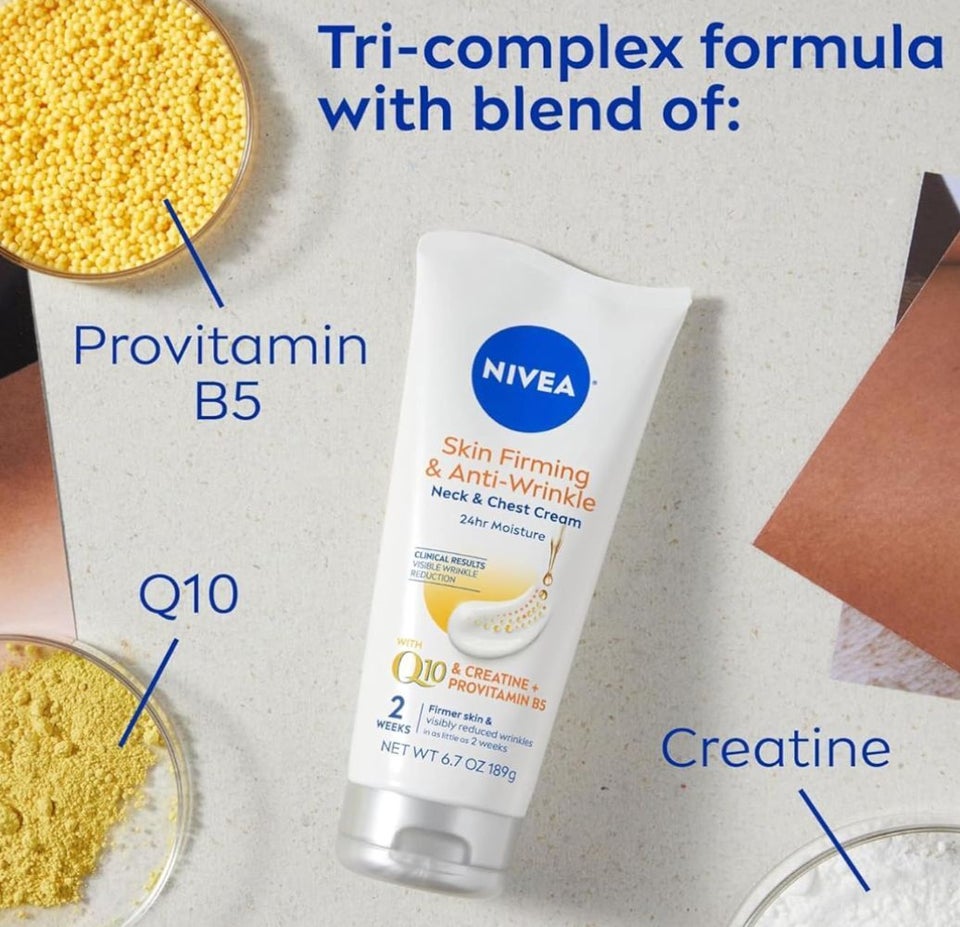
We’ve often heard comparisons made between Nivea’s classic cream formula and the uber-luxe Crème de la Mer, and while we’ve slightly debunked that theory in previous reporting, Nivea’s formulas will always remain trusted and iconic in my heart and bathroom cabinet. This particular drugstore formula features a trio of ingredients to help soften those deep lines you get in between the breasts and on the neck.
Amazon
Advertisement
Amazon
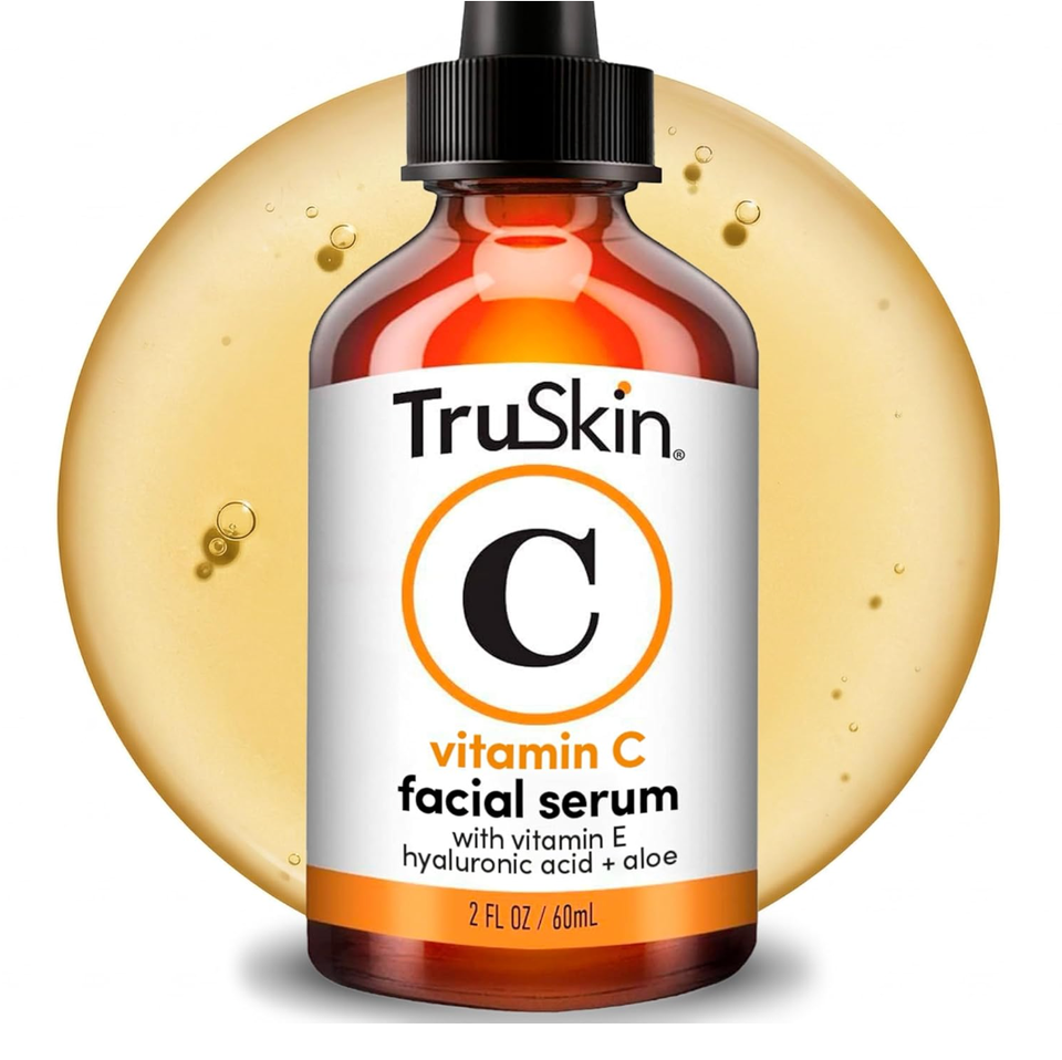
In case you didn’t know, vitamin C is a really valuable player in the skin care game. In fact, after sunscreen and retinol, it’s the most helpful ingredient in defense against premature skin aging, according to a previous HuffPost report. Per the experts, vitamin C applied topically protects skin from external toxins and free radicals that cause premature skin aging, while enhancing collagen production.
This makes this popular serum’s enthusiastic reviews all the more viable. The nourishing formula works to reduce in fine lines, sun damage and aging spots — improving the appearance of skin conditions like melasma and an overall increase in skin brightness and radiance.
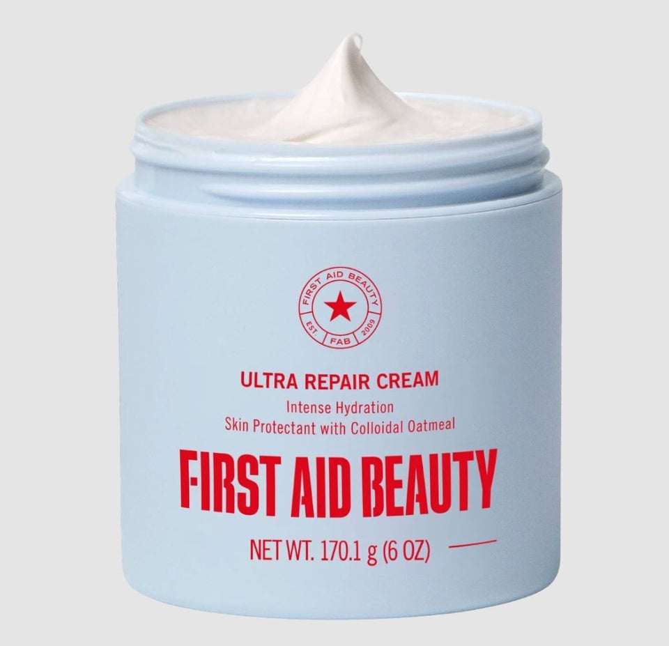
First Aid Beauty
Flores is a fan of a few First Aid Beauty products, like this intensive repair cream. She credits it with saving her itchy, flaky, eczema-prone skin on more than one occasion.
The moisturizing formula was even previously recommended by Dr. Azadeh Shirazi, a board-certified cosmetic and medical dermatologist in San Diego. “[It’s] a must-have for winter!” she said. “It’s a rich cream formulated with colloidal oatmeal, which relieves irritation, and shea butter, which protects and maintains our skin barrier. Allantoin also helps calm and soothe the skin.”
Amazon
This dermatologist-recommended neck tightening cream by StriVectin is a targeted cream containing spilanthol, an ingredient designed to strengthen the skin, along with a patented peptide exclusive to the brand. Peptides can help to improve collagen levels and elastin which can help to fight skin laxity. This neck cream can also help brighten and even skin tone, diminishing the appearance of sunspots. Two different sizes are on sale for Prime Day (the larger 3.4-ounce size is pictured here).
Advertisement
The Real Deal: We use deal trackers and commerce experience to sift through “fake” hike-and-drop deals and other deceptive sales tactics. Products will usually be rated at least 4 stars with a minimum 15% discount. (And when there’s an exception, we’ll tell you why.)

A remarkable fossil discovery inside a cave near Waitomo on New Zealand’s North Island is giving scientists an unprecedented look at a long vanished ecosystem. Researchers from Australia and New Zealand have uncovered the remains of ancient birds and frogs that lived around 1 million years ago, including a previously unknown relative of the iconic kākāpō.
The find marks the first time scientists have recovered a large collection of terrestrial vertebrate fossils from this period in New Zealand’s history. Preserved within the cave were fossils belonging to 12 bird species and four frog species, offering a rare snapshot of a world that existed hundreds of thousands of years before humans reached the islands.
The research, published in Alcheringa: An Australasian Journal of Palaeontology, suggests that New Zealand’s wildlife was already undergoing dramatic changes long before human settlement. Powerful volcanic eruptions and rapid climate shifts repeatedly reshaped habitats, driving extinctions and opening opportunities for new species to evolve.
Ancient Birds Lost to Time
Lead author Associate Professor Trevor Worthy of Flinders University says the fossils reveal a bird community unlike anything seen in New Zealand today.
“This is a newly recognized avifauna for New Zealand, one that was replaced by the one humans encountered a million years later,” says Associate Professor Worthy, from the College of Science and Engineering at Flinders University.
“This remarkable find suggests our ancient forests were once home to a diverse group of birds that did not survive the next million years.”
In biology, the term “avifauna” refers to the collection of bird species living in a particular place and time. The fossils indicate that the birds inhabiting New Zealand a million years ago were substantially different from those present when people eventually arrived.
The study involved paleontologists from Flinders University and Canterbury Museum, as well as volcanologists Joel Baker of the University of Auckland and Simon Barker of Victoria University of Wellington.
According to the researchers, approximately 33-50% of species disappeared during the million years before humans reached Aotearoa New Zealand.
Volcanoes and Climate Change Reshaped Ecosystems
Scientists believe these losses were largely caused by natural environmental upheaval.
“These extinctions were driven by relatively rapid climate shifts and cataclysmic volcanic eruptions,” says co-author Dr. Paul Scofield, Senior Curator of Natural History at Canterbury Museum.
The discovery helps fill one of the largest gaps in New Zealand’s fossil record.
“From our excavations at St Bathans in Central Otago over many years, we have a snapshot of life in Aotearoa between 20 and 16 million years ago. These new findings cast light on the 15 million year period from then to 1 million years ago, which is largely absent from New Zealand’s fossil record,” says Dr. Scofield.
“This wasn’t a missing chapter in New Zealand’s ancient history, it was a missing volume.”
Fossils are often compared to pages in Earth’s history book. In this case, researchers say they have uncovered an entire section of that story that was previously unknown.
A Possible Flying Ancestor of the Kākāpō
One of the most exciting discoveries is a newly identified parrot species called Strigops insulaborealis. It is an ancient relative of the kākāpō, one of New Zealand’s most famous birds.
Today, the kākāpō is the world’s only flightless parrot. It is also one of the heaviest parrots and is known for its unusual nighttime lifestyle. However, the newly discovered ancestor may have been very different.
Analysis of the fossilized bones suggests it had weaker legs than modern kākāpō. Because today’s birds rely heavily on their strong legs and climbing ability, researchers think the ancient species may have spent less time climbing and possibly retained the ability to fly.
Additional research will be needed to determine whether it truly could take to the air.
The cave also contained fossils from an extinct ancestor of the takahē, another distinctive New Zealand bird. Researchers also identified an extinct pigeon species closely related to Australia’s bronzewing pigeons.
“The shifting forest and shrubland habitats forced a reset of the bird populations,” adds Dr. Scofield.
“We believe this was a major driver for the evolutionary diversification of birds and other fauna in the North Island.”
Volcanic Ash Helps Date the Fossils
One reason the discovery is so important is that scientists can determine its age with unusual precision.
The fossils were trapped between two layers of volcanic ash preserved inside the cave. One ash layer came from an eruption about 1.55 million years ago. The second was produced by a massive eruption approximately 1 million years ago.
This natural geological sandwich provides clear age limits for the fossils.
Researchers say the younger eruption likely covered much of the North Island in meters of ash. While rain and erosion eventually removed much of that material, some remained protected inside caves.
The older ash layer also reveals something else remarkable. It shows that the fossil site is the oldest known cave on New Zealand’s North Island.
Rewriting New Zealand’s Natural History
Associate Professor Worthy says the fossils provide a crucial benchmark for understanding how New Zealand’s wildlife evolved.
The fossils “provide a critical, missing baseline for New Zealand’s natural history.”
For many years, scientists focused primarily on the ecological changes that occurred after humans arrived in New Zealand roughly 750 years ago. The new evidence shows that powerful natural forces had already been transforming the islands’ wildlife for hundreds of thousands of years.
“For decades, the extinction of New Zealand’s birds was viewed primarily through the lens of human arrival 750 years ago. This study proves that natural forces like super-volcanoes and dramatic climate shifts were already sculpting the unique identity of our wildlife over a million years ago.”

NASA’s upgraded Cold Atom Lab is back in operation aboard the International Space Station, giving researchers a powerful new way to investigate the fundamental nature of matter and advance the development of future quantum technologies. Taking advantage of the station’s microgravity environment, the facility enables experiments that cannot be performed on Earth.
Quantum science focuses on the behavior of matter and energy at extremely small scales, including atoms, electrons, and particles of light. Although atoms are often pictured as tiny balls colliding with one another, the quantum world is far stranger. Atoms can behave like waves, appear in multiple locations at the same time, and even pass through one another under certain conditions.
NASA’s Cold Atom Lab Studies Matter Near Absolute Zero
About the size of a mini refrigerator and controlled remotely from Earth, the Cold Atom Lab cools atoms to temperatures below minus 459 degrees Fahrenheit (minus 237 degrees Celsius). At temperatures just above absolute zero, atoms can combine into an unusual quantum state known as a Bose-Einstein condensate, or BEC.
A BEC is made up of matter waves and is considered a fifth state of matter in addition to solids, liquids, gases, and plasma. Even though it is much larger than individual subatomic particles, it still follows the laws of quantum mechanics. The microgravity conditions of low Earth orbit allow these matter waves to become even larger than they can on Earth.
“At the coldest temperatures, matter behaves drastically different from anything we have experienced,” said Jason Williams, project scientist for Cold Atom Lab at NASA’s Jet Propulsion Laboratory in Southern California, which built the facility. “The wavelike nature of matter dominates, and ultracold matter can behave in ways that are not only unexpected, but that also enable extremely precise measurements of time, gravity, and motion. The lab has lots of tools — especially with this latest upgrade — to let us probe the nature of the universe.”
The facility currently supports five international research teams studying fundamental physics. It also serves as a testing ground for quantum instruments that could one day support Earth science investigations and future exploration missions.
How the Upgraded Cold Atom Lab Works
At the center of the facility is a sophisticated collection of instruments known as the science module. A newly upgraded version of this module arrived at the space station on April 11 aboard a Commercial Resupply Services mission, expanding the range of experiments scientists can perform.
During an experiment, strips of rubidium or potassium metal are heated to temperatures as high as 750 °F (400 °C), creating a gas inside a vacuum chamber. Researchers then use carefully tuned lasers to remove energy from the atoms. As the atoms lose energy, they slow down and cool dramatically.
After the laser cooling stage, magnetic fields trap the atoms and keep them contained. Additional cooling techniques reduce their energy even further, bringing the atomic cloud close to a complete standstill and allowing scientists to maximize the amount of time it can be studied in microgravity.
Why Quantum Experiments Benefit From Space
Scientists can study ultracold gases in laboratories on Earth, but space offers important advantages. In microgravity, quantum gases can be observed for longer periods and cooled to even lower temperatures.
The low gravity environment also allows larger quantum waves to form and interact with gravity for longer periods of time. To make these experiments possible aboard the station, engineers compressed what would normally be a room-sized atomic physics laboratory filled with lasers and optical equipment into a compact system that fits inside a station experiment rack.
“As the first project to create Bose-Einstein condensates in orbit, we’re demonstrating that we can make quantum technology work reliably in space,” said Ethan Elliott, deputy project scientist for Cold Atom Lab at JPL. “In the previous century, there was a quantum revolution that led to lasers, cellphones, and MRIs for medical imaging. We’re performing quantum 2.0 — direct manipulation of large quantum states — and we hope for similar gains in quantum tech by advancing this science in orbit.”
New Upgrade Expands Quantum Research Capabilities
The latest enhancement is the fourth major upgrade since the Cold Atom Lab was installed on the International Space Station in 2018.
Among the most significant improvements is a redesigned magnetic trap that can alter the shape of quantum gas clouds. This gives researchers new opportunities to investigate the properties and behavior of ultracold atoms. Engineers also introduced redesigned metal atom sources that generate the gas clouds used in experiments.
“It’s the closest thing we have to controlling the boundary of the quantum world,” said Kamal Oudrhiri, project manager of Cold Atom Lab at JPL, referring to those low temperatures. “This new upgrade pushes that boundary even further.”
Oudrhiri added that the new hardware “demonstrates NASA’s ability to maintain U.S. leadership in space-based quantum technologies while maturing future quantum instruments, such as matter-wave interferometers for fundamental physics missions, positioning, navigation, timing, and gravity sensing of Earth, the Moon, and beyond.”
Advancing Quantum Technology in Space
The Cold Atom Lab is managed by Caltech in Pasadena, while NASA’s Jet Propulsion Laboratory designed, built, and operates the facility. The project is sponsored by the Biological and Physical Sciences division within NASA’s Science Mission Directorate in Washington.
The division supports scientific discovery by using the unique conditions of space to conduct experiments that cannot be carried out on Earth. By studying biological and physical processes in extreme environments, researchers gain knowledge that can help humans travel farther and remain in space longer, while also producing benefits for life on Earth.
Watching football is an emotional rollercoaster – but is it good or bad for your health?

A minimally invasive procedure for chronic knee pain is helping some patients find significant relief without undergoing major surgery.
For Cynthia Schraf-Fletcher, 74, the results were “remarkably” successful.
Nearly a year after receiving genicular artery embolization (GAE) on her right knee, Schraf-Fletcher says the improvement is comparable to the total knee replacement she previously underwent on her left knee.
“I couldn’t be more pleased,” says Schraf-Fletcher, who had the procedure performed by Leigh Casadaban, MD, MS, assistant professor of radiology at the University of Colorado Anschutz School of Medicine.
Today, she says everyday activities such as gardening and riding a stationary bicycle are far more enjoyable because of the reduction in pain.
How Genicular Artery Embolization Works
GAE is an outpatient procedure designed to ease chronic knee pain by reducing blood flow to inflamed areas within the joint. By targeting abnormal blood vessels associated with inflammation, the treatment can help decrease swelling and discomfort.
“For treating osteoarthritis in the knees, we often think of medications, physical therapy, maybe a steroid injection, and then on the far end of the spectrum is a total knee replacement. There really hasn’t been anything for patients in between,” Casadaban, a vascular interventional radiologist, says. “GAE is a promising minimally invasive procedure that may fill that spot for people who have failed conservative treatments but are not yet ready to have a major surgery.”
According to Casadaban, people with mild to moderate osteoarthritis tend to benefit the most. Patients with more advanced disease can also undergo the procedure, although the effects are generally less durable.
“We find about 70% of patients have phenomenal results. They cut their pain scores in half, sometimes more. We have a few patients with no pain at all after the procedure,” Casadaban says. “Patients that have tried a lot of other treatments and haven’t had pain relief are happy to get back to their normal activities.”
After experiencing complications from knee replacement surgery, Schraf-Fletcher was eager to explore another option. Looking back, she says choosing GAE was the right decision.
What Happens During the Procedure?
GAE typically takes between one and two hours and is performed under conscious sedation.
During the procedure, an interventional radiology team makes a small incision near the crease of the leg. Using X-ray imaging and contrast dye for guidance, doctors advance a tiny catheter through the femoral artery until it reaches the genicular arteries around the knee.
Once in position, the team releases microscopic beads that block blood flow to the abnormal vessels located in the painful areas identified by the patient.
Patients are monitored for several hours afterward and are usually able to return home the same day. Doctors generally advise taking it easy for a few days during recovery.
Originally developed in Japan a little more than a decade ago, GAE has steadily gained attention worldwide. Since 2021, the FDA has granted “breakthrough device status” to multiple devices related to the procedure in the United States.
Research Suggests Long Lasting Pain Relief
Early and ongoing research continues to produce encouraging results.
“The theory is that GAE reduces inflammation inside the knee joint, and symptom relief can last years,” Casadaban says. “Four-year data published in Japan shows that if you have one outpatient procedure, your pain relief can last for those four years. In the U.S., we now have two-year data, which shows that if you have a good response, pain relief can last two years. That really speaks to the theory that we’re hopefully modifying something in the joint.”
Casadaban is currently leading two clinical trials at CU Anschutz. One study is examining changes in knee fluid among patients receiving GAE. The other is evaluating a temporary arterial treatment device called Nexsphere-F, which blocks small blood vessels in the knee that may contribute to inflammation and pain.
Expanding Beyond Knee Osteoarthritis
Osteoarthritis is a degenerative joint disease that affects millions of people each year and can occur in many different joints throughout the body.
Although GAE is currently used only for knee conditions, Casadaban says researchers and physicians are beginning to explore its use for other painful musculoskeletal disorders, including frozen shoulder, tennis elbow, and plantar fasciitis.
The immunotherpay can give children and adults three extra years before they need to use insulin.
Dr Hilary Cass says she is “absolutely convinced that more children will be harmed if we don’t do the trial than if we do.”
I am 44 years old and I might be the only woman I know who doesn’t mind getting older. In fact, I relish it.
Don’t get me wrong — I’m not fond of everything that comes with it. My hips hurt. My eyesight is getting worse. I’m forced to decide between a glass of wine or a sleepless night. Gray hair, not-so-fine lines and deep wrinkles are certainly humbling. Oh, and my moisturizers are now more expensive than they’ve ever been.
None of this is fun. But aging has given me something that I didn’t even know I needed: delicious invisibility and freedom from unwanted male attention.
Advertisement
I know that many will take issue with my stance. Society is not kind to women who age, and no one knows that better than women themselves. No one enjoys being ignored when they need something, but the world is not kind to girls or women at any stage, and what I’m talking about is something far more sinister than the store clerk who looks right past me.
I remember the first time a man I didn’t know put his hands on me. It happened in broad daylight at a popular spot in Brooklyn, New York, where locals go to enjoy the view of the Hudson River and the Verrazano-Narrows Bridge that connects my borough to Staten Island. I was 15 years old and enjoying summer break. I spent my days biking around the city and often stopped at this spot, which I’d loved since my parents took me there as a small child.

Courtesy of Christina Wyman
Advertisement
I hopped off my bike and was admiring the whitecaps that rushed under the bridge when I felt his hand up my shorts. Then, just as quickly, he sped away on his bicycle while his friend cheered him on. The assault was fast, calculated and breathtakingly scary. I wonder now how many other unsuspecting women and children he groped before — and after — me.
I never saw the face of that man who made me afraid to go to the promenade by myself from that point forward. Nearly 30 years later, I’ve never forgotten the assault. For too many years I blamed myself for what happened: Maybe if I’d stayed on my bike … Maybe if I hadn’t worn such short shorts … Maybe if I hadn’t been so taken by that view …
That man was not the first or last to challenge my relationship with public spaces. The following year, another guy I didn’t know cornered me against a gate and wrapped his leg around my waist before animatedly thrusting his hips into me until I shoved him away with all the force my teenage body could muster. His friends laughed as I hurried off in the opposite direction, glancing over my shoulder until they were out of sight.
Advertisement
A few years before either of those encounters, I was walking to a family member’s house a few blocks from my own home when a man who appeared to be my father’s age pulled up next to me in a van and said, “Hey, little girl, I have a bag of candy for you.” Though this sounds like something out of an after-school special — almost too cliche to be real — the danger I felt in that moment was visceral, and I ran away from him as quickly as my feet would carry me.

Courtesy of Christina Wyman
I know I’m not the only one who has experienced something like this. Many girls and women are often suddenly confronted by a car horn, a whistle or obscenities being leveled at us by men and boys. It seems to get them off, and I would wager that some of them fully intend to instill a bit of fear in their targets.
Advertisement
A few years ago I was on a walk in a rural town when a man approached me on foot, got uncomfortably close and whispered, “Nice tits,” before rubbing himself and walking away. This happened mere feet from a preschool. It was the first time I called the police during an otherwise peaceful walk in broad daylight. I was on high alert the entire way home. I had no way to prove what had just happened, and I’m not sure the police ever came.
Women and girls are targets for unwanted male attention wherever they find themselves. Everything I’m describing has taken place in cities, suburbs and rural towns, and it’s often much worse. It happens in nightclubs. It happens in grocery stores. It’s happening on busy streets in the middle of the day. Women are told to smile. They’re badgered for their numbers. They’re called names if they don’t respond or dare to call out the behavior. It is relentless. It is daunting. And it’s exhausting.
For many of us, this kind of attention begins in our own homes, long before we’re able to make sense of our experiences. I, like many girls, was conditioned by my family to believe that my body was up for criticism, inspection and commentary. When aunts and uncles comment on the length of your legs, or when your parents talk openly about how you’re “filling out” (or not filling out), or when your grandparents admonish you for not “sitting like a lady” before you have any idea what they mean, you come to accept this hyper fixation on your body as normal.
Advertisement

Courtesy of Christina Wyman
To make matters worse, I was told that unwanted male attention was always the fault of the girl or woman receiving it — no matter what. If I was courageous enough to report these confusing, infuriating and terrifying encounters with strange men and boys to my parents, they would summarily dismiss them as my fault.
Our culture does the exact same thing. Just look at Donald Trump (the former president of the United States!) and the way he has treated women — and how our society has normalized his repugnant behavior. It’s 2024 and we’re still considered playthings for men. And if we don’t like it, we’re labeled rude or frigid, and told we can’t take a joke or don’t know our place.
Advertisement
So, I could not be more thrilled to finally be at an age where I’m mostly unseen by men. With each passing year, I find myself navigating unwanted male attention less and less, which means I can now do something as simple as go for a walk without preemptively bracing myself for rude and hypersexualized comments or uninvited physical contact.
It shouldn’t have to be this way. We shouldn’t have to wait half of our lives to feel safe in our own skin. We shouldn’t have to secure the invisibility that aging offers us to exist in the world without fear of who will approach us and what they’ll say or do. Just think of how much mental and emotional energy we waste — and what we could be capable of if we could spend all of that time and energy creating or connecting with others. What’s more, if women were respected — and desired for more than just their bodies — we might not be rendered invisible as we age (which obviously comes with disadvantages too), and I wouldn’t have to be so overjoyed about finally living undetected.
Unfortunately, it’s too late for me — I’ve already aged out — but it may not be too late for the women and girls coming up after me.
Advertisement

Courtesy of Christina Wyman
As an author, I believe that starting conversations about these kinds of experiences can be a powerful part of the solution. I write children’s books about unpalatable topics like bullying, family dysfunction and gender-based harassment involving minors because I do not want these traumas to continue to thrive in silence. If we can teach children how to resist objectification — and teach boys not to objectify in the first place — we can change the dominant narrative about women in our culture. For me, it all begins with a simple idea: No one is entitled to opinions about — or access to — other peoples’ bodies without their consent. Period. And I will not stop talking with children about these difficult topics until society has turned a corner.
I’m not holding my breath. It won’t be easy. It will take courage, dedication, uncomfortable discussions, and a lot of support and understanding. But it’s worth fighting for. Besides, now that I’m invisible, I’ve got all of that extra time and energy on my hands, so I hope I can help make the world a little more palatable for the girls who will soon inherit it.
Advertisement
Christina Wyman is a USA Today bestselling author and teacher living in Michigan. “Slouch,” her highly anticipated middle-grade novel, is about a tall girl navigating friends, family, self-esteem, boundaries and the terrifying realization that her body no longer seems to belong to her. It is available wherever books are sold, including local independent bookstores, starting in October 2024. Her debut novel, “Jawbreaker,” a middle-grade book that follows a seventh grader with a craniofacial anomaly, was named one of Publishers Weekly’s best books of 2023.
Need help? Visit RAINN’s National Sexual Assault Online Hotline or the National Sexual Violence Resource Center’s website.
This piece was previously published on HuffPost and is being shared again now as part of HuffPost Personal’s “Best Of” series.
Advertisement
Do you have a compelling personal story you’d like to see published on HuffPost? Find out what we’re looking for here and send us a pitch at pitch@huffpost.com.