Deadly “red sky” solar storm from 800 years ago discovered in ancient trees

Powerful solar activity can create stunning auroras on Earth, but outside the protection of our planet’s magnetic field, the Sun can become extremely dangerous. Violent eruptions such as solar flares and coronal mass ejections can blast high energy particles through space, creating serious risks for astronauts and spacecraft.

Some of these eruptions produce solar proton events (SPEs), during which charged particles race toward Earth at speeds reaching 90% of the speed of light. In 1972, several SPEs erupted between the Apollo 16 and Apollo 17 Moon missions. If astronauts had been exposed during a lunar mission, they could have faced lethal radiation levels. As space agencies prepare for future Moon exploration, scientists are working to better understand these unpredictable solar events.

Researchers at the Okinawa Institute of Science and Technology (OIST) have now developed a new way to uncover evidence of past SPEs. The team combined medieval historical records with ultra precise carbon 14 measurements taken from buried asunaro trees in northern Japan. Using this method, they identified a solar proton event that likely occurred sometime between the winter of 1200 CE and the spring of 1201 CE, a period marked by unusually intense solar activity. The findings were published in the Proceedings of the Japan Academy, Series B.

Professor Hiroko Miyahara from the OIST Solar-Terrestrial Environment and Climate Unit explained: “Previous studies on historical SPEs have focused on rare, extremely powerful events. Our paper provides a basis for detecting sub-extreme SPEs — events that occur more frequently and are around 10-30% of the size of the most extreme cases, but still hazardous. Sub-extreme SPEs are more challenging to detect, but our method now allows us to efficiently identify them and better understand the conditions under which they are more likely to occur.”

Ancient Trees Preserve Clues About Solar Storms

Earth’s magnetic field blocks most high energy particles released during SPEs. Near the poles, however, magnetic field lines open into space, allowing some particles to enter the atmosphere. During especially powerful events, these particles collide with atmospheric gases and create carbon 14 compounds that spread around the globe and become trapped inside living organisms.

By analyzing carbon 14 levels in preserved organic material such as ancient buried trees, scientists can track changes in solar activity stretching back thousands of years. The OIST team used an ultra precise measurement technique they spent more than a decade refining. This method can detect much smaller carbon 14 fluctuations than conventional techniques, making it possible to identify weaker “sub-extreme” solar proton events that were previously invisible.

Because the carbon 14 analysis is extremely time intensive, the researchers first needed clues about when unusual solar activity may have occurred.

Medieval Japanese Diary Revealed “Red Lights” in the Sky

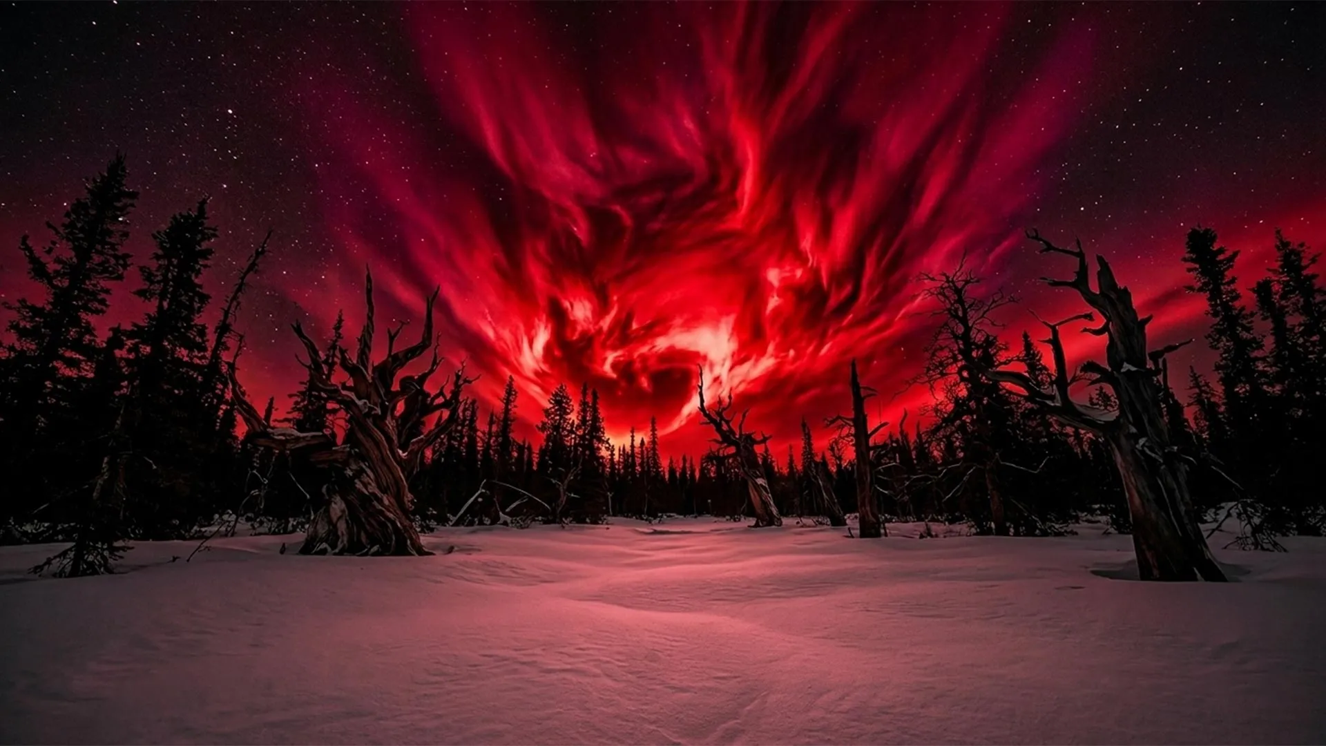
One of the key clues came from Meigetsuki, the diary of the Japanese poet and courtier Fujiwara no Teika (1162-1241). In February 1204 CE, he described seeing “red lights in the northern sky over Kyoto.”

Solar proton events do not directly create auroras, but they are often linked with the same kinds of solar disturbances that do. That historical observation gave researchers a timeframe to investigate more closely.

The scientists then measured carbon 14 levels in buried asunaro wood recovered from Aomori Prefecture in northern Japan. They discovered spikes in carbon 14 that pointed to a sub-extreme solar proton event. By combining those measurements with dendroclimatic studies — that is, a dating method based on comparing patterns of tree-ring growth associated with regional climate — the researchers determined that the event likely occurred sometime between the winter of 1200 CE and the spring of 1201 CE. Historical records from China also described a red aurora visible at unusually low latitudes during that same period.

Evidence of an Exceptionally Active Sun

“The high-precision data not only allowed us to accurately date sub-extreme solar proton events, but it also lets us clearly reconstruct the solar cycles of the period,” said Miyahara. “Today, the Sun’s activity fluctuates over eleven-year-long cycles, but we’ve found that the cycle was just seven to eight years long back then, indicating a very active Sun. The SPE we have dated occurred at the peak of one of these cycles.”

The research helps fill important gaps in the history of solar activity and improves scientists’ understanding of dangerous space weather events. According to Miyahara, carbon 14 analysis alone is not enough. Historical records and other scientific methods are also essential for reconstructing past solar behavior.

“Historical literature provides a candidate time window, and dendroclimatology enables direct intercomparison between detected SPE and reports of sunspots and auroras recorded in literature. Integrated approaches like these are necessary to accurately reconstruct past solar activity, helping us better understand the characteristics of extreme space weather,” concluded Miyahara. “For example, while the SPE we found occurred near the peak of the solar cycle, some of the prolonged low-latitude aurora recorded in the literature seems to fall near the minimum of our reconstructed solar cycle. This is unexpected, and we’re excited to look further into what solar conditions could cause this.”

Share Button

Who are the Japanese? Huge DNA discovery rewrites history

For decades, scientists believed the Japanese population largely descended from two ancient groups: the Jomon hunter-gatherers who lived in the archipelago for thousands of years, and later migrants from East Asia who brought rice farming and new technologies to Japan.

But a major genetic analysis from researchers at RIKEN’s Center for Integrative Medical Sciences suggests the picture is far more complicated.

Using whole-genome sequencing on more than 3,200 people from across Japan, the team found evidence supporting a third ancestral group tied to northeastern Asia and possibly linked to the ancient Emishi people. The findings, published in Science Advances, add powerful support to the increasingly discussed “tripartite origins” theory of Japanese ancestry.

The results also revealed something else surprising: Japan’s population is genetically more diverse than many researchers once assumed.

“The Japanese population isn’t as genetically homogenous as everyone thinks,” said Chikashi Terao, who led the study at RIKEN. “Our analysis revealed Japan’s subpopulation structure on a fine scale, which is very beautifully classified according to geographical locations in the country.”

A Massive DNA Map of Japan

To investigate Japan’s deep genetic history, researchers analyzed DNA samples collected from seven regions stretching from Hokkaido in the north to Okinawa in the south. The project became one of the largest whole-genome studies ever conducted on a non-European population.

Instead of relying on older DNA microarray methods, the team used whole-genome sequencing, which reads nearly all three billion DNA base pairs in a person’s genome. According to the researchers, this provides roughly 3,000 times more information than traditional techniques.

“Whole-genome sequencing gives us the chance to look at more data, which helps us find more interesting things,” Terao explained.

The scientists then combined the genetic information with medical histories, disease diagnoses, family histories, and clinical test results to build a large database known as the Japanese Encyclopedia of Whole-Genome/Exome Sequencing Library (JEWEL).

One especially important focus involved rare genetic variants. These uncommon DNA changes can sometimes preserve clues about ancient migration patterns and long-lost ancestral populations.

“We reasoned that rare variants can sometimes be traced back to specific ancestral populations, and could be informative in revealing fine-scale migration patterns within Japan,” Terao said.

The Hidden Third Ancestor

The analysis uncovered striking regional differences across Japan.

Jomon ancestry appeared strongest in Okinawa, where it was found in 28.5% of samples, while western Japan showed much lower levels at 13.4%. Researchers found that people in western Japan had stronger genetic connections to Han Chinese populations, likely reflecting major migration waves from continental East Asia between 250 and 794 CE. Those migrations also coincided with the spread of Chinese-style government systems, writing, and education throughout Japan.

The newly identified Emishi-related ancestry was concentrated in northeastern Japan and became less common farther west.

The findings build on earlier ancient DNA studies published in 2021 that first proposed the idea that modern Japanese people descend from three major ancestral sources instead of two. Those studies suggested that a third migration connected to the Kofun period played a major role in shaping modern Japan.

Recent follow-up studies have continued strengthening that idea. Researchers analyzing ancient genomes and skeletal remains have found increasing evidence that multiple migration waves entered Japan over centuries, creating a much more layered population history than previously believed.

Ancient Neanderthal and Denisovan DNA Still Affects People Today

The study also explored genetic material inherited from Neanderthals and Denisovans, two ancient human groups that interbred with Homo sapiens tens of thousands of years ago.

Scientists have become increasingly interested in why some of these ancient DNA fragments survived in modern humans while others disappeared. In many cases, the inherited genes appear linked to health, adaptation, or disease risk.

For example, earlier studies showed that Tibetans inherited a Denisovan-related version of the EPAS1 gene that may have helped humans survive in high-altitude environments. Researchers also previously identified Neanderthal-derived DNA associated with severe Covid-19 complications in some populations.

The Japanese genome study identified 44 archaic DNA regions still present in modern Japanese populations, many of them unique to East Asians. One Denisovan-derived region inside the NKX6-1 gene was associated with type 2 diabetes and may influence how some patients respond to semaglutide treatments.

Researchers also found 11 Neanderthal-derived genetic segments connected to conditions including coronary artery disease, prostate cancer, and rheumatoid arthritis.

Toward Personalized Medicine

Beyond tracing ancestry, the researchers believe the work could eventually improve healthcare.

The team identified potentially harmful variants in the PTPRD gene that may be linked to hypertension, kidney failure, and myocardial infarction. They also found common loss-of-function variants in the GJB2 and ABCC2 genes, which are associated with hearing loss and chronic liver disease.

“What we’ve tried to do is to find and catalog loss-of-function gene variants that are very specific to Japanese people, and to understand why they are more likely to have some specific traits and diseases,” Terao said. “We’d like to connect population differences with differences in genetics.”

The study reflects a broader shift happening in genetics research. For years, most large genomic databases heavily focused on people of European ancestry, limiting scientists’ understanding of disease risk in other populations.

Terao hopes expanding JEWEL with more Asian genomic data will help change that.

“It’s quite important to expand this to the Asian population so that in the long run, the results can benefit us too,” he said.

Share Button

Before This $10 Retinol Stick, ‘I Was Convinced The Only Sure Thing To Do For My Wrinkles Was Botox.’

Few places on your body can indicate your age, lifestyle and health changes more than your eyes and the ultra-sensitive skin around them. From insomnia-imposed nights to natural aging to health conditions, the eyes are the windows to a little more than the soul, I’d argue. Luckily, we found a handy retinol eye stick that reviewers are loving for how it addresses everything from fine lines, eye bags, puffiness, dark circles and more. While they’re not saying it works exactly like Botox, they are calling it a “wonder of wonders” that produces impressive results fairly quickly. ” The icing on the cake: It’s freaking only $10 — at least today.

The easy-to-swipe stick is packed with a guest list of ingredients that make one seriously good party for the skin.

Retinol is the guest of honor for restoring smoothness, of course. Meanwhile, bakuchiol shows up like a hype man to help boost the retinol’s efficacy while helping to calm the sensitivity that retinol can create. Hyaluronic acid joins the dance floor with moisture-surging and plumping moves. Then, squalene and vitamin E operate like bouncers to by providing barrier protection, moisturization and skin repair, respectively.

“I don’t look 20 again, but I think it has taken about 10 years off of me,” one reviewer, Susan, shared about the crow’s feet smoothing she’s seeing after using the stick.

People are using it for their undereyes but finding that it’s convenient and effective for other parts of their face and beyond.

People are using it for crow’s feet, undereye bags, their necks, chest wrinkles (often emerging from side-sleeping, by the way) and more! One 78-year-old man, RD, uses it on an enduring “11” that has formed on his forehead over time, and notes that he saw long-lasting improvement after a few fails with other products.

Another 42-year-old reviewer notes that their neck began to smooth out within two weeks of swiping the retinol stick there faithfully.

People previously more resolved about Botox are using it comfortably. 👀

“I was convinced the only sure thing to do for MY wrinkles was to get Botox, which is expensive, at least for me,” shopper Lynette shared.

“But I did try it and I am Amazed… It does what it says it will do and it does what all of the reviews have said it will do… didn’t have to pay upwards of Hundreds of Dollars for something that I wasn’t even sure would help [or] that I would end up throwing away with all of the other products I have tried in the past.”

Another shopper, Cassandra, routinely uses Botox and higher-end beauty products, also notes what a pleasant surprise this stick is. “Smooths fine lines, brightened my eye area,” the Botox fan wrote. “Love this- everyone says my makeup looks great, but it’s the [eye stick].”

Note: We’ll continue to preach that you should always use retinol with sunscreen. Using anti-aging products (or, really, any skin care) without faithfully using a sunscreen is like going fishing with a hole in your boat. You might go somewhere, but it won’t be far.)

Definitely grab the $10 retinol smoothing stick while it’s a whopping 50% off. Still curious about how others are experiencing it? Understandable. Peep more promising reviews below:

“I love that this product is so easy to apply. No messy lotions or serums, you just simply swipe the balm under your eyes for moisturized and smooth under eye skin. I use this every night because it’s quick and effective. You don’t really need to use that much, so it’s a great value for the price. I really did see an improvement in my under eye fine lines and wrinkles after a few weeks of consitent use. This is a keeper in my skin care routine!” — Tami

“I am 74 year old man. I do not have a lot of wrinkles in spite of my age, but I have always had the so called “11” lines between my eyebrows and they give me a “mean” look. I have tried different products for years that claim to fix the problem but nothing worked. One of the instant wrinkle smoothers worked well, but after a very few hours it flaked and the wrinkles were back. I decided to give this Retinol Eye Stick a try and it is working great. I use it on the 11 wrinkles and also on my under eye bags. I began to see improvement in the 11 after around 10 days and the under eye began looking better after two weeks. For the 11 wrinkles I rub the Retinol on the wrinkles and then rub it in with my fingers. The area will appear slightly red for a few minutes then go back to normal. I just ordered and received my second stick. I started using the product around Dec 31 I think. On average the stick lasts about a month. I am still seeing improvement as I use the product and at this rate I believe the 11 will be completely gone or at least not noticeable in another month. I will continue using the product from now on. The under eye looks much better and feels really good. I carry the stick to work and use it about four times a day. I will use it less times per day when the wrinkles are gone. It has no odor that I can detect. Great product! I think that if you try it you will tell the way it feels that it will work.” — RD

“I purchased this in mid June this year and shortly thereafter began using it. I always have 0 expectations for any new product I buy. So, I just used it and forgot about it. Then lately, I guess within the last two weeks or so, my eyes seem to be more alert looking. Taking a closer look, I discovered that my crow’s feet are nearly gone! So, it has been 2 months of use and sure enough, my eyelids look tighter and my skin a bit brighter. I mean, I kept wondering what was going on and then I remembered I was using this eye stick. Note although I am old, I do not naturally have bags or discoloration under my eyes. What I have was just light wrinkling and sagging skin. I don’t look 20 again, but I think it has taken about 10 years off of me.” — Susan

Share Button

Posh sandwich has more salt than nearly five cheeseburgers

Action on Salt & Sugar said people should not be exposed to a “hidden health risk every time they buy lunch”.

Share Button

MPs call for sunbed advertising ban to help prevent skin cancer

A report by a cross-party group of MPs has found the majority of skin cancer cases are preventable.

Share Button

Watch Morning Live here

Watch Morning Live here

Share Button

Sydney dentist’s patients warned of viruses including HIV

Authorities urged patients to get tested due to “poor infection control practices” at the Australian clinic.

Share Button

A rare cancer-fighting plant compound has been decoded

Researchers at UBC Okanagan have uncovered the process plants use to create mitraphylline, a rare natural compound that has attracted attention for its possible cancer fighting properties.

Mitraphylline belongs to a unique class of plant chemicals known as spirooxindole alkaloids. These molecules are recognized for their unusual twisted ring structures and their powerful biological effects, including anti inflammatory and anti tumor activity.

Even though scientists have studied these compounds for years, the exact molecular steps plants use to produce them had remained unknown.

Breakthrough Discovery in Plant Chemistry

That mystery began to unravel in 2023 when Dr. Thu-Thuy Dang’s team in UBC Okanagan’s Irving K. Barber Faculty of Science identified the first known plant enzyme capable of twisting a molecule into the distinctive spiro shape.

Building on that earlier finding, doctoral student Tuan-Anh Nguyen led new research that uncovered two critical enzymes involved in the production of mitraphylline. One enzyme organizes the molecule into the correct three dimensional structure, while the second transforms it into mitraphylline itself.

“This is similar to finding the missing links in an assembly line,” says Dr. Dang, UBC Okanagan Principal’s Research Chair in Natural Products Biotechnology. “It answers a long-standing question about how nature builds these complex molecules and gives us a new way to replicate that process.”

Why Mitraphylline Is So Valuable

Many promising natural compounds are found only in tiny amounts inside plants, making them difficult and expensive to recreate in laboratories. Mitraphylline is one of those rare substances. It exists only in trace quantities in tropical trees such as Mitragyna (kratom) and Uncaria (cat’s claw), both members of the coffee family.

Now that researchers have identified the enzymes responsible for shaping and assembling mitraphylline, they have a clearer path toward producing the compound and related molecules in more sustainable ways.

“With this discovery, we have a green chemistry approach to accessing compounds with enormous pharmaceutical value,” says Nguyen. “This is a result of UBC Okanagan’s research environment, where students and faculty work closely to solve problems with global reach.”

Nguyen also reflected on the experience of contributing to the breakthrough.

“Being part of the team that uncovered the enzymes behind spirooxindole compounds has been amazing,” Nguyen adds. “UBC Okanagan’s mentorship and support made this possible, and I’m excited to keep growing as a researcher here in Canada.”

International Collaboration Fuels the Research

The project brought together Dr. Dang’s laboratory at UBC Okanagan and Dr. Satya Nadakuduti’s research group at the University of Florida.

Funding for the work came from Canada’s Natural Sciences and Engineering Research Council’s Alliance International Collaboration program, the Canada Foundation for Innovation, and the Michael Smith Health Research BC Scholar Program. Additional support was provided by the United States Department of Agriculture’s National Institute of Food and Agriculture.

“We are proud of this discovery coming from UBC Okanagan. Plants are fantastic natural chemists,” Dr. Dang says. “Our next steps will focus on adapting their molecular tools to create a wider range of therapeutic compounds.”

Share Button

Scientists discover a weak spot shared by polio and common cold viruses

Researchers at the University of Maryland, Baltimore County (UMBC) have uncovered a crucial step that enteroviruses use to reproduce inside human cells. The findings, published in Nature Communications, explain how viruses responsible for illnesses such as polio, encephalitis, myocarditis, and even the common cold take control of cellular machinery to copy themselves. Scientists say the discovery could eventually help researchers create a new generation of antiviral drugs capable of targeting many enteroviruses at once.

The study was led by Deepak Koirala, associate professor of chemistry and biochemistry at UMBC, along with recent Ph.D. graduate Naba Krishna Das. Their work helps answer longstanding questions about how these viruses launch replication once they invade a cell.

“My lab has been really motivated to understand how RNA viruses produce their proteins inside the cell and multiply their genome to make more virus particles,” Koirala says. Earlier work from the team identified an important cloverleaf shaped structure within the virus’s RNA. The new study shows how that structure recruits proteins needed to build the viral replication machinery.

How Enteroviruses Reproduce Inside Cells

Enteroviruses carry very small RNA genomes that must perform two jobs at once. The viral RNA has to direct the production of viral proteins while also serving as the template for creating new copies of the virus.

Most of the viral genome contains instructions for structural proteins, but it also encodes several specialized proteins required for replication. One of the most important is a fusion protein called 3CD.

The 3C portion cuts long chains of amino acids into the separate proteins the virus needs. The 3D portion acts as an RNA polymerase, an enzyme that copies viral RNA so the virus can reproduce. Human cells do not naturally contain this type of polymerase, meaning the virus has to supply its own version.

“We previously determined the structure of the RNA alone, and other groups determined the structure of 3C and 3D, but now we’ve captured the structure of the RNA and proteins together, so we know how they are interacting,” Koirala explains. “We found that it’s the 3C domain of 3CD that binds to the RNA in the viral genome, and then it recruits the other components, such as host protein PCBP2, to assemble the replication complex.”

The researchers also found that this molecular complex functions like a switch. When 3CD is attached, the virus copies its RNA genome. When the protein detaches, the RNA becomes available for producing viral proteins instead.

Scientists Resolve a Longstanding Viral Mystery

To examine these interactions in detail, the team used X-ray crystallography to visualize the RNA cloverleaf and the 3CD protein together. They also relied on isothermal titration calorimetry (ITC), which measures the heat released when molecules bind, and biolayer interferometry (BLI), which uses changes in light interference to track how long molecules stay attached.

The experiments helped settle an ongoing scientific debate. The researchers showed that two full 3CD molecules, each carrying its own RNA polymerase, bind side by side on the viral RNA. Earlier research had proposed that the proteins formed a single fused pair instead.

Scientists still do not fully understand why two copies are required, but the new study provides a much clearer picture of how the replication process begins.

Potential for Broad Spectrum Antiviral Drugs

One of the most promising findings was how similar the mechanism appeared across all seven enteroviruses examined in the study. The viruses shared nearly identical RNA cloverleaf structures and binding behavior.

That level of similarity suggests the RNA structure is extremely important to viral survival. Significant mutations would likely disrupt replication, making the structure a potentially stable drug target across many enteroviruses.

Researchers say this raises the possibility of developing broad spectrum antiviral drugs that could work against an entire family of viruses rather than a single pathogen.

Scientists are already developing drugs that interfere with the 3C and 3D proteins, but the new findings reveal another possible strategy.

Drugs disrupting 3C and 3D activity are already in development, but “now we have another layer to test,” Koirala says. “What if we target the RNA, or the RNA-protein interface, so that we break the interaction? That is another opportunity. Now that we have high-resolution structures, you can precisely design drug molecules to target them.”

Koirala says the study highlights how surprisingly sophisticated viruses can be despite their tiny genomes.

“Viruses are so, so clever. Their entire genome is equivalent to about one mRNA sequence in humans, yet they are so effective,” Koirala says. His latest work demonstrates “why we need to investigate this basic science — so that it can be translated into developing drugs targeting pathogens that cause so many harmful diseases.”

Share Button

Scientists make old blood stem cells young again in major anti-aging breakthrough

Scientists at the Icahn School of Medicine at Mount Sinai have successfully reversed aging in blood-forming stem cells in mice by repairing defects in structures known as lysosomes. The findings, published in Cell Stem Cell, point to lysosomal dysfunction and overactivity as major causes of stem cell aging and show that restoring proper lysosomal activity can rejuvenate old stem cells and improve their ability to regenerate blood and immune cells.

Lysosomes function as the cell’s internal recycling centers. They break down proteins, nucleic acids, carbohydrates, and lipids, helping cells dispose of waste and reuse materials for essential biological processes. They also store nutrients that can be released when needed. Because of these roles, lysosomes are critical for maintaining cellular metabolism, including both catabolism (breaking down complex molecules to simple ones) and anabolism (building complex molecules from simpler ones).

The research team focused on hematopoietic stem cells (HSCs), which are rare, long-lasting stem cells found in the bone marrow that generate all blood and immune cells in the body. The study was led by Saghi Ghaffari, MD, PhD, Professor of Cell, Developmental, and Regenerative Biology at the Icahn School of Medicine and a member of the Black Family Stem Cell Institute.

As people grow older, these stem cells gradually lose their ability to repair and replenish the blood system. This decline weakens immune defenses and contributes to the increased vulnerability to infections seen in older adults. Aging HSCs are also linked to clonal hematopoiesis, an asymptomatic condition considered a premalignant state that raises the risk of blood cancers and inflammatory diseases. The condition becomes much more common with age.

According to the American Cancer Society, age and smoking are the two strongest risk factors associated with the five-year risk of developing cancer. Data from the National Cancer Institute’s Surveillance, Epidemiology, and End Results report show that the median age at cancer diagnosis is 67.

Restoring Old Stem Cells to a Youthful State

The researchers discovered that lysosomes in aged HSCs become excessively acidic, damaged, depleted, and abnormally active. These changes disrupt both the metabolic balance and epigenetic stability of the stem cells.

Using single-cell transcriptomics and functional testing, the team found that blocking this excessive lysosomal activity with a vacuolar ATPase inhibitor restored lysosomal health and improved the function of aging blood stem cells.

After treatment, the old stem cells began behaving more like young, healthy cells again. They regained the ability to regenerate effectively, produce balanced blood and immune cells, and generate additional healthy stem cells. The treated cells also showed improved metabolism and mitochondrial performance, healthier epigenetic patterns, reduced inflammation, and fewer harmful inflammatory signals that can damage tissues throughout the body.

“Our findings reveal that aging in blood stem cells is not an irreversible fate. Old blood stem cells have the capacity to revert to a youthful state; they can bounce back,” said Dr. Ghaffari. “By slowing down the lysosomes and reducing their acidity, stem cells became healthier and could make new balanced blood cells and new stem cells much more effectively. By targeting lysosomal hyperactivity, we were able to reset aged stem cells to a younger, healthier state, improving their ability to regenerate blood and immune cells.”

Major Increase in Blood-Forming Capacity

The researchers also tested an ex vivo treatment approach (when cells are removed from the body, modified in a laboratory, and returned to the body). Treating old stem cells with the lysosomal inhibitor increased their blood-forming ability in living animals by more than eightfold, highlighting the powerful regenerative effects of correcting lysosomal dysfunction.

The improvement also reduced damaging inflammatory and interferon-related pathways. According to the researchers, this occurred because healthier lysosomes improved the processing of mitochondrial DNA and lowered activation of the cGAS-STING immune signaling pathway, which appears to play a major role in stem cell inflammation and aging.

Potential for Anti-Aging and Blood Disorder Therapies

The findings could open the door to new treatments aimed at preventing or reversing age-related blood disorders. They may also improve stem cell transplantation outcomes in older patients and enhance conditioning methods used in gene therapy.

“Lysosomal dysfunction emerges as a central driver of stem cell aging,” added Dr. Ghaffari. “Targeting this pathway may one day help maintain healthy blood and immune systems in the elderly, improve their stem cells for transplantation, and reduce the risk of age-associated blood disorders and perhaps have an effect on overall aging.”

The team is now investigating whether lysosomal dysfunction in aging stem cells contributes to the development of leukemic stem cells, potentially connecting normal stem cell aging with cancer formation.

The research involved collaboration with Mickaël Ménager, PhD, and colleagues at the Imagine Institute and INSERM UMR 1163 at Université de Paris Cité in Paris. Funding was provided by the National Institutes of Health, New York State Stem Cell Science, INSERM, and the Agence Nationale de la Recherche.

Share Button