Cosmic voids look empty but they may be tearing the universe apart

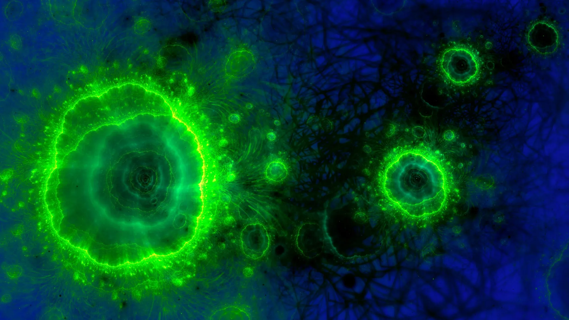
Imagine removing everything from the deepest regions of cosmic voids. Take away ordinary matter, neutrinos, dark matter, cosmic rays, and radiation. What remains appears to be nothing but empty space. It may sound contradictory, but these enormous voids are filled with the vacuum of spacetime. And importantly, that vacuum is not truly nothing.

The vacuum of spacetime contains something fundamental. It is difficult to describe precisely with everyday language, but physicists refer to these underlying ingredients as quantum fields. In quantum field theory, the particles that make up our world such as electrons, top quarks, neutrinos, and even dark matter are not independent objects in the usual sense. What we call a particle is actually a visible expression of something deeper.

These deeper structures are the fields themselves. Every type of particle has a corresponding field. These fields permeate every cubic centimeter of space and time. They have existed since the big bang and extend throughout the entire universe.

When we observe a particle, such as an electron moving through space, we are really detecting a ripple or vibration in its underlying field. The particle is a traveling excitation of that field. Even if all particles were removed, the fields would still remain.

Vacuum Energy and the Origin of Dark Energy

These fields also contain energy. Because of the Heisenberg uncertainty principal, the vacuum cannot be completely devoid of energy. When physicists attempt to calculate how much energy exists in empty space, the results can range from extremely large values to theoretically infinite ones…which is also another episode.

What matters is that this vacuum energy produces a measurable effect. That effect is known as “dark energy,” the name scientists use to describe the accelerated expansion of the universe.

Observations show that the actual amount of vacuum energy is relatively small, though it is not zero. In most environments across the universe, its influence is negligible. Regions filled with matter completely dominate the local behavior of space.

Here on Earth, for example, matter is so dense that dark energy has no noticeable impact. If dark energy suddenly vanished, everyday physics would remain unchanged. The path of a thrown baseball would be identical. Your burrito would still cook in the microwave at exactly the same rate. Nothing about daily life would be different.

Where Dark Energy Dominates the Universe

The same situation applies across much of the cosmos. Galaxies, galaxy clusters, filaments, and walls of the cosmic web are all regions packed with matter. In these environments, dark energy plays almost no role.

Cosmic voids are different.

Voids are enormous regions where matter is largely absent. In these areas, the vacuum of space-time itself becomes the dominant influence. If you could place yourself in the middle of a cosmic void, you would effectively be surrounded by dark energy.

In fact, voids are where dark energy carries out its most important work. The accelerated expansion of the universe does not occur inside dense regions such as galaxies or clusters. Instead, it takes place within the vast empty voids.

Cosmic Voids Are Expanding

Cosmic voids are not just empty gaps between structures in the universe. They are actively growing. As dark energy pushes space outward, the voids expand and press against the surrounding cosmic web.

Over immense spans of time, this process gradually pulls the universe’s large-scale structure apart. The intricate network of galaxies, clusters, and filaments that astronomers see today will not last forever. Over the next 5-10-20 billion years the exact number doesn’t matter the cosmic web will slowly fade as expanding voids stretch everything farther apart.

Why Empty Space Is Never Truly Empty

In that sense, cosmic voids are far from empty. They are filled with the subtle energy of quantum fields. That energy influences the entire universe by driving its accelerating expansion.

Voids are the only regions where this effect becomes dominant, precisely because they contain almost nothing else.

So yes, cosmic voids are empty of matter. That is how astronomers identify and measure them. But their lack of matter means they are filled with dark energy.

Wherever you travel in the universe, whether to a nearby galaxy or to the deepest interior of the emptiest void, you will never truly be alone.


Share Button

Scientists detect a sudden acceleration in global warming

Global warming has been increasing at a faster pace since about 2015, according to a new study from the Potsdam Institute for Climate Impact Research (PIK). By adjusting global temperature records to remove known natural influences, researchers were able to identify a statistically significant rise in the rate of warming for the first time.

During the past decade, global temperatures have climbed at an estimated rate of about 0.35°C per decade, depending on the dataset analyzed. From 1970 through 2015, the average increase was just under 0.2°C per decade. The more recent trend represents the fastest warming observed in any decade since instrumental temperature records began in 1880.

“We can now demonstrate a strong and statistically significant acceleration of global warming since around 2015,” says Grant Foster, a US statistics expert and co-author of the study, which was published today in the scientific journal Geophysical Research Letters.

“We filter out known natural influences in the observational data, so that the ‘noise’ is reduced, making the underlying long-term warming signal more clearly visible,” Foster added.

Removing Natural Climate Variability From Temperature Data

Short term natural events can temporarily raise or lower global temperatures and make it harder to detect changes in long term climate trends. These influences include El Niño events, volcanic eruptions, and variations in solar activity.

To address this challenge, the researchers analyzed measurement data from five widely used global temperature datasets (NASA, NOAA, HadCRUT, Berkeley Earth, ERA5). By adjusting the data to account for these natural factors, the team was able to isolate the underlying warming trend more clearly.

“The adjusted data show an acceleration of global warming since 2015 with a statistical certainty of over 98 percent, consistent across all data sets examined and independent of the analysis method chosen,” explains Stefan Rahmstorf, PIK researcher and lead author of the study.

Statistical Analysis Reveals a Shift in Warming Trends

The study focused on determining whether the pace of warming has changed, rather than identifying the causes behind that shift.

After accounting for the influence of El Niño and the recent solar maximum, the extremely warm years of 2023 and 2024 appear slightly cooler in the adjusted analysis. Even with these corrections, they still rank as the two warmest years recorded since instrumental measurements began. Across all datasets, the faster warming trend becomes visible around 2013 or 2014.

To evaluate whether the warming rate has changed since the 1970s, the researchers applied two statistical techniques: a quadratic trend analysis and a piecewise linear model that identifies when shifts in warming rates occur.

Implications for the Paris Agreement Climate Target

The study does not attempt to determine the specific reasons behind the acceleration in warming. However, the authors note that climate models already allow for the possibility that the rate of warming could increase over time.

“If the warming rate of the past 10 years continues, it would lead to a long-term exceedance of the 1.5°C limit of the Paris Agreement before 2030,” says Stefan Rahmstorf. “How quickly the Earth continues to warm ultimately depends on how rapidly we reduce global CO2 emissions from fossil fuels to zero.”

Share Button

‘I’m in constant pain after vaginal mesh surgery’

Susan McLarnon is travelling to Downing Street later to call for “urgent action” to be taken on redress for those harmed by pelvic mesh implants.

Share Button

Hidden metabolism found operating inside the cell nucleus

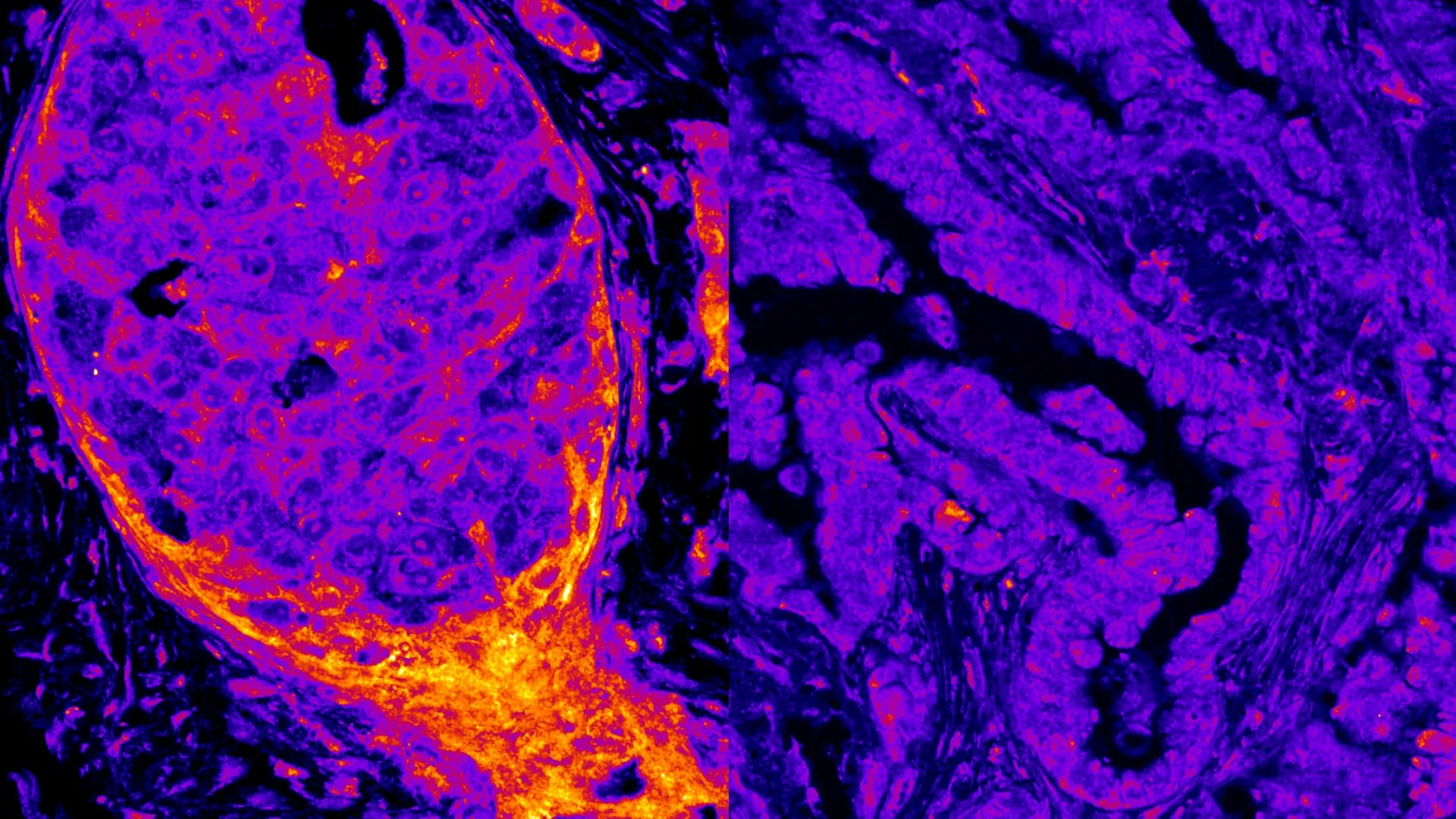
A new study published in Nature Communications has revealed that more than 200 metabolic enzymes can be found directly on human DNA. Many of these enzymes are typically known for producing energy in mitochondria, yet researchers discovered them sitting on chromatin inside the cell nucleus.

The study shows that different cell types, tissues, and cancers each display their own distinctive arrangement of metabolic enzymes within the nucleus. These enzymes interact with DNA in patterns that researchers describe as a “nuclear metabolic fingerprint,” marking the first evidence that human cells may carry such unique nuclear signatures.

Scientists still need to determine the precise role these enzymes play in the nucleus. They could be driving chemical reactions, influencing whether genes are switched on or off, or contributing to structural support. Even so, the findings already provide new insights into how tumors develop, adapt, and sometimes resist therapy.

“Many of these enzymes synthesize essential building blocks of life, and their nuclear localization is associated with DNA repair. Their presence in the nucleus may therefore directly shape how cancer cells respond to genotoxic stress, a hallmark of many chemotherapeutic treatments. It’s an entirely new world to explore,” says Dr. Sara Sdelci, corresponding author of the study and researcher at the Centre for Genomic Regulation.

Studying Proteins Bound to Chromatin

To identify these enzymes, the research team used a technique that isolates proteins physically attached to chromatin, the natural packaging of DNA in human cells. Using this approach, they examined 44 cancer cell lines and 10 healthy cell types collected from ten different tissues.

Metabolism and genome regulation have traditionally been viewed as largely separate biological systems. The nucleus houses the genome, while metabolic enzymes normally produce energy in mitochondria and the cytoplasm.

Because of this assumption, the scale of the discovery surprised the researchers. They found that metabolic enzymes appear to play active roles in nuclear biology. About 7 percent of all proteins attached to chromatin turned out to be metabolic enzymes. This observation suggests that the nucleus may operate its own small metabolic network described by the researchers as a ‘mini metabolism’.

Unexpected Energy Pathways Inside the Nucleus

Some of the enzymes detected were particularly surprising. The team identified proteins involved in oxidative phosphorylation, the cellular process responsible for generating most of a cell’s energy, as regular occupants of the nucleus.

The pattern of these enzymes also varied depending on cancer type. Oxidative phosphorylation enzymes were commonly observed in breast cancer cells but were largely missing in lung cancer cells. When scientists examined tumor samples taken directly from patients, they observed the same trend, confirming that nuclear metabolism varies depending on tissue type and disease.

“We’ve been treating metabolism and genome regulation as two separate universes, but our work suggests they’re talking to each other, and cancer cells might be exploiting these conversations to survive,” says Dr. Savvas Kourtis, first author of the study.

Enzymes Move to Damaged DNA

The researchers also performed experiments to understand what these nuclear enzymes actually do. They focused on a group of enzymes responsible for producing molecules needed for DNA synthesis and repair.

Their experiments showed that these enzymes gather near chromatin when DNA damage occurs. By concentrating in these regions, they appear to assist with repairing the genome.

The team also discovered that the function of an enzyme can depend on its location inside the cell. One enzyme, called IMPDH2, behaved differently depending on where it was located. When researchers forced it to remain inside the nucleus, it helped maintain genome stability. When the same enzyme was restricted to the cytoplasm, it influenced entirely different cellular pathways.

Implications for Cancer Treatment

These findings raise important questions about how cancer treatments work. Some therapies target metabolic processes in cancer cells, while others focus on disrupting DNA repair systems. If these two biological processes are more tightly connected than previously thought, it could change how scientists approach cancer treatment.

“It could help explain why tumors of different origins, even when carrying the same mutations, often respond very differently to chemotherapy, radiotherapy, or targeted inhibitors,” says Dr. Sdelci.

Mapping Nuclear Metabolism

According to the researchers, this study provides the first large scale evidence that metabolic enzymes are widely present inside the nucleus. Over time, mapping where these enzymes are located and understanding their functions could help identify biomarkers for diagnosing cancer or reveal new weaknesses that anti cancer drugs could target.

However, researchers emphasize that much work remains. Scientists still need to determine whether all of the enzymes observed in the nucleus are active and what specific roles each one plays.

“Each enzyme may have its own, unique nuclear function, so this must be addressed one by one,” says Dr. Kourtis.

How Large Enzymes Enter the Nucleus

Another unanswered question involves how these enzymes reach the nucleus in the first place. The nucleus is separated from the cytoplasm by a barrier that normally limits which molecules can pass through nuclear pores.

Many of the enzymes discovered on DNA are significantly larger than the size these pores are believed to allow. Despite this, the bulky proteins still manage to enter the nucleus.

This puzzling observation suggests that cells may use an as yet unknown mechanism to move large enzymes into the nucleus. Understanding how this process works could eventually reveal precise therapeutic targets for controlling nuclear metabolic activity in diseased cells.

Share Button

Light-guided evolution creates proteins that can switch, sense, and compute

Evolution is nature’s way of engineering biological systems. Inside cells, many variations of DNA, RNA, and proteins arise, and natural selection favors the organisms that function most effectively. Humans began harnessing this process long ago. Early farmers influenced evolution by choosing which crops and livestock reproduced, allowing the most productive plants and animals to pass on their traits.

Today, scientists apply similar principles in the lab through a technique known as directed evolution. Researchers use it to improve proteins such as enzymes and antibodies that play important roles in medicine, industrial manufacturing, and even everyday products like laundry detergents.

Limits of Traditional Directed Evolution

Despite its success, standard directed evolution methods have a key limitation. They usually impose a constant selection pressure that favors proteins that remain highly active all the time. However, real biological systems rarely function this way. Many proteins serve as signals, molecular switches, or “logic gates” (proteins that combine multiple inputs to make a yes-or-no decision), meaning they must change states as conditions shift.

For example, a protein might briefly activate, then turn off, and later switch on again. When evolution experiments only reward a single state, other necessary states can degrade. As a result, proteins may lose the ability to switch properly, which can be harmful for cells (e.g. kill a cell). Because of this challenge, creating proteins with complex multi-state behavior has proven difficult with existing directed evolution approaches.

A Light-Based Strategy for Protein Evolution

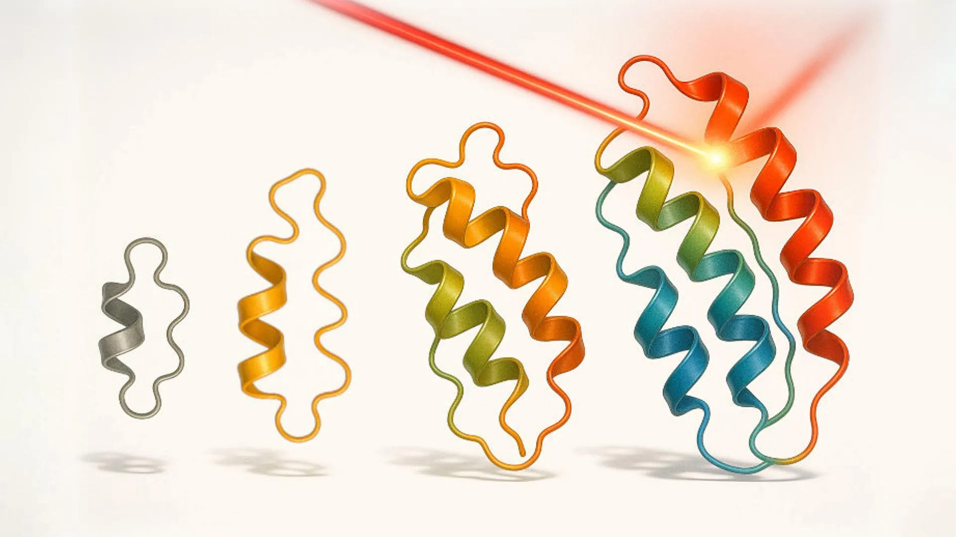
Researchers led by Sahand Jamal Rahi at EPFL’s Laboratory of the Physics of Biological Systems have introduced a new approach called “optovolution.” This method uses light to steer the evolution of proteins that can perform dynamic functions and even carry out simple computational tasks that follow yes-or-no rules.

The study, published in Cell, helps bring directed evolution closer to how cells naturally operate. In living systems, timing and switching between states are just as important as the strength of a signal.

Engineering Yeast Cells to Select the Best Proteins

To build their system, the researchers used the budding yeast Saccharomyces cerevisiae, an organism widely used both in brewing and scientific research. They redesigned the yeast cell cycle so that cell division depended on the behavior of the protein being evolved. The protein needed to switch cleanly between active and inactive states for the cell to survive.

The scientists connected the protein’s output signal to a regulator that controls the cell cycle. This regulator is essential during one stage but becomes toxic during another. If the protein remained on or off for too long, the yeast cell would stall or die. Only cells containing proteins that switched at the correct time continued to divide.

Using Light to Control Evolution in Real Time

Light provided a way to control this process with precision. The researchers used optogenetics, a technique that activates or deactivates genes using light. By delivering timed pulses of light, they forced the protein to alternate between states.

Each yeast cell cycle lasts about 90 minutes, creating a rapid pass or fail test of whether the protein switched at the correct moment. Proteins that performed best allowed the cell to survive and reproduce, while poorly switching variants were eliminated. This allowed optovolution to automatically select proteins with better dynamic behavior without manual screening or repeated adjustments.

New Protein Variants and Expanded Color Sensitivity

Using optovolution, the team evolved several different types of proteins. They first improved a commonly used light controlled transcription factor. The researchers generated 19 new variants that showed greater sensitivity to light, reduced activity in darkness, or the ability to respond to green light rather than only blue light. Engineering proteins that respond to warmer colors than blue has long been considered extremely difficult because of how these proteins absorb light.

The scientists also evolved a red light optogenetic system so that yeast cells no longer required an added chemical cofactor. Evolution produced a mutation that disabled a normal yeast transport protein. This unexpected change allowed the system to use light sensitive molecules already present inside the cell, making the system easier to use in experiments.

Proteins That Act Like Tiny Computers

The study also demonstrated that optovolution can extend beyond light sensing proteins. The researchers evolved a transcription factor that functions like a single protein computer. It activated genes only when two different inputs appeared at the same time – one light signal and one chemical signal.

Dynamic protein behavior is essential for many biological processes, including sensing environmental changes, making decisions inside cells, and controlling cell division. By enabling these behaviors to evolve continuously within living cells, optovolution offers new possibilities for synthetic biology, biotechnology, and fundamental research.

The technique may help scientists design smarter cellular circuits, create optogenetic tools that respond independently to different colors of light, and better understand how complex protein behaviors arise through evolution.

Other contributors

  • EPFL Laboratory of Protein and Cell Engineering
  • University of Bayreuth
  • Lausanne University Hospital (CHUV)
Share Button

Sent 90 miles after giving birth while ‘soaked in urine’

Four days after giving birth, Lizzy Berryman’s psychosis forced her to be taken from York to Derby for specialist care.

Share Button

NHS England pauses new prescriptions of cross-sex hormones for under-18s

The health service said young people who already receive the drugs will continue to do so.

Share Button

Scientists stunned to find signs of ancient life in a place no one expected

Dr. Rowan Martindale, a paleoecologist and geobiologist at the University of Texas at Austin, was hiking through the Dadès Valley in Morocco’s Central High Atlas Mountains when something unusual caught her attention and made her stop.

Martindale and her research team, including Stéphane Bodin of Aarhus University, were exploring the rugged valley to study the ecology of ancient reef systems that once existed there when the area lay beneath the ocean. Reaching those reefs required crossing numerous layers of turbidites, sediments formed by dense underwater debris flows. Ripple patterns often appear on these deposits. However, Martindale noticed small ridges and wrinkles layered on top of the ripples that seemed unusual.

“As we’re walking up these turbidites, I’m looking around and this beautifully rippled bedding plane caught my eye,” says Martindale. “I said, ‘Stéphane, you need to get back here. These are wrinkle structures!'”

What Are Wrinkle Structures

Wrinkle structures are tiny ridges and pits ranging from millimeters to centimeters across. They develop when algae and microbial communities grow in mats across sandy seafloors. These delicate textures are rarely preserved in younger rocks because animals often disturb and destroy them. As a result, wrinkle structures are uncommon in rocks younger than about 540 million years old, when animal life rapidly diversified and began actively stirring ocean sediments.

Today, scientists typically find wrinkle structures in shallow tidal environments where sunlight supports photosynthetic algae.

Why These Wrinkles Should Not Exist

The wrinkle structures Martindale spotted appeared in rocks that formed far below the ocean surface. The turbidites where they were found had been deposited at depths of at least 180 meters, far too deep for sunlight to penetrate. This meant the structures could not have formed from the same sunlight dependent algae that create wrinkle patterns in shallow environments today.

Previous claims of wrinkle structures in deep water turbidite deposits have also been disputed. Another complication was the age of the rocks. At about 180 million years old, they formed during a time when animals were actively disturbing the seafloor worldwide, which normally erases delicate microbial textures. In other words, the wrinkle structures Martindale saw should not have been preserved at all.

Recognizing how unusual the find was, she set out to confirm whether her first impression was correct.

“Let’s go through every single piece of evidence that we can find to be sure that these are wrinkle structures in turbidites,” says Martindale, because wrinkle structures, usually photosynthetic in origin, “shouldn’t be in this deep-water setting.”

Evidence of Chemosynthetic Microbial Life

The research team carefully examined the surrounding rock layers and confirmed that the sediments were indeed turbidites. Next they investigated whether the unusual textures truly formed from biological activity.

Chemical testing provided a key clue. The sediment just beneath the wrinkles contained elevated carbon levels, which often indicate a biological origin. The team also looked to modern ocean environments for comparison. Footage from remotely operated submersibles exploring seafloors far below the photic zone revealed that microbial mats can develop there as well, but they are produced by chemosynthetic bacteria. These microbes obtain energy from chemical reactions instead of sunlight.

How Deep Sea Microbes Created the Wrinkles

By combining geological observations, chemical evidence, and modern examples from the deep ocean, the scientists concluded that they had discovered chemosynthetic wrinkle structures preserved in the rock record.

Turbidite flows likely played a critical role in creating the right conditions. These debris flows transport nutrients and organic material into deep water while also lowering oxygen levels in the surrounding sediments. Such conditions can support communities of chemosynthetic bacteria.

During quieter periods between debris flows, these bacteria can spread across the seafloor and form mats on top of the sediment. As the mats grow, they develop the wrinkled surface patterns that Martindale observed in the rocks of Morocco. In most cases, the next debris flow would erase the mat, but occasionally the structure becomes buried and preserved.

Expanding the Search for Ancient Life

Martindale now hopes to conduct laboratory experiments to better understand how wrinkle structures might develop within turbidite environments. She also hopes the discovery encourages scientists to rethink the long standing assumption that wrinkle structures are created only by photosynthetic microbial mats.

If chemosynthetic mats can also produce these features, geologists may begin searching for wrinkle structures in environments that were previously overlooked in the hunt for ancient life.

“Wrinkle structures are really important pieces of evidence in the early evolution of life,” says Martindale. By ignoring their possible presence in turbidites, “we might be missing out on a key piece of history of microbial life.”

Share Button

Scientists say this simple diet change could transform your gut health

How much fiber you eat can strongly influence your overall health. Fiber supports healthy digestion and has been linked to a lower risk of certain cancers, among many other benefits. That growing awareness may help explain why the “fibermaxxing” trend is gaining attention.

Fibermaxxing refers to consuming at least the recommended daily amount of fiber for your body weight each day. The idea has gained traction across social media and traditional media this year.

Jennifer Lee is a scientist at the Jean Mayer USDA Human Nutrition Research Center on Aging at Tufts University. Her research focuses on how shifts in gut health and differences between sexes affect metabolism throughout a person’s life.

Lee says she is not surprised that fibermaxxing has become popular. In fact, she sees it as a sign that more people are recognizing an important distinction between lifespan and healthspan. Living longer does not necessarily mean living those years in good health, so many people are searching for ways to stay healthier as they age.

“There is a nine-year gap between living to a certain age in good health and then living in poor quality of health at the end of your life,” Lee added. “Behavioral or nutritional strategies that can keep someone healthy are very on trend right now.”

Research shows that consistently low fiber intake can contribute to metabolic and cardiovascular problems, including diabetes and obesity.

“If you’re not consuming a lot of fiber, you’re possibly consuming calories from other macronutrient groups, and they may be high in carbohydrates or fats, which can lead to weight gain,” Lee said. “Then, depending on a number of factors that may impact one’s cancer risk, a fiber deficiency may increase your risk for certain cancers, such as colorectal, breast, and prostate cancer.”

Overall, Lee explained that adding more fiber to your daily diet tends to produce wide ranging health benefits.

How Much Fiber Do I Need?

You can find a detailed recommendation for your personal nutritional intake via the USDA’s National Agriculture Library Dietary Reference Intakes (DRI) calculator.

Meeting Daily Fiber Intake Recommendations

According to the Dietary Guidelines for Americans, 2020-2025, published by the United States Department of Agriculture (USDA) and United States Department of Health and Human Services, adults should consume between 22 and 34 grams of fiber each day, depending on age and sex.

Lee also pointed to a simple guideline. For every 1,000 calories consumed, people should aim for about 14 grams of fiber. As people get older and typically eat fewer calories, their recommended fiber intake decreases accordingly.

“For someone between 19 and 30 years old, a female’s average recommended daily fiber intake would be 28 grams, based on a 2,000-calorie diet,” Lee said. “But for a male in that same age range, the recommended amount of fiber increases to 34 grams because they’re eating a little bit more.”

Soluble vs. Insoluble Fiber

Lee noted that dietary fiber falls into two main categories. Soluble fiber dissolves in water and slows digestion, while insoluble fiber helps move waste through the digestive tract.

“Soluble fiber attracts water into your gut and forms a gel-like substance,” Lee said. “It keeps you full, helps you feel satiated, and once it makes it into the colon, can provide or serve as a substrate for microbiota, meaning your microbiota can metabolize the food that you digest as well. So, this type of fiber serves as a beneficial food source for the microbes.”

Soluble fiber can also help regulate blood sugar by slowing digestion and reducing sudden spikes in glucose levels. It may also help lower cholesterol by preventing some cholesterol from being absorbed into the bloodstream.

Foods rich in soluble fiber include many fruits and vegetables, such as apples, avocados, bananas, cabbage, broccoli, and cauliflower. Legumes, beans, and oatmeal are also good sources. Insoluble fiber is commonly found in whole grains, nuts, and seeds.

“Insoluble fiber, on the other hand, cannot be dissolved and will not contribute to the calories you consume,” Lee said. “The body can’t take up energy from insoluble fiber, but it is critical to consume because it’s the bulk of substrate that helps you have a bowel movement. Because insoluble fiber bulks up your stool, it helps to prevent constipation.”

To maintain a healthy balance, Lee recommends consuming roughly twice as much insoluble fiber as soluble fiber each day. For example, if your daily goal is 30 grams of fiber, about 20 grams should come from insoluble fiber and 10 grams from soluble fiber.

How Can I Eat More Fiber?

The U.S. Centers for Disease Control and Prevention put together a resource on how fiber can help to manage diabetes, which includes tips for adding more fiber to your diet by eating things like fiber-friendly breakfasts.

Fiber Supplements and Potential Side Effects

For people who struggle to get enough fiber through food alone, supplements may help fill the gap. Lee noted that many adults fall short of recommended fiber intake levels, making supplementation a practical option in some cases.

“The majority of adults are not meeting their dietary fiber intake levels, so generally supplementation is a good strategy to meet recommended levels.”

Fiber supplements are available as capsules or powders that can be mixed into drinks. However, Lee cautioned that increasing fiber intake too quickly can cause digestive issues while the body adjusts.

“You could run into the extremes of eating too much, where if you’re not drinking enough water to hydrate and exceed the amount of soluble and insoluble fiber, you can get constipated,” Lee said. “The other extreme is that some people respond differently to fiber and they run the risk of getting diarrhea. You really should check in with your body, since you know how your body is responding to what you’re challenging it with daily.”

Share Button

Satellites are exposing weak bridges in America and around the world

Scientists are using satellites to reveal which bridges around the world may be at risk of failure — and how to catch problems before disaster strikes.

  • Adding satellite monitoring to bridge inspections reduces the number of structures labeled high risk by about one third.
  • Among the bridges that still rank as high risk, roughly half could benefit from ongoing observations from space.
  • The biggest gains could occur in regions such as Africa and Oceania, where bridge monitoring is currently limited or almost nonexistent.

A researcher from the University of Houston is helping identify vulnerable bridges across the planet and offering a new way to address potential failures before they become catastrophic.

In a global analysis of 744 bridges published in Nature Communications, Pietro Milillo and collaborators from several international institutions evaluated the condition of bridges around the world. Their results showed that bridges in North America are generally in the poorest condition, followed by those in Africa. The team also proposed a strategy that could transform how infrastructure is monitored worldwide by using satellites to track bridge stability and detect warning signs early.

Aging Infrastructure and a Growing Risk

Many of the bridges identified in the study are approaching the limits of their intended lifespan. Construction of bridges in North America surged during the 1960s, meaning many of these structures are now decades old and nearing or surpassing their original design life.

To address this challenge, researchers are turning to space based monitoring systems that rely on Synthetic Aperture Radar. This technology captures high resolution images frequently and covers large areas of the planet, while also providing access to extensive historical data.

“Our research shows that spaceborne radar monitoring could provide regular oversight for more than 60 percent of the world’s long-span bridges,” said Milillo, co author of the study and an associate professor of civil and environmental engineering at UH. “By integrating satellite data into risk frameworks, we can significantly lower the number of bridges classified as high-risk, especially in regions where installing traditional sensors is too costly.”

Detecting Tiny Movements From Space

The international research team included Dominika Malinowska from Delft University of Technology (TU Delft) and the University of Bath, Cormac Reale and Chris Blenkinsopp from the University of Bath, and Giorgia Giardina from TU Delft. They relied on a remote sensing method known as Multi-Temporal Interferometric Synthetic Aperture Radar (MT-InSAR).

This technique can complement traditional inspections by identifying extremely small shifts in structures. The system can measure movements as small as a few millimeters caused by slow geological processes such as landslides or ground subsidence. It can also reveal unusual patterns across wide areas that might signal emerging structural issues.

Bridges represent some of the most fragile components of transportation systems, yet the current approaches used to monitor them have clear limitations. Visual inspections carried out in person can be costly and sometimes subjective. They are also typically performed only twice a year, which means early warning signs of deterioration may go unnoticed between inspections.

Structural Health Monitoring (SHM) sensors provide a more continuous way to track structural performance. However, these systems are usually installed only on newer bridges or structures already known to have issues. According to the study, fewer than 20% of the world’s long span bridges are equipped with these sensors, leaving many structures without consistent monitoring.

A Satellite Based Monitoring Solution

“Remote sensing offers a complement to SHM sensors, can reduce maintenance costs, and can support visual inspections, particularly when direct access to a structure is challenging,” said Millilo. “For bridges specifically, MT-InSAR allows for more frequent deformation measurements across the entire infrastructure network, unlike traditional inspections, which typically occur only a few times per year and require personnel on the ground.”

Said Malinowska. “While using MT-InSAR to monitor bridges is well-established in academic circles, it has yet to be routinely adopted by the authorities and engineers responsible for them. Our work provides the global-scale evidence showing this is a viable and effective tool that can be deployed now.”

The researchers found that adding MT-InSAR data to bridge risk evaluations can improve accuracy. The technique analyzes satellite pixels known as persistent scatterers (PS), which have stable radar reflections. Using these signals reduces uncertainty and allows engineers to better prioritize which bridges require maintenance or closer inspection.

The approach proposed by the research team combines monitoring information from SHM sensors with satellite observations from systems such as the European Space Agency’s Sentinel-1 and the recently launched NASA NISAR mission. Integrating these data sources into a bridge’s structural vulnerability score allows engineers to receive more frequent updates than traditional inspection schedules provide.

With more consistent monitoring, authorities can gain a clearer picture of a bridge’s condition and make better decisions about maintenance and risk management.

Share Button