Why multiple sclerosis slowly steals balance and movement

Multiple sclerosis (MS) affects about 2.3 million people worldwide. In roughly 80% of cases, the disease involves inflammation in the cerebellum, a region of the brain essential for balance and coordinated movement. Damage in this area can trigger tremors, unsteady motion, and difficulty controlling muscles. Over time, these symptoms often intensify as healthy tissue in the cerebellum is gradually lost.

New research from the University of California, Riverside offers fresh insight into why this decline occurs. The study, published in the Proceedings of the National Academy of Sciences, points to malfunctioning mitochondria as a major contributor to the progressive breakdown of cerebellar neurons known as Purkinje cells. The loss of these cells appears closely tied to worsening movement problems in people with MS.

Inflammation, Myelin Loss, and Energy Failure

MS is defined by ongoing inflammation and demyelination within the central nervous system. Demyelination is the process in which the myelin sheath — a protective, insulating layer surrounding nerve fibers in the brain and spinal cord — is damaged or destroyed. Without this insulation, electrical signals struggle to travel efficiently along nerves, leading to a wide range of neurological symptoms.

Mitochondria play a different but equally critical role. These structures supply most of a cell’s energy, which is why they are often called the “powerhouses” of the cell.

“Our study, conducted by my graduate student Kelley Atkinson, proposes that inflammation and demyelination in the cerebellum disrupt mitochondrial function, contributing to nerve damage and Purkinje cell loss,” said Seema Tiwari-Woodruff, a professor of biomedical sciences in the UC Riverside School of Medicine, who led the research team. “We observed a significant loss of the mitochondrial protein COXIV in demyelinated Purkinje cells, suggesting that mitochondrial impairment contributes directly to cell death and cerebellar damage.”

Why Purkinje Cells Matter

Everyday movements such as walking, reaching, or maintaining balance rely on tight coordination between muscles, sensory organs, and multiple brain regions. The cerebellum plays a central role in this process.

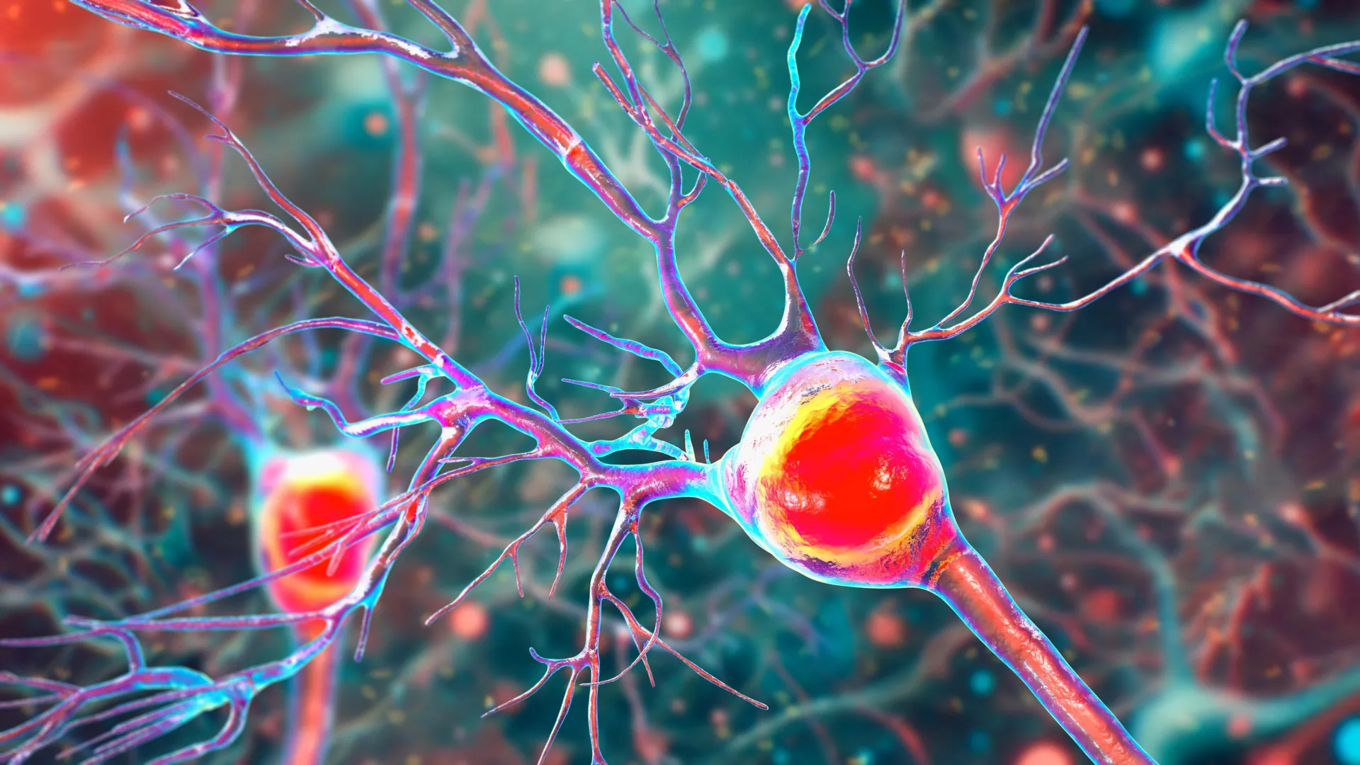
“Inside the cerebellum are special cells called Purkinje neurons,” Tiwari-Woodruff said. “These large, highly active cells help coordinate smooth, precise movements — like dancing, throwing a ball, or even just walking. They’re essential for balance and fine motor skills.”

In MS and related neurological diseases, damage to the cerebellum often leads to the gradual death of Purkinje cells. As these neurons disappear, people may develop ataxia, a condition marked by poor coordination and unstable movement.

“Our research looked at brain tissue from MS patients and found major issues in these neurons: they had fewer branches, were losing myelin, and had mitochondrial problems — meaning their energy supply was failing,” Tiwari-Woodruff said. “Because Purkinje cells play such a central role in movement, their loss can cause serious mobility issues. Understanding why they’re damaged in MS could help us find better treatments to protect movement and balance in people with the disease.”

Evidence From an MS Mouse Model

To better understand how these changes unfold, the researchers also studied experimental autoimmune encephalomyelitis (EAE) — a mouse model that develops MS-like symptoms. This allowed them to track mitochondrial changes as the disease progressed.

Over time, the mice experienced a steady decline in Purkinje cells, mirroring what is seen in human MS.

“The remaining neurons don’t work as well because their mitochondria, the energy-producing parts, start to fail,” Tiwari-Woodruff said. “We also saw that the myelin breaks down early in the disease. These problems — less energy, loss of myelin, and damaged neurons — start early, but the actual death of the brain cells tends to happen later, as the disease becomes more severe. The loss of energy in brain cells seems to be a key part of what causes damage in MS.”

Although the EAE model does not replicate every feature of MS, its similarities to the human condition make it a powerful tool for studying neurodegeneration and testing new therapeutic approaches.

Targeting Mitochondria as a Treatment Strategy

“Our findings offer critical insights into the progression of cerebellar dysfunction in MS,” Tiwari-Woodruff said. “Targeting mitochondrial health may represent a promising strategy to slow or prevent neurological decline and improve quality of life for people living with MS. This research brings us a step closer to understanding the complex mechanisms of MS and developing more effective, targeted treatments for this debilitating disease.”

What Comes Next

The research team is now exploring whether mitochondrial damage extends beyond Purkinje cells to other cerebellar cell types, including oligodendrocytes, which help form white matter, and astrocytes, which support overall brain function.

“To answer this, one of our ongoing research projects is focused on studying mitochondria in specific types of brain cells in the cerebellum,” Tiwari-Woodruff said. “Such research can open the door to finding ways to protect the brain early on — like boosting energy in brain cells, helping them repair their protective myelin coating, or calming the immune system before too much damage is done. This is especially important for people with MS who struggle with balance and coordination, as these symptoms are tied to damage in the cerebellum.”

The Importance of Continued Research

Tiwari-Woodruff stressed the broader importance of sustained investment in medical research.

“Cutting funding to science only slows progress when we need it most,” she said. “Public support for research matters now more than ever.”

The study was conducted by Tiwari-Woodruff and Atkinson alongside Shane Desfor, Micah Feria, Maria T. Sekyia, Marvellous Osunde, Sandhya Sriram, Saima Noori, Wendy Rincóna, and Britany Belloa.

Researchers analyzed postmortem cerebellar tissue from individuals with secondary progressive MS and compared it with tissue from healthy donors. The samples were obtained from the National Institutes of Health’s NeuroBioBank and the Cleveland Clinic.

Funding for the study was provided by the National Multiple Sclerosis Society.

The research paper is titled “Decreased mitochondrial activity in the demyelinating cerebellum of progressive multiple sclerosis and chronic EAE contributes to Purkinje cell loss.”


Share Button

Tiny 3D-printed light cages could unlock the quantum internet

Storing quantum information is essential for the future of both quantum computing and a global quantum internet. Today’s quantum communication systems struggle with signal loss over long distances, which limits how far quantum information can travel. Quantum memories help solve this problem by making quantum repeaters possible, allowing information to hop across a network through entanglement swapping rather than fading away.

A new study published in Light: Science & Applications reports a major advance in this area. Researchers from the Humboldt-Universität zu Berlin, the Leibniz Institute of Photonic Technology, and the University of Stuttgart have introduced a new type of quantum memory built from 3D-nanoprinted structures known as “light cages” filled with atomic vapor. By bringing both light and atoms together on a single chip, the team has created a platform designed for scalability and seamless integration into quantum photonic systems.

What Makes Light Cages Different

Light cages are hollow-core waveguides engineered to tightly guide light while still allowing access to the space inside. This design offers a key advantage over conventional hollow-core fibers, which can take months to fill with atomic vapor. In contrast, the open structure of light cages lets cesium atoms diffuse into the core much more quickly, cutting the filling process down to just a few days without sacrificing optical performance.

The structures are fabricated using two-photon polymerization lithography with commercial 3D printing systems. This approach allows researchers to directly print intricate hollow-core waveguides onto silicon chips with extremely high precision. To protect the devices from chemical reactions with cesium, the waveguides are coated with a protective layer. Tests showed no signs of degradation even after five years of operation, highlighting the system’s long-term stability.

“We created a guiding structure that allows quick diffusion of gases and fluids inside its core, with the versatility and reproducibility provided by the 3D-nanoprinting process. This enables true scalability of this platform, not only for intra-chip fabrication of the waveguides but also inter-chip, for producing multiple chips with the same performance,” explained the research team.

Turning Light Into Stored Quantum Information

Inside the light cages, incoming light pulses are efficiently converted into collective excitations of the surrounding atoms. After a chosen storage time, a control laser reverses this process and releases the stored light exactly when needed. In a key demonstration, the researchers successfully stored very weak light pulses containing only a few photons for several hundred nanoseconds. They believe this approach can eventually be extended to store single photons for many milliseconds.

Another major milestone was the integration of multiple light cage memories on a single chip placed inside a cesium vapor cell. Measurements showed that different light cages with the same design delivered nearly identical storage performance across two separate devices on the same chip. This level of consistency is essential for building scalable quantum systems.

The strong reproducibility comes from the precision of the 3D-nanoprinting process. Variations within a single chip were kept below 2 nanometers, while differences between chips remained under 15 nanometers. Such tight control is critical for spatial multiplexing, a technique that could dramatically increase the number of quantum memories operating together on one device.

Implications for Quantum Networks and Computing

Light cage quantum memories address several long-standing challenges in quantum technology. In quantum repeater networks, they could synchronize multiple single photons at the same time, greatly boosting the efficiency of long-distance quantum communication. In photonic quantum computing, the memories provide controlled delays that are needed for feed-forward operations in measurement-based quantum computing systems.

The platform also stands out for its practicality. Unlike many competing technologies, it operates slightly above room temperature and does not require cryogenic cooling or complex atom-trapping setups. This makes the system easier to deploy while also offering higher bandwidth per memory mode. The ability to produce many identical quantum memories on a single chip opens a clear path toward large-scale quantum photonic integration.

Thanks to its flexible fabrication process, the technology can potentially be combined with direct fiber coupling and existing photonic components. These advantages position light cage quantum memories as a strong candidate for future quantum communication infrastructure.

A Scalable Path Forward

The development of light cage quantum memories marks a significant step in quantum photonic research. By merging advanced 3D-nanoprinting with core principles of quantum optics, the researchers have created a compact, scalable system that could speed the arrival of practical quantum networks and more powerful quantum computers.

Share Button

A simple drug pair may succeed where liver fibrosis treatments failed

Researchers have found that a pair of existing drugs is far more effective when used together than when either is taken alone. The findings point to a practical and potentially fast route toward a long-awaited treatment for liver fibrosis.

Liver fibrosis is a common but often overlooked condition that affects hundreds of millions of people worldwide. Over time, it can worsen into cirrhosis or liver cancer. Even after decades of scientific effort, there are still no antifibrotic drugs approved for clinical use.

The disease develops when repeated or long-term liver damage — caused by viral hepatitis, excessive alcohol use, metabolic disorders, toxins, or autoimmune disease — triggers an overactive healing response. A major driver of this process is the activation of hepatic stellate cells (HSCs). Under normal conditions, these cells remain inactive. When injury occurs, they switch into collagen-producing cells that build up scar tissue in the liver.

This harmful transformation is controlled by several overlapping signaling systems, including TGF-β, PDGF, and Wnt/β-catenin pathways. Because fibrosis involves many biological routes at once, drugs that target only one pathway often have limited success. This complexity has fueled growing interest in combination treatments that can block multiple drivers of disease at the same time.

A New Use for Two Familiar Drugs

A study published in Targetome on December 15, 2025 by Hong Wang’s & Haiping Hao’s team, China Pharmaceutical University, reports that a fixed-dose combination of silybin and carvedilol can strongly suppress hepatic stellate cell activation. By targeting Wnt4/β-catenin signaling, the drug pair was able to reverse liver fibrosis in experimental models, offering a promising strategy for a disease that currently lacks approved therapies.

To better understand silybin’s potential and its limitations, the researchers combined laboratory experiments, animal studies, phenotype-based drug screening, and molecular analysis. Early tests focused on liver cell injury models triggered by ActD/TNFα, tBHP, and TNFα. These experiments showed that silybin effectively protected liver cells by restoring viability, lowering harmful reactive oxygen species, and reducing inflammatory gene activity. It also showed strong antiapoptotic, antioxidative, and anti-inflammatory effects without detectable toxicity.

However, when researchers examined whether silybin could directly stop fibrosis, the results were less impressive. In human LX-2 and rat HSC-T6 stellate cells stimulated with TGFβ1, silybin only slightly lowered key fibrosis-related markers such as COL1A1, COL1A2, ACTA2, and TGFB. Similar patterns appeared in mice with liver fibrosis caused by carbon tetrachloride exposure. While silybin led to modest improvements in liver enzymes, collagen buildup, and fibrotic gene expression, its benefits appeared to come mainly from protecting liver cells rather than directly blocking stellate cell activation.

Finding the Right Partner Drug

To overcome this limitation, the research team screened 397 FDA-approved drugs using a COL1A1-luciferase reporter system to identify compounds that could enhance silybin’s antifibrotic effect. Carvedilol emerged as the strongest synergistic partner.

When used together, silybin and carvedilol sharply reduced collagen production and stellate cell activation in human and rat cell cultures, as well as in primary hepatic stellate cells. In every case, the combination outperformed either drug on its own.

Further testing in animals showed that a fixed-dose ratio of 50:1 (silybin to carvedilol) produced the most consistent and powerful results. This optimized pairing significantly reduced liver injury, inflammation, and fibrosis severity in mice. The effects increased with dose and were stronger than those seen with obeticholic acid.

How the Drug Duo Stops Liver Scarring

Mechanistic studies revealed why the combination works so well. Together, silybin and carvedilol shut down the Wnt/β-catenin signaling pathway more effectively than either drug alone. This includes suppressing the Wnt ligand Wnt4 and reducing downstream β-catenin activity. These findings provide a clear molecular explanation for the combination’s strong antifibrotic effects.

A Fast Track Toward Clinical Use

The study highlights a realistic treatment strategy based on drug repurposing and carefully designed combination therapy. Both silybin and carvedilol are already widely used in clinical practice, have established safety records, and are low in cost. As a result, their combined use could move quickly into clinical testing and help address a major unmet medical need.

Beyond liver fibrosis, the research also demonstrates how phenotype-based screening can reveal powerful and unexpected drug partnerships that may be hiding in plain sight.

Funding and Support

This work was supported by the Major State Basic Research Development Program of China (2022YFA1303800 and 2021YFA1301300); the National Natural Science Foundation of China (82373946, 82073926, 82321005, 82530122, and 81930109); Major Science and Technology Project of Jiangsu Province (BG2024045); Overseas Expertise Introduction Project for Discipline Innovation (G20582017001); the Project of State Key Laboratory of Natural Medicines, China Pharmaceutical University (SKLNMZZ202402); and the Project Program of Basic Science Research Center Base (Pharmaceutical Science) of Yantai University (P202404).

Share Button

Parents say babies failed by delayed diagnosis like Jesy Nelson’s twins

After Little Mix star Jesy Nelson’s revelation that her twins have SMA, parents speak about their children’s late diagnoses.

Share Button

Our children waited too long for the same diagnosis as Jesy Nelson’s twins

After Little Mix star Jesy Nelson’s revelation that her twins have SMA, parents speak about their children’s late diagnoses.

Share Button

New NHS online hospital to focus on eyes, menopause and prostates

NHS Online due to launch in England next year will provide eye, prostate and menopause care.

Share Button

Nestle recalls some baby formula products over toxin fears

Nestle said it had received no reports of problems and was recalling the products “out of an abundance of caution”.

Share Button

US cuts universal childhood vaccine recommendations, including covid and hepatitis

The overhaul is the latest in a host of sweeping changes made under the Trump administration by health secretary Robert F Kennedy Jr.

Share Button

Everyday chemicals are quietly damaging beneficial gut bacteria

Scientists have completed a large laboratory analysis of widely used human-made chemicals and found that 168 of them are harmful to bacteria that normally live in a healthy human gut. These substances slow or stop the growth of microbes that play an important role in supporting overall health.

Many of the chemicals identified are ones people are likely to encounter through everyday exposure, including food, drinking water, and the environment. Until now, most were not believed to interfere with bacteria at all.

Links to Antibiotic Resistance Raise New Concerns

When gut bacteria are exposed to these chemical pollutants, some change how they function in an attempt to survive. In certain cases, this adaptation also makes the bacteria resistant to antibiotics such as ciprofloxacin. If similar changes occur inside the human body, infections could become more difficult to treat.

The study was led by researchers at the University of Cambridge and involved testing 1076 different chemical contaminants on 22 species of gut bacteria under laboratory conditions.

Pesticides and Industrial Chemicals Among the Most Harmful

The chemicals shown to damage gut bacteria include pesticides such as herbicides and insecticides commonly applied to crops. Industrial compounds used in products like flame retardants and plastics were also found to be toxic to these microbes.

The human gut microbiome contains roughly 4,500 different types of bacteria that help keep the body functioning properly. When this delicate system is disrupted, it can contribute to a wide range of health problems, including digestive issues, obesity, weakened immune function, and effects on mental health.

Why Chemical Safety Testing Misses Gut Health

Current chemical safety evaluations typically do not account for the gut microbiome. This is because chemicals are designed to target specific organisms or processes, for example insecticides should target insects.

Using the data from their experiments, the researchers developed a machine learning model to help predict whether industrial chemicals — whether already in use, or in development — are likely to harm human gut bacteria. The findings and the new model were published in the journal Nature Microbiology.

Researchers Call for a New Approach to Chemical Safety

Dr. Indra Roux, a researcher at the University of Cambridge’s MRC Toxicology Unit and the study’s first author, said: “We’ve found that many chemicals designed to act only on one type of target, say insects or fungi, also affect gut bacteria. We were surprised that some of these chemicals had such strong effects. For example, many industrial chemicals like flame retardants and plasticizers — that we are regularly in contact with — weren’t thought to affect living organisms at all, but they do.”

Professor Kiran Patil, senior author of the study and also based at the University of Cambridge’s MRC Toxicology Unit, added: “The real power of this large-scale study is that we now have the data to predict the effects of new chemicals, with the aim of moving to a future where new chemicals are safe by design.”

Dr. Stephan Kamrad, another researcher involved in the work, said: “Safety assessments of new chemicals for human use must ensure they are also safe for our gut bacteria, which could be exposed to the chemicals through our food and water.”

What Scientists Still Don’t Know About Real-World Exposure

There is currently limited information about how environmental chemicals directly affect the gut microbiome and, in turn, human health. The researchers say it is likely that gut bacteria are frequently exposed to many of the chemicals tested, but the exact amounts that reach the digestive system remain unclear. To better understand the risks, future studies will need to track chemical exposure throughout the body.

Patil said: “Now we’ve started discovering these interactions in a laboratory setting it’s important to start collecting more real-world chemical exposure data, to see if there are similar effects in our bodies.”

Until more is known, the researchers recommend simple steps to reduce exposure, such as washing fruits and vegetables before eating them and avoiding the use of pesticides in home gardens.

Share Button

This CRISPR breakthrough turns genes on without cutting DNA

Scientists at UNSW Sydney have developed a new form of CRISPR technology that could make gene therapy safer while also resolving a decades-long debate about how genes are switched off. The research shows that small chemical markers attached to DNA actively silence genes, rather than simply appearing as harmless byproducts in inactive regions of the genome.

For years, researchers have questioned whether methyl groups, tiny chemical clusters that collect on DNA, merely show up where genes are already turned off or whether they are the direct cause of gene suppression.

In a study published recently in Nature Communications, researchers from UNSW, working with colleagues at the St Jude Children’s Research Hospital (Memphis), demonstrated that removing these chemical tags causes genes to become active again. When the tags were added back, the genes shut down once more. The results confirm that DNA methylation directly controls gene activity.

“We showed very clearly that if you brush the cobwebs off, the gene comes on,” says study lead author Professor Merlin Crossley, UNSW Deputy Vice-Chancellor Academic Quality.

“And when we added the methyl groups back to the genes, they turned off again. So, these compounds aren’t cobwebs — they’re anchors.”

How CRISPR Technology Has Evolved

CRISPR, short for Clustered Regularly Interspaced Short Palindromic Repeats, is the foundation of modern gene-editing technology. It allows scientists to locate specific DNA sequences and make targeted changes, often replacing faulty genetic code with healthy versions.

The system is based on a natural defense mechanism found in bacteria, which use CRISPR to recognize and cut up the DNA of invading viruses.

Early versions of CRISPR tools worked by cutting DNA to disable malfunctioning genes. Later versions became more precise, allowing scientists to correct individual letters in the genetic code. However, both approaches rely on breaking DNA strands, which can lead to unintended changes and increase the risk of serious side effects.

The latest version, known as epigenetic editing, takes a different approach. Instead of cutting DNA, it targets chemical markers attached to genes inside the nucleus of each cell. By removing methyl groups from genes that have been silenced, researchers can restore gene activity without altering the underlying DNA sequence.

New Possibilities for Treating Sickle Cell Disease

The team believes this approach could lead to safer treatments for Sickle Cell-related diseases. These inherited conditions affect the shape and function of red blood cells, often causing severe pain, organ damage, and shortened life expectancy.

“Whenever you cut DNA, there’s a risk of cancer. And if you’re doing a gene therapy for a lifelong disease, that’s a bad kind of risk,” Prof. Crossley says.

“But if we can do gene therapy that doesn’t involve snipping DNA strands, then we avoid these potential pitfalls.”

Rather than cutting DNA, the new technique uses a modified CRISPR system to deliver enzymes that remove methyl groups. This process releases the genetic brakes that keep certain genes switched off. One key target is the fetal globin gene, which helps deliver oxygen before birth. Reactivating this gene after birth could help bypass defects in the adult globin gene that cause Sickle Cell diseases.

“You can think of the fetal globin gene as the training wheels on a kid’s bike,” says Prof. Crossley. “We believe we can get them working again in people who need new wheels.”

What the Research Shows So Far

So far, all experiments have been carried out in laboratory settings using human cells at UNSW and in Memphis.

Study co-author Professor Kate Quinlan says the findings could have far-reaching implications beyond Sickle Cell disease. Many genetic conditions involve genes that are improperly turned on or off, and adjusting methyl groups may provide a way to correct those problems without damaging DNA.

“We are excited about the future of epigenetic editing as our study shows that it allows us to boost gene expression without modifying the DNA sequence. Therapies based on this technology are likely to have a reduced risk of unintended negative effects compared to first or second generation CRISPR,” she says.

Looking ahead, the researchers describe how the therapy might one day work in practice. Doctors would collect a patient’s blood stem cells, which produce red blood cells. In the lab, epigenetic editing would be used to remove methyl tags from the fetal globin gene, reactivating it. The edited cells would then be returned to the patient, where they could settle into the bone marrow and begin producing healthier blood cells.

The Next Steps in Epigenetic Editing

The research teams at UNSW and St Jude plan to test the approach in animal models and continue exploring additional CRISPR-based tools.

“Perhaps the most important thing is that it is now possible to target molecules to individual genes,” Prof. Crossley says.

“Here we removed or added methyl groups but that is just the beginning, there are other changes that one could make that would increase our abilities to alter gene output for therapeutic and agricultural purposes. This is the very beginning of a new age.”

Share Button