Watch Morning Live here


Watch Morning Live here
Authorities urged patients to get tested due to “poor infection control practices” at the Australian clinic.

Researchers at UBC Okanagan have uncovered the process plants use to create mitraphylline, a rare natural compound that has attracted attention for its possible cancer fighting properties.
Mitraphylline belongs to a unique class of plant chemicals known as spirooxindole alkaloids. These molecules are recognized for their unusual twisted ring structures and their powerful biological effects, including anti inflammatory and anti tumor activity.
Even though scientists have studied these compounds for years, the exact molecular steps plants use to produce them had remained unknown.
Breakthrough Discovery in Plant Chemistry
That mystery began to unravel in 2023 when Dr. Thu-Thuy Dang’s team in UBC Okanagan’s Irving K. Barber Faculty of Science identified the first known plant enzyme capable of twisting a molecule into the distinctive spiro shape.
Building on that earlier finding, doctoral student Tuan-Anh Nguyen led new research that uncovered two critical enzymes involved in the production of mitraphylline. One enzyme organizes the molecule into the correct three dimensional structure, while the second transforms it into mitraphylline itself.
“This is similar to finding the missing links in an assembly line,” says Dr. Dang, UBC Okanagan Principal’s Research Chair in Natural Products Biotechnology. “It answers a long-standing question about how nature builds these complex molecules and gives us a new way to replicate that process.”
Why Mitraphylline Is So Valuable
Many promising natural compounds are found only in tiny amounts inside plants, making them difficult and expensive to recreate in laboratories. Mitraphylline is one of those rare substances. It exists only in trace quantities in tropical trees such as Mitragyna (kratom) and Uncaria (cat’s claw), both members of the coffee family.
Now that researchers have identified the enzymes responsible for shaping and assembling mitraphylline, they have a clearer path toward producing the compound and related molecules in more sustainable ways.
“With this discovery, we have a green chemistry approach to accessing compounds with enormous pharmaceutical value,” says Nguyen. “This is a result of UBC Okanagan’s research environment, where students and faculty work closely to solve problems with global reach.”
Nguyen also reflected on the experience of contributing to the breakthrough.
“Being part of the team that uncovered the enzymes behind spirooxindole compounds has been amazing,” Nguyen adds. “UBC Okanagan’s mentorship and support made this possible, and I’m excited to keep growing as a researcher here in Canada.”
International Collaboration Fuels the Research
The project brought together Dr. Dang’s laboratory at UBC Okanagan and Dr. Satya Nadakuduti’s research group at the University of Florida.
Funding for the work came from Canada’s Natural Sciences and Engineering Research Council’s Alliance International Collaboration program, the Canada Foundation for Innovation, and the Michael Smith Health Research BC Scholar Program. Additional support was provided by the United States Department of Agriculture’s National Institute of Food and Agriculture.
“We are proud of this discovery coming from UBC Okanagan. Plants are fantastic natural chemists,” Dr. Dang says. “Our next steps will focus on adapting their molecular tools to create a wider range of therapeutic compounds.”

Researchers at the University of Maryland, Baltimore County (UMBC) have uncovered a crucial step that enteroviruses use to reproduce inside human cells. The findings, published in Nature Communications, explain how viruses responsible for illnesses such as polio, encephalitis, myocarditis, and even the common cold take control of cellular machinery to copy themselves. Scientists say the discovery could eventually help researchers create a new generation of antiviral drugs capable of targeting many enteroviruses at once.
The study was led by Deepak Koirala, associate professor of chemistry and biochemistry at UMBC, along with recent Ph.D. graduate Naba Krishna Das. Their work helps answer longstanding questions about how these viruses launch replication once they invade a cell.
“My lab has been really motivated to understand how RNA viruses produce their proteins inside the cell and multiply their genome to make more virus particles,” Koirala says. Earlier work from the team identified an important cloverleaf shaped structure within the virus’s RNA. The new study shows how that structure recruits proteins needed to build the viral replication machinery.
How Enteroviruses Reproduce Inside Cells
Enteroviruses carry very small RNA genomes that must perform two jobs at once. The viral RNA has to direct the production of viral proteins while also serving as the template for creating new copies of the virus.
Most of the viral genome contains instructions for structural proteins, but it also encodes several specialized proteins required for replication. One of the most important is a fusion protein called 3CD.
The 3C portion cuts long chains of amino acids into the separate proteins the virus needs. The 3D portion acts as an RNA polymerase, an enzyme that copies viral RNA so the virus can reproduce. Human cells do not naturally contain this type of polymerase, meaning the virus has to supply its own version.
“We previously determined the structure of the RNA alone, and other groups determined the structure of 3C and 3D, but now we’ve captured the structure of the RNA and proteins together, so we know how they are interacting,” Koirala explains. “We found that it’s the 3C domain of 3CD that binds to the RNA in the viral genome, and then it recruits the other components, such as host protein PCBP2, to assemble the replication complex.”
The researchers also found that this molecular complex functions like a switch. When 3CD is attached, the virus copies its RNA genome. When the protein detaches, the RNA becomes available for producing viral proteins instead.
Scientists Resolve a Longstanding Viral Mystery
To examine these interactions in detail, the team used X-ray crystallography to visualize the RNA cloverleaf and the 3CD protein together. They also relied on isothermal titration calorimetry (ITC), which measures the heat released when molecules bind, and biolayer interferometry (BLI), which uses changes in light interference to track how long molecules stay attached.
The experiments helped settle an ongoing scientific debate. The researchers showed that two full 3CD molecules, each carrying its own RNA polymerase, bind side by side on the viral RNA. Earlier research had proposed that the proteins formed a single fused pair instead.
Scientists still do not fully understand why two copies are required, but the new study provides a much clearer picture of how the replication process begins.
Potential for Broad Spectrum Antiviral Drugs
One of the most promising findings was how similar the mechanism appeared across all seven enteroviruses examined in the study. The viruses shared nearly identical RNA cloverleaf structures and binding behavior.
That level of similarity suggests the RNA structure is extremely important to viral survival. Significant mutations would likely disrupt replication, making the structure a potentially stable drug target across many enteroviruses.
Researchers say this raises the possibility of developing broad spectrum antiviral drugs that could work against an entire family of viruses rather than a single pathogen.
Scientists are already developing drugs that interfere with the 3C and 3D proteins, but the new findings reveal another possible strategy.
Drugs disrupting 3C and 3D activity are already in development, but “now we have another layer to test,” Koirala says. “What if we target the RNA, or the RNA-protein interface, so that we break the interaction? That is another opportunity. Now that we have high-resolution structures, you can precisely design drug molecules to target them.”
Koirala says the study highlights how surprisingly sophisticated viruses can be despite their tiny genomes.
“Viruses are so, so clever. Their entire genome is equivalent to about one mRNA sequence in humans, yet they are so effective,” Koirala says. His latest work demonstrates “why we need to investigate this basic science — so that it can be translated into developing drugs targeting pathogens that cause so many harmful diseases.”

Scientists at the Icahn School of Medicine at Mount Sinai have successfully reversed aging in blood-forming stem cells in mice by repairing defects in structures known as lysosomes. The findings, published in Cell Stem Cell, point to lysosomal dysfunction and overactivity as major causes of stem cell aging and show that restoring proper lysosomal activity can rejuvenate old stem cells and improve their ability to regenerate blood and immune cells.
Lysosomes function as the cell’s internal recycling centers. They break down proteins, nucleic acids, carbohydrates, and lipids, helping cells dispose of waste and reuse materials for essential biological processes. They also store nutrients that can be released when needed. Because of these roles, lysosomes are critical for maintaining cellular metabolism, including both catabolism (breaking down complex molecules to simple ones) and anabolism (building complex molecules from simpler ones).
The research team focused on hematopoietic stem cells (HSCs), which are rare, long-lasting stem cells found in the bone marrow that generate all blood and immune cells in the body. The study was led by Saghi Ghaffari, MD, PhD, Professor of Cell, Developmental, and Regenerative Biology at the Icahn School of Medicine and a member of the Black Family Stem Cell Institute.
As people grow older, these stem cells gradually lose their ability to repair and replenish the blood system. This decline weakens immune defenses and contributes to the increased vulnerability to infections seen in older adults. Aging HSCs are also linked to clonal hematopoiesis, an asymptomatic condition considered a premalignant state that raises the risk of blood cancers and inflammatory diseases. The condition becomes much more common with age.
According to the American Cancer Society, age and smoking are the two strongest risk factors associated with the five-year risk of developing cancer. Data from the National Cancer Institute’s Surveillance, Epidemiology, and End Results report show that the median age at cancer diagnosis is 67.
Restoring Old Stem Cells to a Youthful State
The researchers discovered that lysosomes in aged HSCs become excessively acidic, damaged, depleted, and abnormally active. These changes disrupt both the metabolic balance and epigenetic stability of the stem cells.
Using single-cell transcriptomics and functional testing, the team found that blocking this excessive lysosomal activity with a vacuolar ATPase inhibitor restored lysosomal health and improved the function of aging blood stem cells.
After treatment, the old stem cells began behaving more like young, healthy cells again. They regained the ability to regenerate effectively, produce balanced blood and immune cells, and generate additional healthy stem cells. The treated cells also showed improved metabolism and mitochondrial performance, healthier epigenetic patterns, reduced inflammation, and fewer harmful inflammatory signals that can damage tissues throughout the body.
“Our findings reveal that aging in blood stem cells is not an irreversible fate. Old blood stem cells have the capacity to revert to a youthful state; they can bounce back,” said Dr. Ghaffari. “By slowing down the lysosomes and reducing their acidity, stem cells became healthier and could make new balanced blood cells and new stem cells much more effectively. By targeting lysosomal hyperactivity, we were able to reset aged stem cells to a younger, healthier state, improving their ability to regenerate blood and immune cells.”
Major Increase in Blood-Forming Capacity
The researchers also tested an ex vivo treatment approach (when cells are removed from the body, modified in a laboratory, and returned to the body). Treating old stem cells with the lysosomal inhibitor increased their blood-forming ability in living animals by more than eightfold, highlighting the powerful regenerative effects of correcting lysosomal dysfunction.
The improvement also reduced damaging inflammatory and interferon-related pathways. According to the researchers, this occurred because healthier lysosomes improved the processing of mitochondrial DNA and lowered activation of the cGAS-STING immune signaling pathway, which appears to play a major role in stem cell inflammation and aging.
Potential for Anti-Aging and Blood Disorder Therapies
The findings could open the door to new treatments aimed at preventing or reversing age-related blood disorders. They may also improve stem cell transplantation outcomes in older patients and enhance conditioning methods used in gene therapy.
“Lysosomal dysfunction emerges as a central driver of stem cell aging,” added Dr. Ghaffari. “Targeting this pathway may one day help maintain healthy blood and immune systems in the elderly, improve their stem cells for transplantation, and reduce the risk of age-associated blood disorders and perhaps have an effect on overall aging.”
The team is now investigating whether lysosomal dysfunction in aging stem cells contributes to the development of leukemic stem cells, potentially connecting normal stem cell aging with cancer formation.
The research involved collaboration with Mickaël Ménager, PhD, and colleagues at the Imagine Institute and INSERM UMR 1163 at Université de Paris Cité in Paris. Funding was provided by the National Institutes of Health, New York State Stem Cell Science, INSERM, and the Agence Nationale de la Recherche.
The tablet – orforglipron – is available in the US and could soon launch in the UK.
Swindon’s new mental health support facility for women will be an “alternative” to hospitalisation.
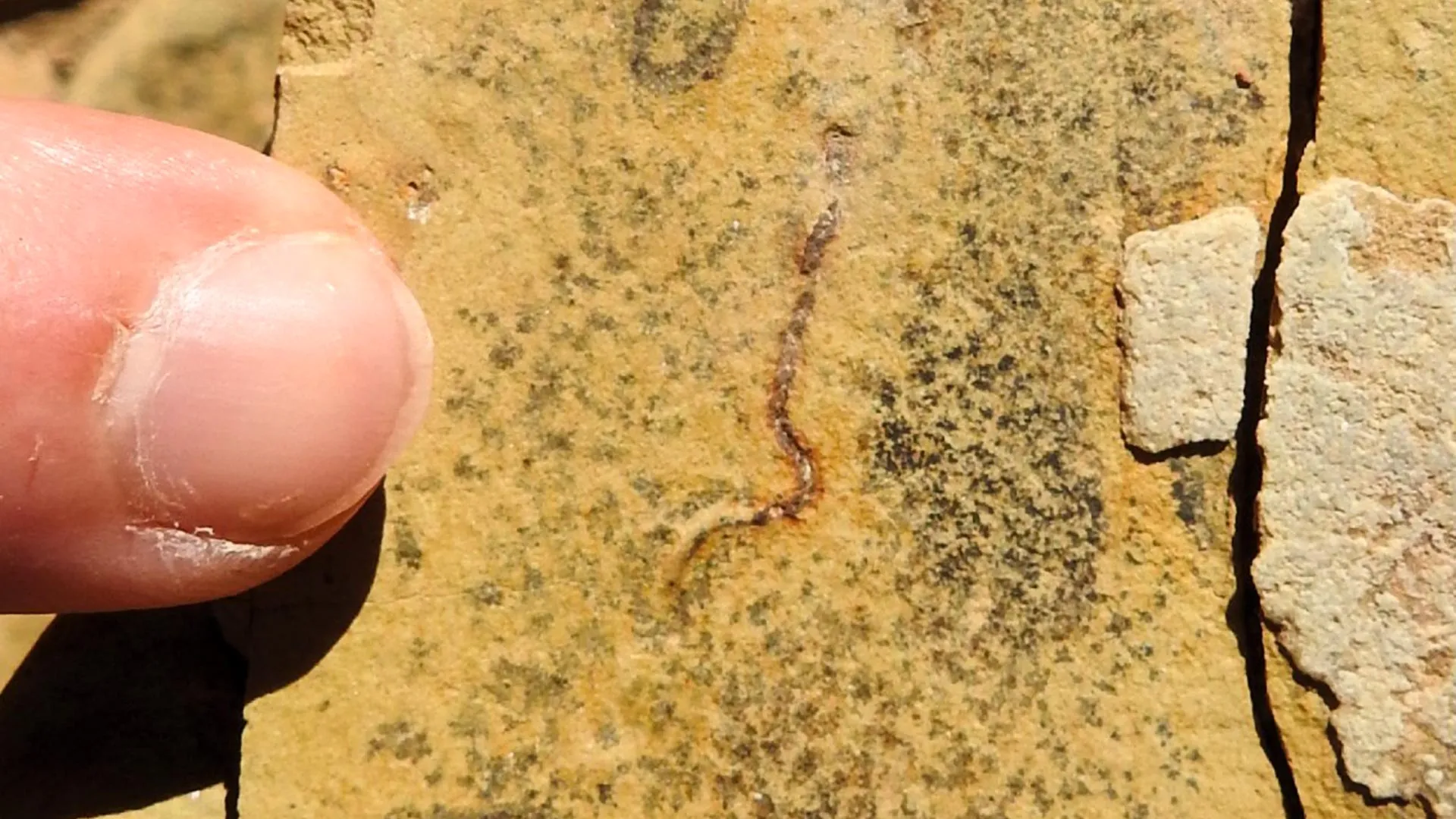
Scientists studying ancient microfossils from Brazil have discovered that structures once believed to be traces left behind by tiny animals were actually formed by communities of microscopic bacteria and algae. The findings challenge previous ideas about when small animals first appeared on Earth and suggest oxygen levels in ancient oceans may still have been too low to support certain forms of animal life around 540 million years ago.
The research focused on fossils found in the Brazilian state of Mato Grosso do Sul and was published in the journal Gondwana Research. Earlier studies had interpreted the marks as evidence of wormlike creatures or other tiny marine animals moving through seafloor sediment during the Ediacaran period, which came just before the Cambrian explosion.
“Using microtomography and spectroscopy techniques, we observed that the microfossils have cellular structures — sometimes with preserved organic material — consistent with bacteria or algae that existed during that period. These aren’t traces of animals that may have passed through the area,” says Bruno Becker-Kerber, the first author of the study. He carried out the research during postdoctoral work at the Institute of Geosciences at the University of São Paulo (USP) and at the Brazilian Center for Research in Energy and Materials (CNPEM), with support from FAPESP.
Becker-Kerber, who is now conducting postdoctoral research at Harvard University, explains that if the marks had truly been left by animals, they would represent evidence of meiofauna during the Ediacaran period. Meiofauna are tiny invertebrates measuring less than one millimeter long. Finding them in rocks this old would have pushed back the fossil record for these organisms significantly.
Ancient Oceans Before the Cambrian Explosion
The Ediacaran period occurred before the Cambrian explosion, a major evolutionary turning point when rising oxygen levels helped complex organisms diversify rapidly across Earth’s oceans. Fossil evidence clearly shows meiofauna existed during the Cambrian, but the new findings suggest they may not have been present earlier in the way some scientists proposed.
The project forms part of the “Rio de la Plata Craton and Western Gondwana” study supported by FAPESP and coordinated by Miguel Angelo Stipp Basei, a professor at IGc-USP and coauthor of the paper.
Another coauthor, Lucas Warren of São Paulo State University (IGCE-UNESP) in Rio Claro, also received support from FAPESP.
Researchers reexamined fossils collected in Corumbá and also analyzed newly studied material from Bonito in the Serra da Bodoquena region. Both sites are located in Mato Grosso do Sul within the Tamengo geological formation.
These rocks formed in a shallow marine environment along a continental shelf during the final stages of Gondwana’s formation, before the supercontinent eventually split apart to form regions that became South America and Africa.
The same research group previously identified what may be the oldest known lichen fossil, also discovered in Mato Grosso do Sul and younger than the bacteria and algae described in the current study.
High Resolution Fossil Imaging Revealed Hidden Structures
To investigate the fossils in greater detail, the team used the MOGNO beamline at Sirius, CNPEM’s particle accelerator facility in Campinas. The technology allowed researchers to study fossils ranging from only a few micrometers to a few millimeters in size.
The scientists used both microtomography and nanotomography, techniques capable of generating images at extremely small scales, including micrometers (one-thousandth of a millimeter) and nanometers (one-billionth of a meter).
“When you have a large sample and want to image a structure inside it, the resolution obtained is often insufficient. The MOGNO beamline is one of the few in the world that performs so-called zoom tomography, in which we focus on something inside the sample and analyze it at the nanoscale without destroying the sample,” says Becker-Kerber.
He notes that the earlier study interpreting the structures as animal traces did not have access to this level of imaging technology.
Researchers also used Raman spectroscopy to examine the fossils’ chemical makeup. The technique identified organic material within fossil cell walls, strengthening the interpretation that the structures were preserved microbial bodies rather than marks left behind by passing animals.
Giant Ancient Bacteria and Algae
Some fossil samples contained pyrite, a mineral made of iron and sulfur. Based on the shapes and chemistry of the specimens, researchers believe some may represent sulfur-oxidizing bacteria, organisms that use sulfur in their metabolism.
“This group of bacteria is surprising. Some of the largest ever recorded belong precisely to this category. Unlike the common image we have of microscopic bacteria, certain species can reach diameters larger than a strand of hair and are visible to the naked eye,” says Becker-Kerber.
Although the fossils do not preserve enough detail to identify exact species, the researchers found preserved cells, divisions within cell walls, and traces of organic matter across multiple collection sites. According to the team, these features would not exist if the structures were simply disturbances created by moving animals.
The fossils also appear in three different size ranges, suggesting several species may have lived together in microbial communities. The largest forms resemble green or red algae, while the smaller fossils may represent algae, cyanobacteria, or sulfur-oxidizing bacteria.
“There are concave and convex partitions, coiled filaments, cells without sediment but containing organic matter. This evidence is much closer to bacteria or algae than to mere marks of disturbance caused by animals,” the researcher concludes.
The findings provide scientists with a clearer picture of the world before the Cambrian explosion and may help researchers better understand the environmental conditions that paved the way for the rise of complex animal life.

For decades, scientists searching for life beyond Earth have focused on one central challenge: identifying the right molecules to look for on distant planets and moons.
But new research published in Nature Astronomy suggests the answer may lie not in the molecules themselves, but in the hidden patterns that connect them.
“We’re showing that life does not only produce molecules,” said Fabian Klenner, UC Riverside assistant professor of planetary sciences and co-author of the study. “Life also produces an organizational principle that we can see by applying statistics.”
Hidden Chemical Patterns May Reveal Life
The researchers discovered that amino acids found in living systems tend to be both more varied and more evenly distributed than amino acids formed through nonbiological processes. Fatty acids showed the opposite trend, with nonliving chemical processes producing more even distributions than biological ones.
According to the team, this is the first study to show that this underlying signature of life can be detected through statistics alone, without relying on any single specialized instrument. That means the approach could potentially work using data already being collected by current and future space missions.
The findings arrive at a time when planetary exploration is advancing rapidly. Missions studying Mars, Europa, Enceladus, and other worlds are producing increasingly detailed measurements of organic chemistry. However, interpreting those chemical signals remains a major challenge.
Many molecules linked to life on Earth, including amino acids and fatty acids, can also form naturally without biology. Scientists have found them in meteorites and created them in laboratory experiments designed to mimic space environments. Because of that, simply detecting these compounds is not considered strong enough evidence to confirm life.
“Astrobiology is fundamentally a forensic science,” said Gideon Yoffe, postdoctoral researcher at the Weizmann Institute of Science in Israel and first author of the study. “We’re trying to infer processes from incomplete clues, often with very limited data collected by missions that are extraordinarily expensive and infrequent.”
Borrowing a Tool From Ecology
To tackle the problem, the researchers adapted a statistical method commonly used in ecology. Ecologists measure biodiversity using two main concepts: richness, which describes how many different species are present, and evenness, which measures how uniformly they are distributed.
Yoffe first encountered this framework during doctoral studies in statistics and data science, where diversity metrics were used to uncover patterns in complicated datasets, including research involving ancient human cultures.
The team then applied the same statistical logic to chemistry associated with possible extraterrestrial life.
Using roughly 100 existing datasets, the scientists examined amino acids and fatty acids from microbes, soils, fossils, meteorites, asteroids, and synthetic laboratory samples. Again and again, biological materials displayed distinct organizational patterns that separated them from nonliving chemistry.
Fossils Still Carried Signs of Ancient Life
One of the most surprising findings was how effective the method remained despite its simplicity.
By analyzing samples through this statistical lens, the researchers could reliably distinguish biological samples from abiotic ones. They also observed that biological materials formed a continuum ranging from well preserved to heavily degraded.
“That was genuinely surprising,” Klenner said. “The method captured not only the distinction between life and nonlife, but also degrees of preservation and alteration.”
Even samples that had undergone significant degradation still preserved traces of this organizational structure. Fossilized dinosaur eggshells included in the study, for example, continued to show detectable statistical patterns connected to ancient biological activity.
A New Tool for Future Space Missions
The researchers caution that no single technique will be enough to prove the existence of extraterrestrial life.
“Any future claim of having found life would require multiple independent lines of evidence, interpreted within the geological and chemical context of a planetary environment,” Klenner said.
Even so, the team believes this framework could become a valuable addition to future planetary missions searching for evidence of life beyond Earth.
“Our approach is one more way to assess whether life may have been there,” Klenner said. “And if different techniques all point in the same direction, then that becomes very powerful.”

NASA’s Hubble Space Telescope has captured the most detailed visible light images ever taken of the largest known protoplanetary disk surrounding a young star. The enormous structure appears far more chaotic and turbulent than astronomers expected, with huge wisps of gas and dust extending high above and below the disk. Even more unusual, the longest filament-like structures can only be seen on one side.
The discovery, published in The Astrophysical Journal, offers scientists a rare look at how planets may form in extreme cosmic environments and highlights Hubble’s continuing role in exploring the universe.
Giant Planet-Forming Disk Unlike Any Seen Before
The system, known as IRAS 23077+6707 and nicknamed “Dracula’s Chivito,” is located about 1,000 light-years from Earth. The giant disk stretches nearly 400 billion miles across, making it about 40 times wider than our solar system out to the Kuiper Belt.
At the center of the disk is a young star hidden by thick clouds of dust and gas. Researchers think the object may be a single massive star or possibly two stars orbiting each other. Besides being the largest planet-forming disk ever identified, scientists say it may also be one of the strangest.
“The level of detail we’re seeing is rare in protoplanetary disk imaging, and these new Hubble images show that planet nurseries can be much more active and chaotic than we expected,” said lead author Kristina Monsch of the Center for Astrophysics | Harvard & Smithsonian (CfA). “We’re seeing this disk nearly edge-on and its wispy upper layers and asymmetric features are especially striking. Both Hubble and NASA’s James Webb Space Telescope have glimpsed similar structures in other disks, but IRAS 23077+6707 provides us with an exceptional perspective — allowing us to trace its substructures in visible light at an unprecedented level of detail. This makes the system a unique, new laboratory for studying planet formation and the environments where it happens.”
The unusual nickname reflects the backgrounds of the researchers involved. One scientist is from Transylvania, while another is from Uruguay, where a chivito is a popular sandwich. Viewed edge-on, the disk resembles a hamburger with a dark center surrounded by glowing layers of dust and gas above and below it.
Mysterious One-Sided Filaments
Scientists were especially intrigued by the disk’s uneven appearance. Hubble’s images revealed towering filament-like structures extending from only one side of the disk, while the opposite side appears sharply defined and lacks similar features.
Researchers believe this strange asymmetry could be caused by active processes within the system, such as fresh material falling into the disk or interactions with nearby surroundings.
“We were stunned to see how asymmetric this disk is,” said co-investigator Joshua Bennett Lovell, also an astronomer at the CfA. “Hubble has given us a front row seat to the chaotic processes that are shaping disks as they build new planets — processes that we don’t yet fully understand but can now study in a whole new way.”
Clues to How Planetary Systems Form
Planetary systems develop from massive disks of gas and dust surrounding young stars. Over time, some of the material falls into the star while the remaining matter gradually forms planets.
Scientists estimate the mass of IRAS 23077+6707 may equal 10 to 30 times the mass of Jupiter, providing more than enough material to create several giant planets. Researchers say the system could resemble an oversized version of the early solar system.
“In theory, IRAS 23077+6707 could host a vast planetary system,” said Monsch. “While planet formation may differ in such massive environments, the underlying processes are likely similar. Right now, we have more questions than answers, but these new images are a starting point for understanding how planets form over time and in different environments.”
Hubble’s Continuing Discoveries
The Hubble Space Telescope has operated for more than 30 years and continues to deliver major discoveries that expand scientists’ understanding of the cosmos. Hubble is a joint project between NASA and ESA (European Space Agency). NASA’s Goddard Space Flight Center in Greenbelt, Maryland, oversees telescope and mission operations, while Lockheed Martin Space in Denver also supports operations. The Space Telescope Science Institute in Baltimore, operated by the Association of Universities for Research in Astronomy, manages Hubble’s science operations for NASA.