A Christmas tree 80 light-years wide appears in space

NGC 2264 is a large and active region of space where new stars are forming, located about 2,700 light years from Earth. It lies within the faint but imaginative constellation Monoceros, which represents a unicorn in star maps. Astronomers assign catalog numbers like NGC 2264 to help identify and study deep space objects, especially those made of gas, dust, and young stars. This region is positioned near the celestial equator and close to the flat disk of the Milky Way, which makes it visible from many locations on Earth during certain seasons.

Glowing Nebulae and Dark Cosmic Dust

The scene is filled with enormous clouds of interstellar gas and dust, the raw ingredients needed to form stars. As young stars ignite within these clouds, they release intense energy that causes surrounding hydrogen gas to glow red. These glowing regions are known as emission nebulae. Dark dust clouds thread through the area as well, blocking light from stars behind them and creating dramatic shadows. In places where this dust lies close to hot, newly formed stars, it reflects their light instead of absorbing it, producing soft blue regions called reflection nebulae.

The Christmas Tree Star Cluster

Near the center of NGC 2264 is S Monocerotis, a bright variable star whose brightness changes over time. This star is surrounded by a noticeable blue glow caused by reflected starlight from nearby dust. Above S Monocerotis, a group of young stars forms a simple triangular pattern. Because of this distinctive shape, the cluster has become widely known as the Christmas Tree star cluster.

The Cone Nebula and the Fox Fur Nebula

At the top of this star filled scene sits the Cone Nebula, a tall structure of gas and dust shaped by powerful radiation from nearby young stars. Beneath it spreads a tangled and glowing cloud called the Fox Fur Nebula, named for its textured, fur like appearance. These features are constantly being reshaped as energetic starlight pushes and sculpts the surrounding material.

Immense Size on a Galactic Scale

When viewed through a telescope, the entire region stretches about 1.5 degrees from top to bottom, which is roughly the width of three full moons lined up in the sky. At a distance of 2,700 light years, that apparent size corresponds to a real span of nearly 80 light years. This immense scale highlights just how vast and dynamic this stellar nursery truly is.

Share Button

Scientists reverse Alzheimer’s in mice and restore memory

A study reveals that restoring the brain’s energy balance may not just slow Alzheimer’s — but actually reverse it.

  • For more than a century, Alzheimer’s disease has been widely viewed as permanent and untreatable once it begins. As a result, most research has focused on preventing the disease or slowing its progression rather than attempting to reverse it.
  • By studying multiple mouse models of Alzheimer’s alongside human Alzheimer’s brain tissue, researchers identified a critical biological problem at the center of the disease. They found that the brain’s inability to maintain healthy levels of a vital cellular energy molecule called NAD+ plays a major role in driving Alzheimer’s.
  • In animal models, maintaining normal brain NAD+ levels prevented Alzheimer’s from developing. Even more striking, restoring NAD+ balance after the disease was already advanced allowed the brain to repair damage and fully restore cognitive function.
  • These results suggest that treatments aimed at restoring the brain’s energy balance could potentially move Alzheimer’s therapy beyond slowing decline and toward meaningful recovery.
  • The findings also open the door to further research, including the exploration of complementary strategies and carefully designed clinical trials to determine whether these results can translate to patients.

A Longstanding View of Alzheimer’s Is Being Questioned

For more than 100 years, Alzheimer’s disease (AD) has been widely viewed as a condition that cannot be undone. Because of this belief, most scientific efforts have focused on preventing the disease or slowing its progression, rather than attempting to restore lost brain function. Even after decades of research and billions of dollars in investment, no drug trial for Alzheimer’s has ever been designed with the goal of reversing the disease and recovering cognitive abilities.

That long-held assumption is now being challenged by researchers from University Hospitals, Case Western Reserve University, and the Louis Stokes Cleveland VA Medical Center. Their work set out to answer a bold question: can brains already damaged by advanced Alzheimer’s recover?

New Study Targets Brain Energy Failure

The research was led by Kalyani Chaubey, PhD, of the Pieper Laboratory and published on December 22 in Cell Reports Medicine. By examining both human Alzheimer’s brain tissue and multiple preclinical mouse models, the team identified a key biological failure at the center of the disease. They found that the brain’s inability to maintain normal levels of a critical cellular energy molecule called NAD+ plays a major role in driving Alzheimer’s. Importantly, maintaining proper NAD+ balance was shown to not only prevent the disease but also reverse it in experimental models.

NAD+ levels naturally decline throughout the body, including the brain, as people age. When NAD+ drops too low, cells lose the ability to carry out essential processes needed for normal function and survival. The researchers discovered that this decline is far more severe in the brains of people with Alzheimer’s. The same pattern was seen in mouse models of the disease.

How Alzheimer’s Was Modeled in the Lab

Although Alzheimer’s occurs only in humans, scientists study it using specially engineered mice that carry genetic mutations known to cause the disease in people. In this study, researchers used two such models. One group of mice carried multiple human mutations affecting amyloid processing, while the other carried a human mutation in the tau protein.

Amyloid and tau abnormalities are among the earliest and most significant features of Alzheimer’s. In both mouse models, these mutations led to widespread brain damage that closely mirrors the human disease. This included breakdown of the blood-brain barrier, damage to nerve fibers, chronic inflammation, reduced formation of new neurons in the hippocampus, weakened communication between brain cells, and extensive oxidative damage. The mice also developed severe memory and cognitive problems similar to those seen in people with Alzheimer’s.

Testing Whether Alzheimer’s Damage Could Be Reversed

After confirming that NAD+ levels dropped sharply in both human and mouse Alzheimer’s brains, the team explored two possibilities. They tested whether maintaining NAD+ balance before symptoms appeared could prevent Alzheimer’s, and whether restoring that balance after the disease had already progressed could reverse it.

This approach built on the group’s earlier work published in Proceeding of the National Academy of Sciences USA, which showed that restoring NAD+ balance led to both structural and functional recovery after severe, long-lasting traumatic brain injury. In the current study, the researchers used a well-characterized pharmacologic compound called P7C3-A20, developed in the Pieper laboratory, to restore NAD+ balance.

Full Cognitive Recovery Observed in Advanced Disease

The results were striking. Preserving NAD+ balance protected mice from developing Alzheimer’s, but even more surprising was what happened when treatment began after the disease was already advanced. In those cases, restoring NAD+ balance allowed the brain to repair the major pathological damage caused by the genetic mutations.

Both mouse models showed complete recovery of cognitive function. This recovery was also reflected in blood tests, which showed normalized levels of phosphorylated tau 217, a recently approved clinical biomarker used to diagnose Alzheimer’s in people. These findings provided strong evidence of disease reversal and highlighted a potential biomarker for future human trials.

Researchers Express Cautious Optimism

“We were very excited and encouraged by our results,” said Andrew A. Pieper, MD, PhD, senior author of the study and Director of the Brain Health Medicines Center, Harrington Discovery Institute at UH. “Restoring the brain’s energy balance achieved pathological and functional recovery in both lines of mice with advanced Alzheimer’s. Seeing this effect in two very different animal models, each driven by different genetic causes, strengthens the idea that restoring the brain’s NAD+ balance might help patients recover from Alzheimer’s.”

Dr. Pieper also holds the Morley-Mather Chair in Neuropsychiatry at UH and the CWRU Rebecca E. Barchas, MD, DLFAPA, University Professorship in Translational Psychiatry. He serves as Psychiatrist and Investigator in the Louis Stokes VA Geriatric Research Education and Clinical Center (GRECC).

A Shift in How Alzheimer’s Is Viewed

The findings suggest a fundamental change in how Alzheimer’s could be approached in the future. “The key takeaway is a message of hope — the effects of Alzheimer’s disease may not be inevitably permanent,” said Dr. Pieper. “The damaged brain can, under some conditions, repair itself and regain function.”

Dr. Chaubey added, “Through our study, we demonstrated one drug-based way to accomplish this in animal models, and also identified candidate proteins in the human AD brain that may relate to the ability to reverse AD.”

Why This Approach Differs From Supplements

Dr. Pieper cautioned against confusing this strategy with over the counter NAD+-precursors. He noted that such supplements have been shown in animal studies to raise NAD+ to dangerously high levels that promote cancer The method used in this research relies instead on P7C3-A20, a pharmacologic agent that helps cells maintain healthy NAD+ balance during extreme stress, without pushing levels beyond their normal range.

“This is important when considering patient care, and clinicians should consider the possibility that therapeutic strategies aimed at restoring brain energy balance might offer a path to disease recovery,” said Dr. Pieper.

Next Steps Toward Human Trials

The research also opens the door to additional studies and eventual testing in people. The technology is currently being commercialized by Glengary Brain Health, a Cleveland-based company co-founded by Dr. Pieper.

“This new therapeutic approach to recovery needs to be moved into carefully designed human clinical trials to determine whether the efficacy seen in animal models translates to human patients,” Dr. Pieper explained. “Additional next steps for the laboratory research include pinpointing which aspects of brain energy balance are most important for recovery, identifying and evaluating complementary approaches to Alzheimer’s reversal, and investigating whether this recovery approach is also effective in other forms of chronic, age-related neurodegenerative disease.”


Share Button

Flu cases falling ahead of Christmas, experts say

UK health agency says drop is encouraging news, but warns flu could still bounce back in new year.

Share Button

What you eat could decide the planet’s future

eople, the holidays often bring joyful indulgence, followed by regret and ambitious New Year’s resolutions to eat better.

A recent study from the University of British Columbia suggests moderation should not be a seasonal goal but a long-term one. The research found that 44 percent of the global population would need to change their eating habits to keep global warming below 2 °C.

The study was led by Dr. Juan Diego Martinez while he was a doctoral student at UBC’s Institute for Resources, Environment and Sustainability. He explains what the research uncovered and outlines practical diet changes that could make a real difference.

What did you find?

Half of us globally and at least 90 percent of Canadians need to change our diets to prevent severe planetary warming. And that number is conservative, because we used 2012 data. Since then, emissions and the world’s population have both increased. Looking ahead to 2050, we found that 90 percent of us will need to be eating differently.

We looked at data from 112 countries, accounting for 99 percent of food-related greenhouse gas emissions globally, and divided each country’s population into 10 income groups. We calculated a food emissions budget for each person by combining emissions from food consumption, global food production and supply chains, and compared these emissions to the total the world can afford if we want to stay below 2 °C of warming.

Why focus on dietary changes rather than, say, flying less?

The world’s food systems are responsible for more than one-third of all human greenhouse gas emissions.

We found that the 15 percent of people who emitted the most account for 30 percent of total food emissions, equaling the contribution of the entire bottom 50 percent. This select group consists of the wealthiest people in high emissions countries, including the Central African Republic, Brazil and Australia.

Even though this group is emitting a lot, there is a much higher number of people whose diets are above that cap. This is why half, not just the richest, of the global population needs to change diets. In Canada, all 10 income groups are above the cap.

Debates around flying less, driving electric and buying fewer luxury goods are valid: We need to reduce emissions any way we can. However, food emissions are not just a problem for the richest — we all need to eat, so we can all make a change. For people who are both flying frequently and eating lots of beef, it’s not an either/or: Try to reduce both.

What changes can we make to our diets?

Eat only what you need. Repurpose what you don’t. Less wasted food means fewer emissions, less cooking and more easy, tasty leftovers.

Eliminate or reduce your beef consumption — 43 percent of food-related emissions from the average Canadian come from beef alone. We could have had our beef and eaten it too if we’d followed the agreements laid out in the Kyoto Protocol, but we’re now at a point where food emissions also need to fall to avoid the worst of climate change.

I grew up in Latin America where eating a lot of beef is part of the culture, so I get how much of an ask this is. But we just can’t deny the data anymore.

Vote with your fork. This is a first step to demand change from your political leaders. The more we talk about our own dietary changes and what matters to us, the more politicians will begin to care about policies that bring positive changes to our food systems.

Share Button

Why consciousness can’t be reduced to code

Today’s arguments about consciousness often get stuck between two firm camps. One is computational functionalism, which says thinking can be fully described as abstract information processing. If a system has the right functional organization (regardless of the material it runs on), it should produce consciousness. The other is biological naturalism, which argues the opposite. It says consciousness cannot be separated from the special features of living brains and bodies because biology is not just a container for cognition, it is part of cognition itself. Both views capture real insights, but the deadlock suggests an important piece is still missing.

In our new paper, we propose a different approach: biological computationalism. The label is meant to be provocative, but also to sharpen the conversation. Our main argument is that the standard computational framework is broken, or at least poorly suited to how brains actually work. For a long time, it has been tempting to picture the mind as software running on neural hardware, with the brain “computing” in roughly the way a conventional computer does. But real brains are not von Neumann machines, and forcing that comparison leads to shaky metaphors and fragile explanations. If we want a serious account of how brains compute, and what it would take to build minds in other substrates, we first need a broader definition of what “computation” can be.

Biological computation, as we describe it, has three core features.

Hybrid Brain Computation in Real Time

First, biological computation is hybrid. It mixes discrete events with continuous dynamics. Neurons fire spikes, synapses release neurotransmitters, and networks shift through event-like states. At the same time, these events unfold within constantly changing physical conditions such as voltage fields, chemical gradients, ionic diffusion, and time-varying conductances. The brain is not purely digital, and it is not simply an analog machine either. Instead, it works as a multi-layered system where continuous processes influence discrete events, and discrete events reshape the continuous background, over and over, in an ongoing feedback loop.

Why Brain Computation Cannot Be Separated by Scale

Second, biological computation is scale-inseparable. In conventional computing, it is often possible to cleanly separate software from hardware, or a “functional level” from an “implementation level.” In the brain, that kind of separation breaks down. There is no neat dividing line where you can point to the algorithm on one side and the physical mechanism on the other. Cause and effect run across many scales at once, from ion channels to dendrites to circuits to whole-brain dynamics, and these levels do not behave like independent modules stacked in layers. In biological systems, changing the “implementation” changes the “computation,” because the two are tightly intertwined.

Metabolism and Energy Constraints Shape Intelligence

Third, biological computation is metabolically grounded. The brain operates under strict energy limits, and those limits shape its structure and function everywhere. This is not just an engineering detail. Energy constraints influence what the brain can represent, how it learns, which patterns remain stable, and how information is coordinated and routed. From this perspective, the tight coupling across levels is not accidental complexity. It is an energy optimization strategy that supports robust, flexible intelligence under severe metabolic limits.

The Algorithm Is the Substrate

Taken together, these three features point to a conclusion that can feel strange if you are used to classical computing ideas. Computation in the brain is not abstract symbol manipulation. It is not simply about moving representations around according to formal rules while the physical medium is treated as “mere implementation.” In biological computation, the algorithm is the substrate. The physical organization does not just enable the computation, it is what the computation consists of. Brains do not merely run a program. They are a specific kind of physical process that computes by unfolding through time.

What This Means for AI and Synthetic Minds

This view also exposes a limitation in how people often describe modern AI. Even powerful systems mostly simulate functions. They learn mappings from inputs to outputs, sometimes with impressive generalization, but the computation is still a digital procedure running on hardware built for a very different style of computing. Brains, by contrast, carry out computation in physical time. Continuous fields, ion flows, dendritic integration, local oscillatory coupling, and emergent electromagnetic interactions are not just biological “details” that can be ignored while extracting an abstract algorithm. In our view, these are the computational primitives of the system. They are the mechanisms that enable real-time integration, resilience, and adaptive control.

Not Biology Only, But Biology Like Computation

This does not mean we think consciousness is somehow restricted to carbon-based life. We are not arguing “biology or nothing.” Our claim is narrower and more practical. If consciousness (or mind-like cognition) depends on this kind of computation, then it may require biological-style computational organization, even if it is built in new substrates. The key issue is not whether the substrate is literally biological, but whether the system instantiates the right kind of hybrid, scale-inseparable, metabolically (or more generally energetically) grounded computation.

A Different Target for Building Conscious Machines

That reframes the goal for anyone trying to build synthetic minds. If brain computation cannot be separated from how it is physically realized, then scaling digital AI alone may not be enough. This is not because digital systems cannot become more capable, but because capability is only part of the puzzle. The deeper risk is that we may be optimizing the wrong thing by improving algorithms while leaving the underlying computational ontology unchanged. Biological computationalism suggests that building truly mind-like systems may require new kinds of physical machines whose computation is not organized as software on hardware, but spread across levels, dynamically linked, and shaped by the constraints of real-time physics and energy.

So if we want something like synthetic consciousness, the central question may not be, “What algorithm should we run?” It may be, “What kind of physical system must exist for that algorithm to be inseparable from its own dynamics?” What features are required, including hybrid event-field interactions, multi-scale coupling without clean interfaces, and energetic constraints that shape inference and learning, so that computation is not an abstract description layered on top but an intrinsic property of the system itself?

That is the shift biological computationalism calls for. It moves the challenge from finding the right program to finding the right kind of computing matter.

Share Button

What is happening to flu this winter – and should you buy a vaccine?

Flu has come early this year with a new mutated version of the virus circulating.

Share Button

Christmas can be a nightmare for misophonia sufferers like me

Christmas is a difficult time if you suffer from a reduced tolerance to sounds, but there are ways to make it easier.

Share Button

Scientists find a weak spot in deadly fungus that shut down hospital intensive care units

Scientists have identified a genetic mechanism that could point to new ways of treating a rare but deadly fungal infection that has forced multiple hospital intensive care units to shut down. The finding offers early hope against a pathogen that has been difficult to control and nearly impossible to treat once it spreads.

Candida auris is especially dangerous for people who are already critically ill, leaving hospitals highly exposed to outbreaks. Although the fungus can exist on the skin without causing symptoms, patients who depend on ventilators face a much greater risk. Once infection occurs, about 45 percent of patients die, and the fungus is resistant to all major types of antifungal drugs. This resistance makes treatment extremely challenging and allows the pathogen to persist in hospital wards.

A global health threat with mysterious origins

The infection was first detected in 2008, and its origin is still unknown. Since then, outbreaks have been reported in more than 40 countries, including the UK. Candida auris, also called Candidozyma auris, is now recognized as a serious global health threat and appears on the World Health Organization’s critical priority fungal pathogens list. In the UK, reported cases have continued to rise steadily.

Studying infection in a living model

Researchers at the University of Exeter have now taken a major step forward by examining how genes are activated during Candida auris infection. This marks the first time such genetic activity has been studied in a living host using an approach based on fish larvae. The study was published in the Nature portfolio journal Communications Biology and was supported by Wellcome, the Medical Research Council (MRC), and the National Center for Replacement, Reduction and Refinement (NC3Rs).

The researchers say the results could help identify a biological target for new antifungal treatments or even allow existing drugs to be reused, if the same genetic behavior is confirmed during infection in humans.

The project was co-led by NIHR Clinical Lecturer Hugh Gifford of the University of Exeter’s MRC Center for Medical Mycology (CMM). He said: “Since it emerged, Candida auris has wreaked havoc where it takes hold in hospital intensive care units. It can be deadly for vulnerable patients, and health trusts have spent millions on the difficult job of eradication. We think our research may have revealed an Achilles heel in this lethal pathogen during active infection, and we urgently need more research to explore whether we can find drugs that target and exploit this weakness.”

Why traditional research models fell short

One of the biggest obstacles in studying Candida auris has been its ability to survive high temperatures. When combined with its unusually strong tolerance for salt, this has led some researchers to suggest it may have originated in tropical oceans or marine animals. These traits also made it difficult to study using conventional laboratory models.

To overcome this, the Exeter team developed a new infection model using Arabian killifish. The eggs of this species can survive at temperatures similar to the human body, making them suitable for observing infection in conditions that closely resemble real illness.

Genetic activity reveals possible vulnerabilities

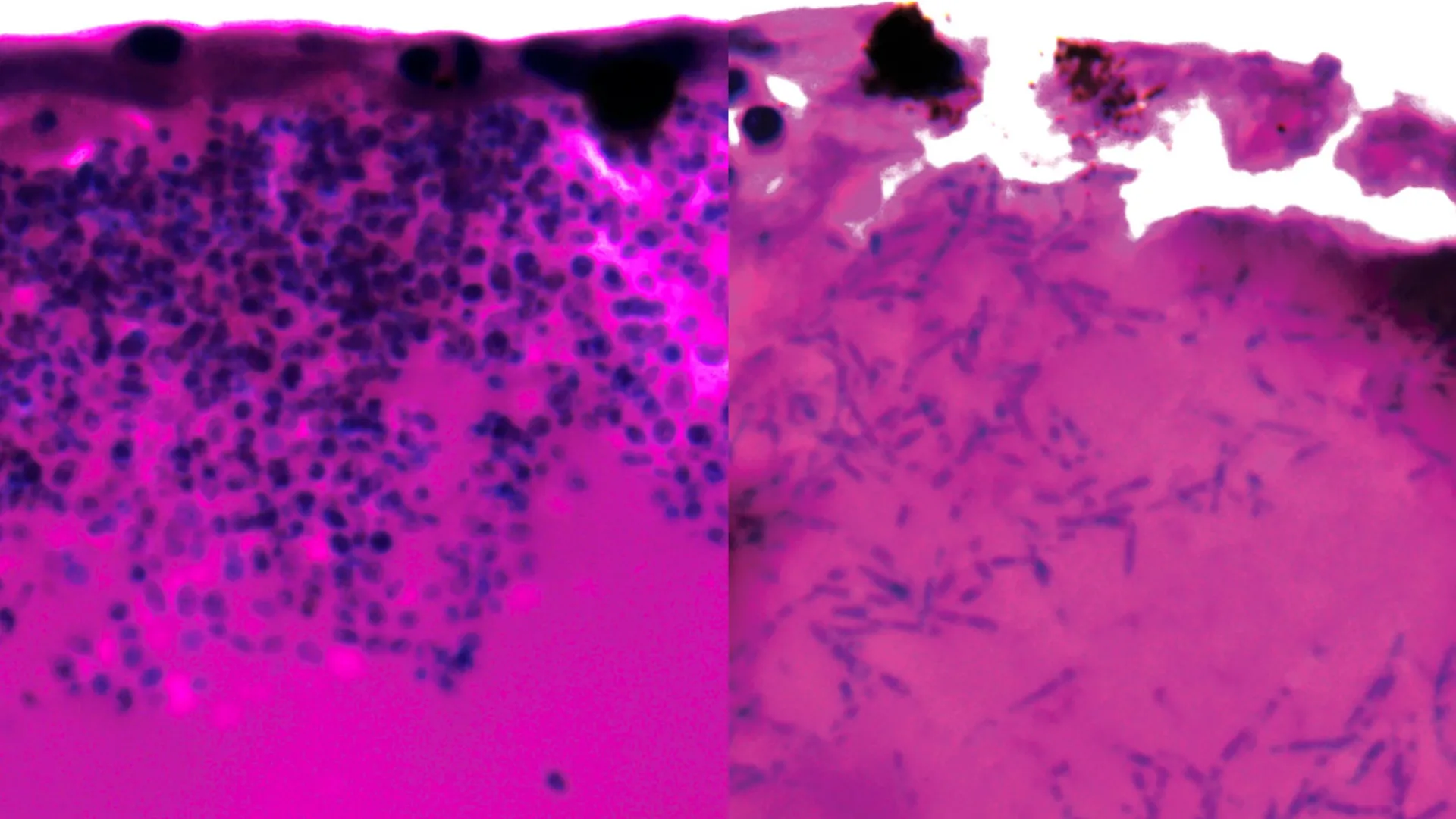
During the experiments, researchers observed that Candida auris can change its shape by forming elongated fungal structures known as filaments. These structures may help the fungus search for nutrients while infecting a host.

The team also analyzed which genes were activated or switched off during infection to identify possible weak points. Several of the genes that became active are responsible for producing nutrient pumps that capture iron-scavenging molecules and transport iron into fungal cells. Because iron is essential for survival, this process may represent a critical vulnerability.

Co-senior author Dr. Rhys Farrer of the University of Exeter’s MRC Centre for Medical Mycology said: “Until now, we’ve had no idea what genes are active during infection of a living host. We now need to find out if this also occurs during human infection. The fact that we found genes are activated to scavenge iron gives clues to where Candida auris may originate, such as an iron-poor environment in the sea. It also gives us a potential target for new and already existing drugs.”

Hope for future treatments

Dr. Gifford, who also works as a resident physician in intensive care and respiratory medicine at the Royal Devon & Exeter Hospital, emphasized the clinical importance of the findings. He said: “While there are a number of research steps to go through yet, our finding could be an exciting prospect for future treatment. We have drugs that target iron scavenging activities. We now need to explore whether they could be repurposed to stop Candida auris from killing humans and closing down hospital intensive care units.”

The Arabian killifish larvae model was developed with support from an NC3Rs project grant as an alternative to using mouse and zebrafish models, which are commonly used to study interactions between pathogens and their hosts. Dr. Katie Bates, NC3Rs Head of Research Funding, said: “This new publication demonstrates the utility of the replacement model to study Candida auris infection and enable unprecedented insights into cellular and molecular events in live infected hosts. This is a brilliant example of how innovative alternative approaches can overcome key limitations of traditional animal studies.”

The paper is titled ‘Xenosiderophore transporter gene expression and clade-specific filamentation in Candida auris killifish (Aphanius dispar) infection’ and is published in the Nature portfolio journal Communications Biology.

Share Button

5 Live News Specials

That Time of the Month with Naga Munchetty, Dr Nighat Arif and Dr Christine Ekechi

Share Button

This ultra-sensitive imaging system can spot cancer earlier

Scientists have developed a compact Raman imaging system that can reliably tell cancerous tissue apart from normal tissue. The approach could support earlier cancer detection and help move advanced molecular imaging tools beyond research laboratories and into more practical clinical settings.

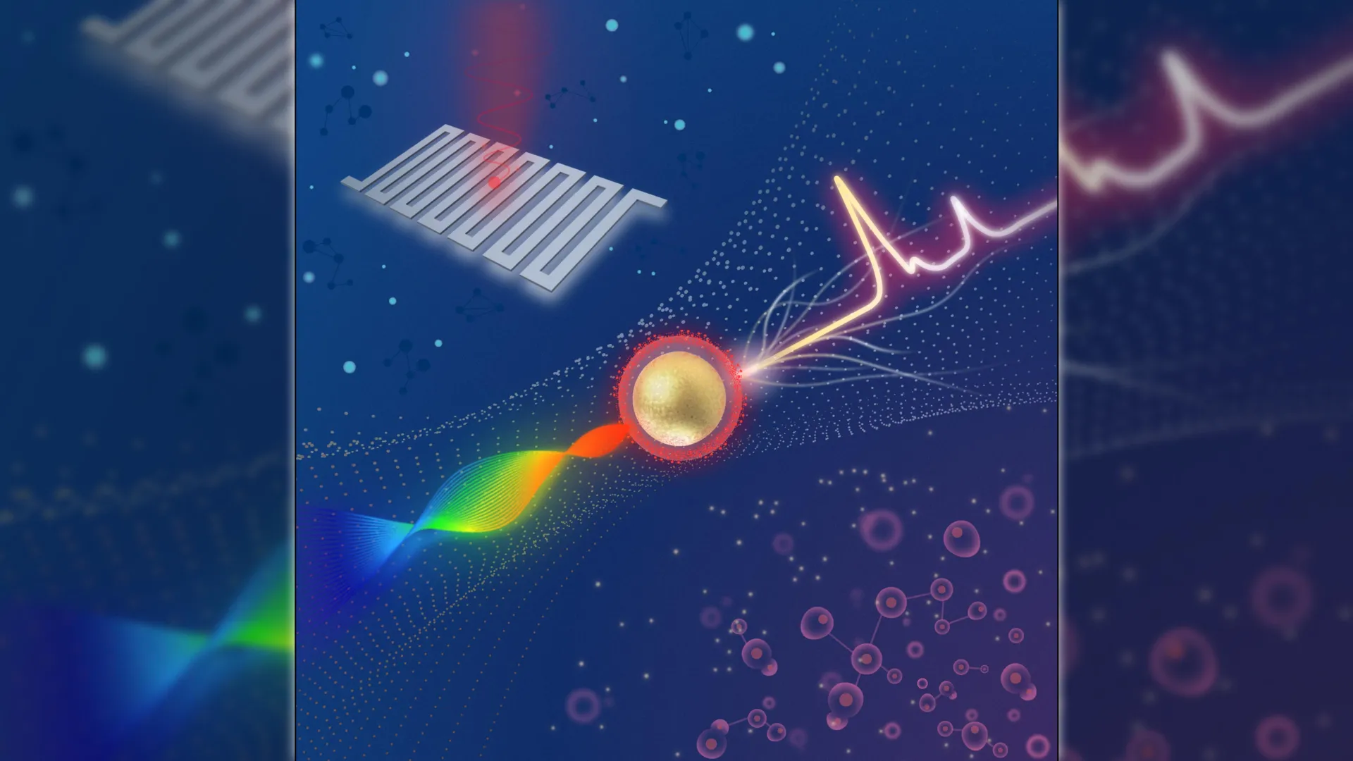
The imaging system is designed to detect extremely weak signals from surface-enhanced Raman scattering (SERS) nanoparticles that are engineered to attach to tumor markers. Once these nanoparticles are applied to a sample or to the area being examined, the system reads their Raman signal and automatically highlights regions that are more likely to contain tumor tissue.

“Traditional methods for cancer-related diagnosis are time-consuming and labor-intensive because they require staining tissue samples and having a pathologist look for any abnormalities,” said research team leader Zhen Qiu from the Institute for Quantitative Health Science and Engineering (IQ), Michigan State University. “While our system would not immediately replace pathology, it could serve as a rapid screening tool to accelerate diagnosis.”

Published results show major gains in sensitivity

In Optica, Optica Publishing Group’s journal for high-impact research, Qiu and colleagues report that their system can distinguish cancerous cells from healthy ones while detecting Raman signals that are about four times weaker than those measured by a comparable commercial system. This improved sensitivity comes from combining a swept-source laser — which changes wavelength during analysis — with an ultra-sensitive detector called a superconducting nanowire single-photon detector (SNSPD).

“This technology could eventually enable portable or intraoperative devices that enable clinicians to detect cancers at earlier stages, improve the accuracy of biopsy sampling and monitor disease progression through less invasive testing,” said Qiu. “Ultimately, such advances could enhance patient outcomes and reduce diagnostic delays, accelerating the path from detection to treatment.”

Pushing detection limits with superconducting detectors

Qiu’s lab studies how SNSPDs can be used to enhance a range of imaging technologies. SNSPDs rely on a superconducting wire that can detect individual particles of light, allowing the system to capture extremely weak optical signals at high speed while keeping background noise very low.

For this project, the researchers aimed to build a platform that could measure Raman signals far fainter than those detected by existing Raman systems. Raman imaging works by mapping a sample’s chemical composition through the unique light-scattering fingerprints of its molecules. These signals can be strengthened by using SERS nanoparticles.

“Combining this advanced detector with a swept-source Raman architecture that replaces a bulky camera and collects light more efficiently resulted in a system with a detection limit well beyond that of comparable commercial systems,” said Qiu. “Also, the fiber coupling configuration and compact design facilitate system miniaturization and future clinical translation.”

Strong tumor contrast across multiple sample types

To test the system, the team used SERS nanoparticles coated with hyaluronan acid, which enables the particles to bind to CD44, a surface protein found on many tumor cells. Initial experiments with simple nanoparticle solutions showed that the system could reach femtomolar sensitivity. The researchers then applied the imaging platform to cultured breast cancer cells, mouse tumors, and healthy tissue samples.

“The SERS signals were strongly concentrated in tumor samples, with only minimal background detected in healthy tissue,” said Qiu. “This demonstrates both the system’s exceptional sensitivity and its ability to provide reliable tumor-versus-healthy contrast. Moreover, by adjusting or substituting the targeting molecule, this method could be adapted for other cancer types.”

Next steps toward clinical use

According to the researchers, additional work is needed before the system can be used in clinical settings. Future improvements will focus on increasing readout speed and expanding validation studies. The team is exploring faster laser sources, including VCSELs, and testing whether narrowing the sweep range can further improve performance. They also plan multiplexing experiments that use different nanoparticles to target multiple biomarkers at the same time.

The researchers acknowledge industry collaborator Quantum Opus, which provided the SNSPD devices used in this work.

Share Button