An 11-year-old needed two new organs and doctors made history

Children’s Hospital Colorado (Children’s Colorado) has completed its first-ever dual heart and liver transplant, marking a major milestone for the hospital. The complex procedure involved dozens of specialists working across 25 multidisciplinary care teams. Nationwide, only 38 pediatric patients have previously received both a heart and liver transplant.

“Performing Children’s Colorado’s first-ever heart and liver dual organ transplant is an amazing accomplishment for our Pediatric Transplant Program,” said Dr. Megan Adams, surgical director of the Pediatric Liver Transplant and Kidney Transplant Programs. “Thanks to years of dedication and a team committed to being the trusted leaders in pediatric transplant across our seven-state region, we’re grateful to provide this level of care to even more kids who need complex organ transplants to treat life-threatening illnesses and help them live healthy and happy lives.”

Years of Preparation Lead to a Life-Saving Moment

Care teams at Children’s Colorado had spent years preparing for the possibility of a dual heart and liver transplant. Close coordination among specialists in surgery, cardiology, hepatology, and other fields, along with strong backing from hospital leadership, ensured the team was ready when 11-year-old Gracie Greenlaw and her family needed help.

Gracie was born with hypoplastic left heart syndrome (HLHS), a condition in which her heart developed with only one functioning pumping chamber. Before turning three, she underwent three major surgeries, the Norwood, the Glenn and the Fontan, to allow her heart to circulate blood effectively. Although many children with HLHS now survive into adulthood, the condition and its treatments can lead to serious long-term complications, including liver damage and liver failure.

Managing the Long-Term Effects of Congenital Heart Disease

To address these ongoing challenges, Children’s Colorado established the Fontan Multidisciplinary Clinic in 2016 as part of its Single Ventricle Program. The clinic focuses on caring for patients with HLHS and other single ventricle conditions, such as tricuspid atresia and unbalanced common atrioventricular canal, by providing coordinated, whole-patient care.

Through this program, Gracie received continuous monitoring and treatment for both her heart and liver. Her care team included experts like cardiologist Dr. Kathleen Simpson and hepatologist Dr. Dania Brigham, who worked together to manage her condition until a transplant became the best option.

“The Fontan is a lifesaving surgery, but the longer someone lives after the procedure, there is an increased chance of developing comorbidities,” Simpson said. “Our care team worked to keep her healthy and living a typical day-to-day life as long as possible before we determined a dual organ transplant would give her the best long-term quality of life.”

Preparing for a Complex Dual Organ Transplant

For years, Gracie lived with plastic bronchitis, a condition that causes thick, protein-like material to build up in the airways. Over the past year, her symptoms worsened, and signs of liver failure began to appear. Her medical team concluded that moving forward with a dual transplant was necessary, and she was placed on the transplant waitlist in April.

In preparation, dozens of specialists met regularly to plan for the surgery. They carefully accounted for the challenges of transplanting two organs at once, including differences in blood volume needs and electrolyte management during the operation.

A Carefully Orchestrated 16-Hour Surgery

Less than a month after joining the waitlist, compatible donor organs became available, made possible by another family’s decision to donate. Because the heart can only remain viable for a short time, the surgical team began with the heart transplant. Dr. Matthew Stone, surgical director of the Pediatric Heart Transplant Program, and congenital heart surgeon Dr. Emily Downs led the nine-hour procedure.

While the heart surgery was underway, the donor liver was maintained on a TransMedics Organ Care System — a specialized device designed to replicate normal liver function. This technology preserved the liver and allowed the heart surgeons the time they needed to complete their work. Dr. Adams and transplant surgeon Dr. Kendra Conzen then performed the liver transplant, which took an additional seven hours. Throughout the process, close coordination with anesthesiology teams was essential to protect Gracie’s health.

Recovery and a Return to Everyday Life

The surgery was successful. Gracie left the cardiac progress care unit just over a month later. Seven months after the transplant, she continues to attend monthly follow-up visits, but she has returned to school and is back home with her dogs.

Like other pediatric heart transplant recipients, Gracie will need another heart transplant later in life. Her transplanted liver, however, is expected to last for the rest of her lifetime.

“This procedure showcases the expertise, talent and level of care Children’s Colorado provides to our patients, including those with complex medical needs,” said Dr. Duncan Wilcox, Surgeon in Chief. “As the top-ranked pediatric hospital in Colorado and the Rocky Mountain region, we are proud of our leading-edge transplant care and look forward to supporting more patients who need dual organ transplants in the future.”

Share Button

Young people will feel burden of UK’s ageing society, report suggests

The House of Lords said raising the state pension age and increasing immigration would not be a solution.

Share Button

Neurons aren’t supposed to regrow but these ones brought back vision

For decades, neuroscientists have taught that neurons do not regenerate once they are damaged or destroyed. This belief has shaped how brain injuries are understood and treated. Yet people often regain at least some lost abilities after trauma, raising an important question: if neurons do not grow back, how does recovery happen?

A new JNeurosci paper offers insight into this puzzle. Athanasios Alexandris and colleagues at Johns Hopkins University used mice to study what happens inside the visual system after traumatic brain injury. The visual system includes cells in the eye that send information to the brain, allowing animals and humans to see. Damage to this system can disrupt communication between the eye and the brain, leading to vision problems.

Surviving Cells Rebuild Eye to Brain Connections

After injury, the researchers closely tracked the connections between cells in the eye and neurons in the brain. Instead of finding widespread regrowth of new cells, they observed something different. The cells that survived the injury began to adapt.

These surviving cells grew extra branches, which allowed them to connect with more neurons in the brain than before. This process, known as sprouting, helped compensate for cells that were lost due to injury. Over time, the number of connections between the eye and the brain returned to levels similar to those seen before the injury occurred.

Importantly, these rebuilt connections were not just structural. Measurements of brain activity showed that the new pathways were working properly and could transmit signals effectively. In practical terms, this means the visual system was able to function again despite the damage.

Sex Differences in Visual System Recovery

The study also revealed a significant difference between male and female mice. While male mice showed strong recovery through this compensatory sprouting process, female mice experienced slower or incomplete repair. The eye to brain connections in females did not always fully return to preinjury levels.

According to the authors, these findings point to a recovery mechanism that operates differently depending on sex. As Alexandris explains, “We didn’t expect to see sex differences, but this aligns with clinical observations in humans. Women experience more lingering symptoms from concussion or brain injury than men. Understanding the mechanism behind the branch sprouting we observed — and what delays or prevents this mechanism in females — could eventually point toward strategies to promote recovery from traumatic or other forms of neural injury.”

The research team plans to continue investigating why this repair process differs between females and males. By uncovering the biological factors that influence neural recovery, they hope to identify new ways to improve healing after brain injuries, including concussions and other forms of trauma.

Share Button

Has flu peaked? What the figures tell us

NHS remains on high alert over flu, health bosses say, but there are signs infections are levelling off.

Share Button

Astronomers just watched a black hole twist spacetime

The universe has delivered a rare breakthrough for researchers chasing one of the hardest effects to catch in the night sky.

In findings reported in Science Advances, scientists describe the first observations of a spiraling swirl in spacetime linked to a fast spinning black hole.

First evidence of black hole frame dragging

This phenomenon is called Lense-Thirring precession or frame-dragging. It refers to the way a rotating black hole twists the spacetime around it, tugging on nearby matter such as stars and causing their paths to wobble.

The research team was led by the National Astronomical Observatories at the Chinese Academy of Sciences, with support from Cardiff University. They focused on AT2020afhd, a tidal disruption event (TDE) where a star was ripped apart by a supermassive black hole.

As the star was destroyed, its remains formed a spinning disk around the black hole. From this disk, intense jets of material were launched at nearly the speed of light.

A 20 day cosmic wobble seen in X rays and radio

By tracking repeating patterns in both X ray and radio signals from the event, the researchers found that the disk and the jet were wobbling together. The motion repeated on a 20 day cycle.

Einstein first proposed the idea behind this effect in 1913, and it was later put into mathematical form by Lense and Thirring in 1918. These new measurements support a key prediction of general relativity and could help scientists investigate black hole spin, accretion physics, and how jets form.

Dr. Cosimo Inserra, a Reader in the School of Physics and Astronomy at Cardiff University and one of the paper’s co-authors, said: “Our study shows the most compelling evidence yet of Lense-Thirring precession — a black hole dragging space time along with it in much the same way that a spinning top might drag the water around it in a whirlpool.

“This is a real gift for physicists as we confirm predictions made more than a century ago. Not only that, but these observations also tell us more about the nature of TDEs — when a star is shredded by the immense gravitational forces exerted by a black hole.

“Unlike previous TDEs studied, which have steady radio signals, the signal for AT2020afhd showed short-term changes, which we were unable to attribute to the energy release from the black hole and its surrounding components. This is further confirmed the dragging effect in our minds and offers scientists a new method for probing black holes.”

Swift and VLA data plus spectroscopy

To pin down the frame dragging signal, the team analyzed X ray observations from the Neil Gehrels Swift Observatory (Swift) and radio measurements from the Karl G. Jansky Very Large Array (VLA).

They also examined the composition, structure and behavior of the material involved using electromagnetic spectroscopy, which helped them describe and identify the effect.

“By showing that a black hole can drag space time and create this frame-dragging effect, we are also beginning to understand the mechanics of the process,” explains Dr. Inserra.

“So, in the same way a charged object creates a magnetic field when it rotates, we’re seeing how a massive spinning object — in this case a black hole — generates a gravitomagnetic field that influences the motion of stars and other cosmic objects nearby.

“It’s a reminder to us, especially during the festive season as we gaze up at the night sky in wonder, that we have within our grasp the opportunity to identify ever more extraordinary objects in all the variations and flavors that nature has produced.”

The paper, ‘Detection of disk-jet coprecession in a tidal disruption event’, is published in Science Advances.

Share Button

The gear meant to protect firefighters may carry hidden dangers

A new study published on Dec. 16 in Environmental Science & Technology Letters reports that some firefighter protective gear contains brominated flame retardants, chemicals that may pose health risks to firefighters.

The research is the first U.S. study to formally examine and document the use of brominated flame retardants in firefighter turnout gear, which is worn during emergency responses. The findings could play a role in how fire departments decide whether to continue using existing equipment or invest in replacements.

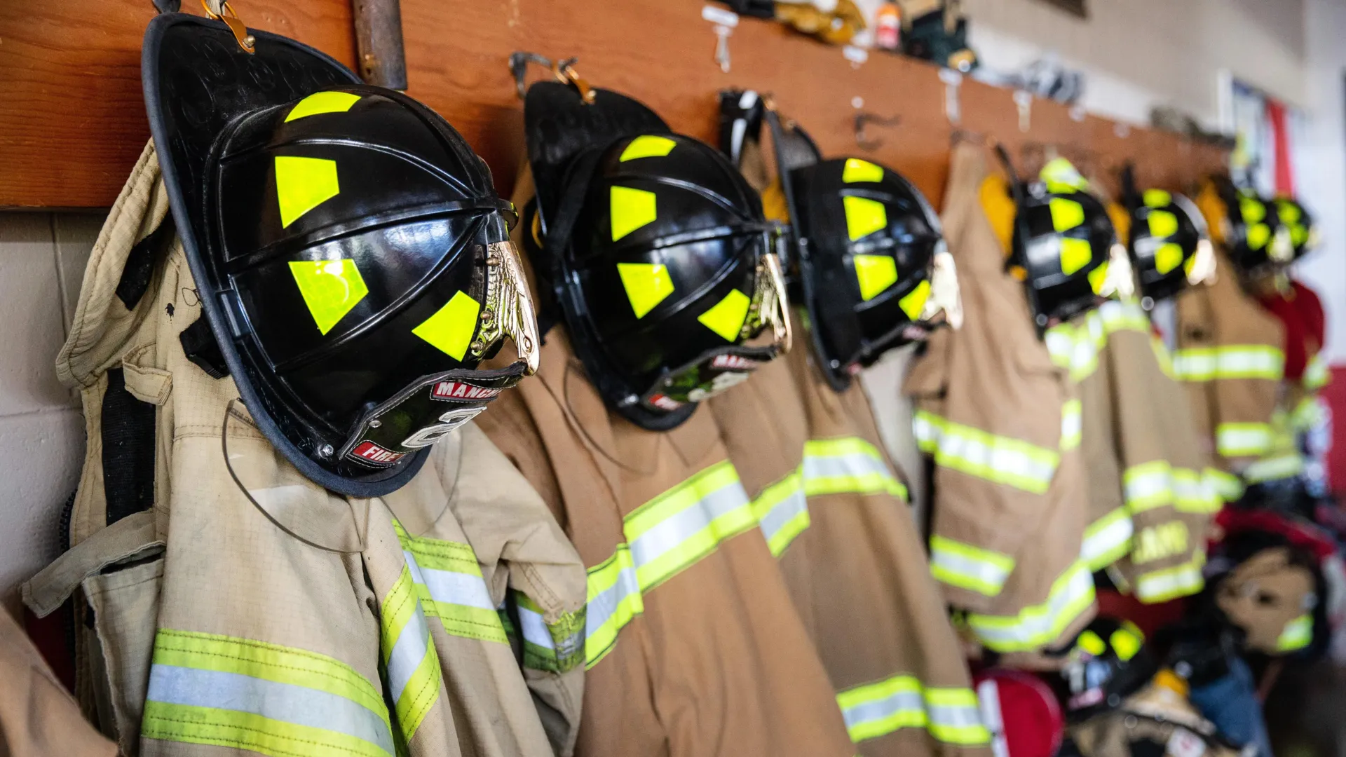
Structural firefighters — those working in the built environment — rely on turnout gear made up of three distinct layers. These include a flame-resistant outer shell, a middle moisture barrier that blocks germs while allowing airflow, and an inner lining designed to help regulate body temperature.

According to lead author Heather Stapleton, Ronie-Richele Garcia-Johnson Distinguished Professor at Duke University’s Nicholas School of the Environment, manufacturers apply chemical treatments to these garments to ensure they meet strict safety standards set by the National Fire Protection Association.

PFAS Concerns Raise New Questions

For several years, firefighters have expressed concern about the presence of PFAS in turnout gear. These chemicals are used to repel oil and water and can also contribute to flame resistance. Numerous studies involving humans and animals have linked PFAS exposure to health problems, including certain cancers.

While no studies have directly connected PFAS treated turnout gear to health outcomes in firefighters, manufacturers have begun moving away from these chemicals. In addition, multiple states have passed laws that will ban the purchase of PFAS treated turnout gear starting in 2027.

As PFAS are phased out, attention has turned to what substances may be taking their place. Chemical ingredients used in turnout gear treatments are not typically disclosed by manufacturers.

“There was a rumor that one of the turnout gear manufacturers might be using brominated flame retardants in the non-PFAS treated textiles,” Stapleton said. “Because some brominated flame retardants have known toxicity, I requested a sample of the gear in question to test.'”

Why Brominated Flame Retardants Matter

Brominated flame retardants are commonly added to fabrics and other products to reduce how easily they ignite. Exposure to these chemicals has been linked to health concerns including cancer, thyroid disease, and problems with brain development.

Stapleton’s initial testing confirmed the presence of brominated flame retardants in the turnout gear sample. That finding led to a broader study conducted in collaboration with researchers at North Carolina State University’s Wilson College of Textiles and the International Association of Fire Fighters. The goal was to examine how often PFAS and brominated flame retardants appeared in turnout gear made during different time periods.

Peeling Back the Layers

The research team analyzed nine sets of used turnout gear manufactured between 2013 and 2020, along with three sets produced in 2024 that were marketed as non-PFAS treated. Using two analytical techniques, the scientists tested each layer of the gear for both PFAS and brominated flame retardants. This approach allowed them to measure total chemical content as well as the portion that could transfer during use, referred to as “extractable” levels.

“We wanted to know which chemicals were intentionally applied during manufacturing, and we wanted to know what was likely to leach out over time, which could raise the risk of exposure through skin absorption or inhalation,” Stapleton said.

As expected, PFAS were detected in all turnout gear produced between 2013 and 2020. In contrast, gear manufactured in 2024 showed only low or non-detectable extractable PFAS levels, indicating the garments had not been treated with these chemicals, consistent with manufacturer claims. The small amounts detected were likely picked up from the surrounding environment during use, the authors noted.

Every set of turnout gear tested also contained brominated flame retardants, with extractable levels generally higher than those measured for PFAS.

Higher Levels in PFAS Free Gear

The highest extractable concentrations of brominated flame retardants were found in gear marketed as non-PFAS treated, particularly within the moisture barrier. Stapleton said this suggests manufacturers intentionally added brominated flame retardants to meet flammability requirements, likely replacing a PFAS compound previously used in that layer.

Among the chemicals identified, decabromodiphenyl ethane, or DBDPE, appeared at the highest extractable levels.

Although no U.S. studies have examined health effects linked to DBDPE exposure, the researchers pointed to a 2019 study of workers at a chemical manufacturing plant in China. That study found associations between DBDPE exposure, altered thyroid hormone levels, and signs of thyroid disease.

“I was really surprised that the manufacturers used DBDPE in turnout gear,” Stapleton said. “It has similar properties as a toxic chemical called decaBDE that has been largely phased out globally, raising questions about its safety.”

Fire Exposure Versus Manufacturing Choices

For turnout gear produced between 2013 and 2020, the outer shell typically contained higher extractable levels of brominated flame retardants than the moisture barrier or inner lining. Stapleton said this pattern likely reflects buildup from smoke and soot encountered during fires.

“When building materials burn, they can release brominated flame retardants into the air that stick to gear and don’t wash out very well,” she explained.

At the same time, the presence of these chemicals in internal layers suggests that some manufacturers had incorporated brominated flame retardants into turnout gear treatments for years, even before PFAS began to be phased out, according to the authors.

Weighing the Costs

While researchers have not yet determined firefighters’ exact exposure levels or the long-term health effects associated with these chemicals, the study provides new information for fire departments evaluating protective gear.

“Turnout gear is really expensive — one set costs thousands of dollars — and firefighters often use these garments for many years. Fire departments must consider both the financial and personal safety costs of keeping or replacing gear,” said coauthor R. Bryan Ormond, an associate professor at the Wilson College of Textiles and director of NC State’s Milliken Textile Protection and Comfort Center, who studies trade-offs in gear performance.

Stapleton noted that some manufacturers now offer turnout gear that avoids both PFAS and brominated flame retardants. She encouraged fire departments to push for clearer disclosure about chemical treatments used in protective equipment.

“We know firefighters receive higher exposure to multiple chemicals from all the hazards they face during their duty, and they shouldn’t have to worry about receiving additional chemical exposures from their gear,” said Stapleton, who also leads a study on cancer incidence in firefighters. “These first responders are a critically important component of our public safety and deserve to be respected and protected.”

Study Support and Funding

This project was supported by the North Carolina Collaboratory at the University of North Carolina at Chapel Hill, with funding appropriated by the North Carolina General Assembly (to HMS and BO). HMS also wishes to thank Michael and Annie Falk for establishing the Falk Exposomics Laboratory.

Share Button

A hidden T cell switch could make cancer immunotherapy work for more people

Over the past ten years, T cell immunotherapy has emerged as one of the most promising developments in cancer treatment. These therapies work by training a patient’s own immune system to detect and destroy dangerous cells. Despite their success, scientists have struggled to fully explain how these treatments function at a molecular level. This lack of understanding has slowed progress, especially since T cell therapies work well for only a small number of cancer types and fail in most others, for reasons that have remained unclear. Gaining insight into their modus operandi could help make these therapies effective for far more patients.

Scientists at The Rockefeller University have now uncovered crucial details about the T cell receptor (TCR), a protein complex embedded in the cell membrane that plays a central role in T cell therapies. Using cryo-EM, researchers from the Laboratory of Molecular Electron Microscopy studied the receptor in a biochemical setting designed to closely resemble its native milieu. They discovered that the TCR behaves like a jack-in-the-box, staying compact until it encounters an antigen or another suspicious particle, at which point it rapidly opens. This behavior contradicts what earlier cryo-EM studies of the receptor had shown.

The findings, published in Nature Communications, could help researchers improve and expand the use of T cell immunotherapies.

“This new fundamental understanding of how the signaling system works may help re-engineer that next generation of treatments,” says first author Ryan Notti, an instructor in clinical investigation in Walz’s lab and a special fellow in the Department of Medicine at Memorial Sloan Kettering Cancer Center, where he treats patients with sarcomas, or cancers that arise in soft tissue or bone.

“The T cell receptor is really the basis of virtually all oncological immunotherapies, so it’s remarkable that we use the system but really have had no idea how it actually works — and that’s where basic science steps in,” says Walz, a world expert in cryo-EM imaging. “This is some of the most important work to ever come out of my lab.”

How T Cells Detect Threats

Walz’s lab focuses on producing detailed images of macromolecular complexes, especially proteins found in cell membranes that help cells communicate with their surroundings. The TCR is one such complex. Made up of multiple proteins, it enables T cells to recognize antigens displayed by human leukocyte antigen (HLA) complexes on other cells. This recognition process is what T cell therapies rely on to mobilize the immune system against cancer.

Although scientists have known the individual parts of the TCR for many years, the earliest steps that trigger its activation have remained elusive. Notti, who works as both a physician and a researcher, found this gap especially troubling because many of his sarcoma patients were not benefiting from T cell immunotherapies.

“Determining that would help us understand how the information gets from outside the cell, where those antigens are being presented by HLAs, to the inside of the cell, where signaling turns on the T cell,” he says.

Notti earned his Ph.D. in structural microbiology at Rockefeller before moving into oncology, and he suggested to Walz that they investigate this unanswered question together.

Rebuilding the TCR’s Natural Environment

Walz’s team is known for creating custom membrane environments that closely mimic the natural surroundings of membrane proteins. “We can change the biochemical composition, the thickness of the membrane, the tension and curvature, the size — all kinds of parameters that we know have an influence on the embedded protein,” Walz says.

For this study, the researchers set out to observe the TCR in conditions that closely resemble those inside a living cell. They placed the receptor into a nanodisc, a tiny disc-shaped section of membrane held in solution by a scaffold protein wrapped around its edge. Assembling the full receptor was difficult, and “getting all eight of these proteins properly assembled into the nanodisc was challenging,” Notti says.

Previous structural studies of the TCR had relied on detergent, which often strips away the surrounding membrane. Walz notes that this was the first time the receptor complex had been restored to a membrane environment for detailed imaging.

Seeing the Receptor Switch On

Once the TCR was embedded in the nanodisc, the researchers used cryo-EM to visualize it. The images showed that the receptor remains closed and compact when inactive. When it encounters an antigen-presenting molecule, however, the structure opens and extends outward, resembling a wide-reaching motion.

The result surprised the team. “The data that were available when we began this research depicted this complex as being open and extended in its dormant state,” Notti explains. “As far as anyone knew, the T cell receptor didn’t undergo any conformational changes when binding to these antigens. But we found that it does, springing open like a sort of jack-in-the-box.”

The researchers believe two factors made this discovery possible. First, they carefully recreated the TCR’s in vivo membrane environment using the right lipid mixture. Second, they reinserted the receptor into a membrane using nanodiscs before conducting cryo-EM imaging. They found that an intact membrane keeps the receptor in a closed position until activation occurs. In earlier studies, detergent may have removed this restraint, allowing the receptor to open prematurely.

“It was important that we used a lipid mixture that resembled that of the native T cell membrane,” says Walz. “If we had just used a model lipid, we wouldn’t have seen this closed dormant state either.”

Implications for Cancer Therapies and Vaccines

The team believes their findings could help improve treatments that rely on T cell receptors. “Re-engineering the next generation of immunotherapies tops the charts in terms of unmet clinical needs,” Notti says. “For example, adoptive T cell therapies are being used successfully to treat certain very rare sarcomas, so one could imagine using our insights to re-engineer the sensitivity of those receptors by tuning their activation threshold.”

Walz also sees potential applications beyond cancer therapy. “This information may be used for vaccine design as well,” he adds. “People in the field can now use our structures to see refined details about the interactions between different antigens presented by HLA and T cell receptors. Those different modes of interaction might have some implication for how the receptor functions — and ways to optimize it.”

Share Button

Flu: How are hospitals in your area affected?

Use our interactive tool to explore the latest flu numbers in your area

Share Button

People fake weight to obtain skinny jabs, says GP

A doctor from Chelmsford calls for more checks to stop the drugs being sold inappropriately online.

Share Button

A quantum mystery that stumped scientists for decades is solved

A global research team led by Rice University physicist Pengcheng Dai has verified the presence of emergent photons and fractionalized spin excitations in an unusual quantum spin liquid. Reported in Nature Physics, the work points to the crystal cerium zirconium oxide (Ce2Zr2O7) as a clean three-dimensional example of this exotic state of matter.

Quantum spin liquids have fascinated physicists for years because they could eventually support transformative technologies, including quantum computing and dissipationless energy transmission. Unlike ordinary magnets that settle into an orderly pattern, these materials avoid conventional magnetic order. Instead, their magnetic moments remain strongly quantum-entangled and in constant collective motion at temperatures close to absolute zero, producing behavior that resembles emergent quantum electrodynamics.

“We’ve answered a major open question by directly detecting these excitations,” said Dai, the Sam and Helen Worden Professor of Physics and Astronomy. “This confirms that Ce2Zr2O7 behaves as a true quantum spin ice, a special class of quantum spin liquids in three dimensions.”

Cleaner Measurements With Polarized Neutron Scattering

To pin down these elusive signatures, the researchers relied on advanced polarized neutron scattering. This approach helped them isolate the magnetic scattering they cared about while filtering out other signals, even as the system approached the zero temperature limit.

Their measurements also revealed emergent photon signals near zero energy — a defining trait that separates quantum spin ice from more familiar phases found in conventional magnets. Additional evidence came from specific heat measurements, which supported the idea that these predicted emergent photons follow a dispersion resembling the way sound moves through a solid.

Earlier attempts to confirm this kind of behavior were often undermined by technical noise and incomplete data. The Rice-led team addressed those challenges through improved sample preparation and high-precision instruments, supported by an international effort involving major laboratories across Europe and North America.

First-of-Its-Kind Observation With Big Implications

In this three-dimensional candidate material, the researchers observed both emergent photons and spinons — key hallmarks of quantum spin ice. The result resolves a long-running debate in condensed matter physics and gives scientists a strong platform for studying next-generation quantum phenomena and potential technology pathways.

Bin Gao, a research scientist in Rice’s Department of Physics and Astronomy and the study’s first author, said the findings back up decades of theoretical expectations.

“This surprising result encourages scientists to look deeper into such unique materials, potentially changing how we understand magnets and the behavior of materials in the extreme quantum regime,” Gao said.

Research Team and Funding

Co-authors of this study include Félix Desrochers and Yong Baek Kim of the University of Toronto; Rice alumnus David Tam of the Paul Scherrer Institut; Silke Paschen, Diana Kirschbaum and Duy Ha Nguyen of Vienna University of Technology; Paul Steffens and Arno Hiess of the Institut Laue-Langevin; Yixi Su of Jülich Centre of Heinz Maier-Leibnitz Zentrum; and Sang-Wook Cheong of Rutgers University.

The U.S. Department of Energy, the Gordon and Betty Moore Foundation and the Robert A. Welch Foundation supported this study.

Share Button