Satellites spot rapid “Doomsday Glacier” collapse

Thwaites Glacier in West Antarctica — widely known as the “Doomsday Glacier” — is changing more quickly than almost any other ice-ocean system on the planet. Its future behavior remains one of the biggest unknowns in forecasts of global sea-level rise. One part of this system, the Thwaites Eastern Ice Shelf (TEIS), floats on the ocean and is partly held in place by a pinning point at its northern edge. Over the past twenty years, this shelf has developed increasing fractures around a major shear zone located upstream of that pinning point.

A new study published in the Journal of Geophysical Research: Earth Surface (AGU, 2025) offers the most detailed account yet of how this slow breakdown has unfolded. The research was produced at the Centre for Earth Observation Sciences and led by Debangshu Banerjee, a recent graduate student from the Centre for Earth Observation Science (CEOS), along with Dr. Karen Alley (Assistant Professor, CEOS) and Dr. David Lilien (Assistant Professor, Indiana University Bloomington and former Research Associate at CEOS). Their work contributes to the TARSAN (Thwaites-Amundsen Regional Survey and Network) project, one of the programs within the International Thwaites Glacier Collaboration (ITGC) — a major U.S.-U.K. effort to understand the forces driving change in Thwaites Glacier. Several well-known glaciologists, including Dr. Ted Scambos, Dr. Martin Truffer, Dr. Adrian Luckman, and Dr. Erin Pettitt, also contributed to this research.

Fracture Growth, Ice Dynamics, and a Strengthening Feedback Loop

Using twenty years (2002-2022) of satellite observations, ice-flow speed measurements, and in-situ GPS data, the team documented how fractures within the TEIS shear zone formed and evolved. Their analysis shows that the gradual growth of these fractures weakened the shelf’s connection to the pinning point. As this attachment deteriorated, the ice upstream began to flow more quickly, reducing the shelf’s mechanical stability.

The researchers identified four clear stages in this weakening and highlighted two major findings. The first is that the fractures expanded in two distinct phases: long fractures aligned with the direction of ice flow appeared first, followed later by shorter fractures that cut across the flow. The second is evidence of a positive feedback cycle in which these fractures increased ice acceleration, which in turn caused further damage. This accelerating loop played a significant role in the shelf’s recent decline.

A Shift From Stabilizing Force to Source of Weakness

The study notes that the pinning point, once a key factor holding the TEIS in place, has slowly shifted into a feature that now contributes to its instability. This four-stage pattern of structural decline may be a signal for other Antarctic ice shelves that appear to be entering similar phases of weakness. If these floating shelves continue to deteriorate, the Antarctic Ice Sheet could contribute even more to future sea-level rise.

Share Button

Flu jabs offered in pub in vaccination drive

Regular pop-up vaccination clinics are available at The Bevy community pub in Brighton.

Share Button

A lost Amazon world just reappeared in Bolivia

In September 2021, a team of specialists traveled to one of the least-documented areas of the Bolivian Amazon: the Great Tectonic Lakes of Exaltación in the department of Beni. The expedition, organized by the Grupo de Trabajo para los Llanos de Moxos (GTLM), brought together experts from the Wildlife Conservation Society, the National Museum of Natural History, the Institute of Ecology, the Biodiversity and Environment Research Center, the Aquatic Resources Research Center, and the Department of Anthropology of the Americas at the University of Bonn.

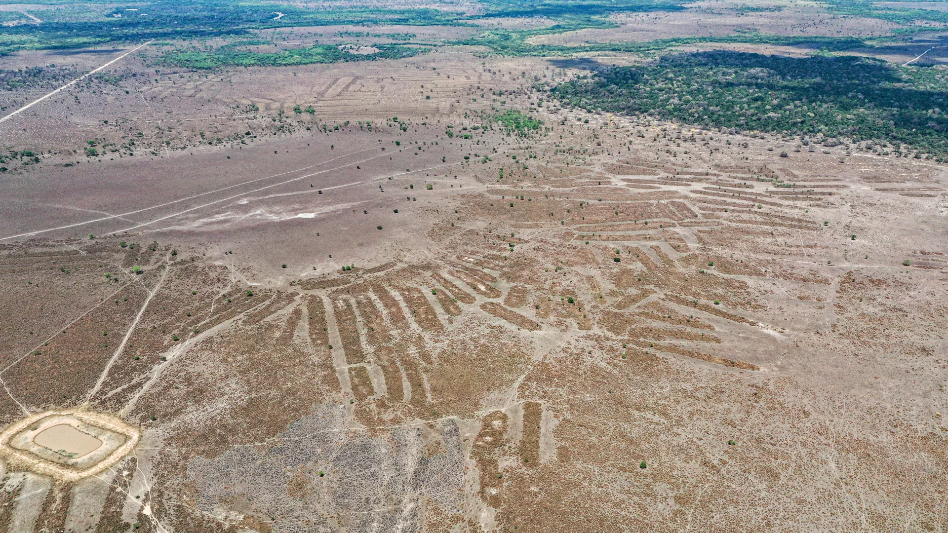
In the southwestern Amazon, the tectonic Lakes Rogaguado and Ginebra reveal a landscape shaped by generations of human innovation. Beneath the open grasslands and shallow waters lie the remains of extensive earthworks, raised agricultural fields, and interconnected canals that reflect a long history of people adapting to a dynamic environment.

These lakes sit within the Municipal Protected Area of Grandes Lagos Tectónicos de Exaltación and form part of the Río Yata Ramsar wetland complex, which is recognized by UNESCO for both its ecological and cultural value. Set in the Llanos de Moxos, a vast network of savannas, gallery forests, and floodplains that make up the largest wetland system in the Amazon basin, this area has inspired curiosity for centuries. Ethnohistorical accounts even linked it to the legendary ‘Land of Paititi.’ Only recently has its deep human past begun to come into clearer view.

Mapping a Long History of Settlement

Using a combination of surveys, excavations, and LiDAR imaging, the research team documented several archaeological sites: Paquío, Coquinal, Isla del Tesoro, and Jasschaja. Each site represents a different stage in the long sequence of habitation across this region.

Radiocarbon dates reveal repeated occupations from roughly 600 to 1400 CE. Findings at Paquío show an early settlement beginning around 600 CE, followed by a more intensive occupation between 1000 and 1200 CE. This later phase included shell middens, dense ceramic refuse, and a sophisticated system of canals and raised fields connected to maize-based farming. Jasschaja, which dates from 1300 to 1400 CE, reflects broader landscape alterations and greater botanical diversity, suggesting intensified forest and crop management during its occupation.

Innovations in Water Management and Agriculture

The terrain of the Llanos de Moxos is filled with geometric forms that reveal themselves on closer inspection. Circular and rectangular ditches, drainage channels, raised planting platforms, and clusters of mounds create a complex network designed for water control and cultivation. These structures were built to regulate seasonal flooding, guide water flow, and create stable areas for living and farming within a wetland that changes dramatically throughout the year.

Their many shapes, ranging from geometric enclosures to long elevated fields, indicate that there was no single standardized design. Instead, they represent centuries of experimentation as communities responded to shifting ecological and social conditions. Together, these features highlight the cultural diversity and long-term resilience of the people who developed them.

Excavations at Paquío and Jasschaja also revealed details about a varied pre-Hispanic diet that relied on wetland resources. Fish such as wolf fish, peacock bass, and South American lungfish were especially common, accompanied by reptiles including caimans and turtles, and mammals such as capybaras, pacas, and armadillos. Plant remains show the use of maize, legumes, and multiple palm species — moriche palm, corozo palm, cumare palm, totai palm, palmita, and peach palm. Together, these remains point to a balanced subsistence strategy that combined fishing, hunting, gathering, and farming.

Biocultural Knowledge and Indigenous Leadership

The Cayubaba and Movima communities continue to live in these landscapes, where rich biodiversity is deeply connected to cultural heritage. Their long-standing presence and environmental knowledge help maintain a unique form of biocultural heritage in which ecological and cultural diversity have developed side by side over many generations.

During the post-Covid-19 field seasons, collaboration with local communities was rooted in open communication and mutual respect. Representatives of the Cayubaba Indigenous Council, which includes 21 Cayubaba and Movima communities, assisted researchers in identifying areas for study, providing access to culturally important places, and specifying sites that should not be disturbed. While interactions were limited for health reasons, this partnership ensured that the research reflected community priorities and contributed to a fuller understanding of the living heritage of the region.

Through the GTLM, Indigenous leaders and scientists are working together to link archaeological and ecological research with conservation initiatives. These efforts emphasize that the Llanos de Moxos is not only a center of biodiversity but also a landscape shaped through long human histories, and they support continued management of the Yata River Ramsar site and the protected areas connected to it.

Lessons From the Past for a Changing Amazon

As deforestation, expanding agriculture, and climate change put increasing pressure on the Amazon, the landscapes around Lakes Rogaguado and Ginebra highlight the importance of sustainable land-use traditions. Archaeological evidence shows that past communities developed flexible ways of living that combined farming, fishing, and forest management. Rather than seeking to control or overexploit the environment, they adapted to its seasonal cycles and used periodic flooding as an opportunity.

Although raised-field agriculture eventually ended — likely because of population decline and social upheaval after European colonization — this does not diminish the effectiveness of these systems. For centuries, communities maintained productive landscapes by working with the region’s natural rhythms. Their practices challenge modern assumptions about what counts as “development” and remind us that resilience often emerges from diversity: of species, of knowledge, and of cultural traditions.

Protecting this biocultural heritage is now a global responsibility. The wetlands of the Llanos de Moxos continue to store carbon, moderate water systems, and support a wide range of species. Conservation efforts must also respect the people who have cared for these landscapes for generations. In this way, archaeology becomes more than a study of the past; it becomes a means of reconnecting ancient knowledge with today’s urgent debates about sustainability and environmental justice.

The Llanos de Moxos demonstrate that the Amazon has always been a place where people and nature have shaped one another. Its monumental earthworks, forest islands, and living cultural traditions suggest that part of our shared future may depend on listening more closely to these landscapes that remember.


Share Button

New 3D scan reveals a hidden network of moai carvers on Easter Island

A new study published November 26, 2025 in the open-access journal PLOS One reports that the well-known stone figures of Rapa Nui (Easter Island) were created by many separate carving groups. The research team, led by Carl Philipp Lipo of Binghamton University, New York and colleagues, examined how these communities contributed to the making of the island’s famous monuments.

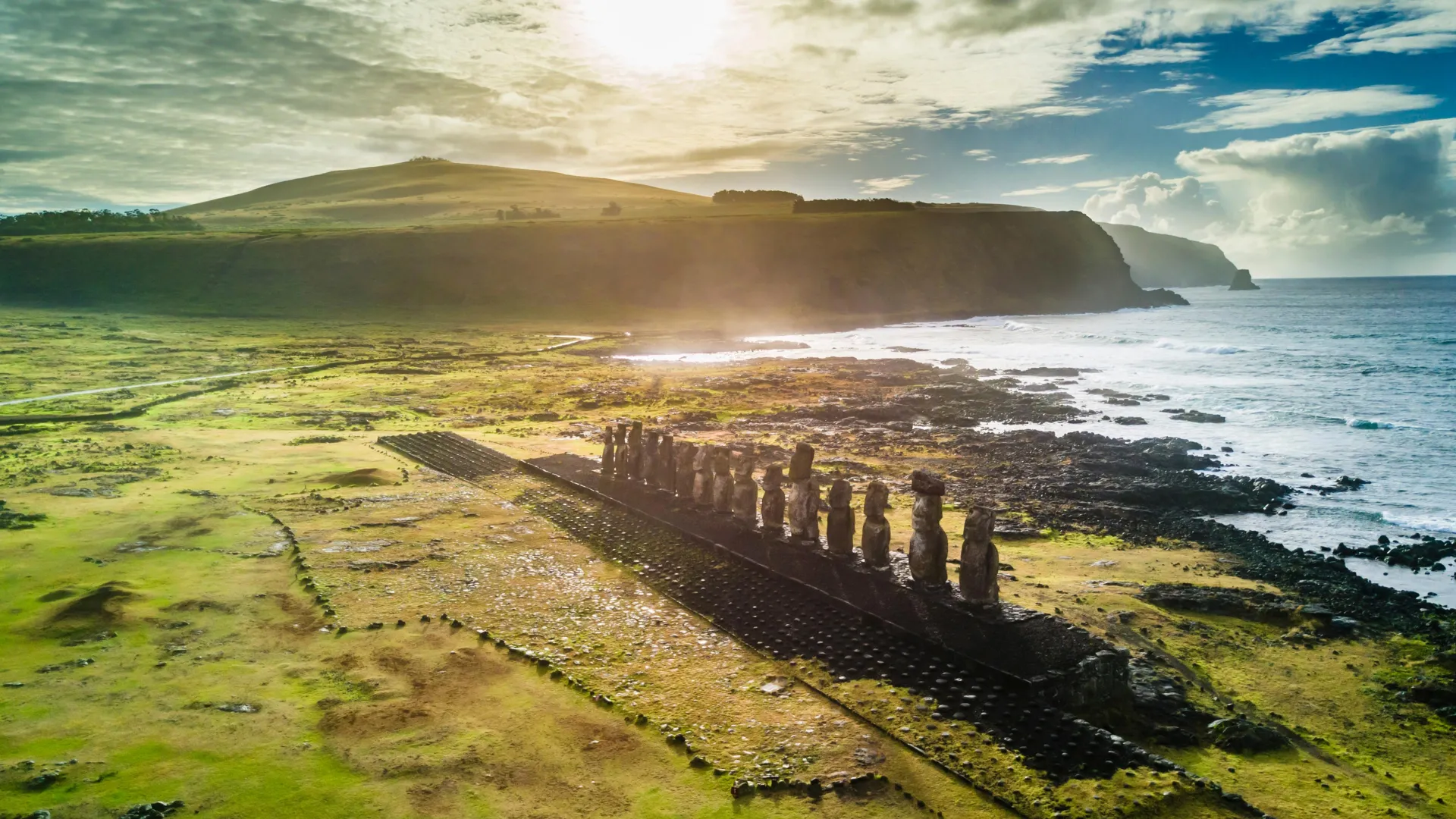
Rapa Nui is widely recognized for its hundreds of stone statues (moai), crafted by Polynesian settlers beginning in the 13th century. Archaeological work has repeatedly shown that the island was home to many small family groups rather than a unified political system. This background has prompted researchers to explore whether the carving of moai followed the same decentralized structure.

High-Resolution 3D Modeling Reveals 30 Quarry Work Zones

For this study, scientists gathered more than 11,000 photographs of Rano Raraku, the primary moai quarry. These images were merged into a detailed 3D reconstruction that captured hundreds of moai preserved in different stages of production. After analyzing the model, the team identified 30 distinct quarrying areas, each showing unique carving approaches. Additional clues indicate that completed or partially shaped moai were moved away from the quarry along several different paths. Taken together, these patterns suggest that statue creation reflected the island’s broader social organization, with carving efforts carried out independently rather than through centralized oversight.

New Evidence Challenges Long-Held Assumptions

The findings call into question the idea that projects of this scale require strict hierarchy or a single coordinating authority. Similarities between moai appear to come from the sharing of cultural knowledge instead of coordinated, joint labor. The new quarry model also provides a valuable dataset that can support future investigations and guide cultural management at this UNESCO World Heritage site. The same methods used here can also be applied to study other archaeological locations.

The authors explain: “Much of the so-called “mystery” of Rapa Nui (Easter Island) comes from the lack of openly available, detailed evidence that would allow researchers to evaluate hypotheses and construct explanations. Here, we present the first high-resolution 3D model of the moai quarry at Rano Raraku, the central quarry for nearly 1,000 statues, offering new insights into the organizational and manufacturing processes of these giant megalithic figures.”

Fieldwork for this research was supported by a National Science Foundation grant (Award #2218602). The funders had no involvement in study design, data collection and analysis, decisions related to publication, or manuscript preparation.

Share Button

Scientists discover a hidden brain circuit that rewrites vision

Vision guides an animal’s actions, but new research from MIT shows that the relationship goes both ways. The study, published November 25 in Neuron, reports that behavior and internal conditions directly influence how visual information is processed. In mice, the brain’s prefrontal cortex, which serves as a major center for executive control, sends tailored signals to regions involved in vision and movement. These signals adjust how those regions operate depending on factors such as how alert the mouse is and whether it is actively moving.

“That’s the major conclusion of this paper: There are targeted projections for targeted impact,” said senior author Mriganka Sur, Paul and Lilah Newton Professor in The Picower Institute for Learning and Memory and MIT’s Department of Brain and Cognitive Sciences.

Investigating Customized Prefrontal Signals

Scientists have long proposed, including Sur’s colleague Earl K. Miller at MIT, that the prefrontal cortex can guide the activity of more posterior areas of the brain. While anatomical evidence has supported this idea, the goal of the new study was to determine whether the prefrontal cortex sends one broad type of signal or instead crafts distinct messages for different target regions. Lead author and Sur Lab postdoctoral researcher Sofie Ährlund-Richter also sought to identify which specific neurons receive these signals and how the communication influences downstream processing.

Different Prefrontal Regions Serve Different Roles

The team identified a number of new insights. Two areas in the prefrontal cortex, the orbitofrontal cortex (ORB) and the anterior cingulate area (ACA), were found to relay information about both arousal and movement to two other regions: the primary visual cortex (VISp) and the primary motor cortex (MOp). These messages appear to have unique effects. For example, higher arousal increased ACA’s tendency to help VISp sharpen its visual representations. ORB, however, became influential only when arousal was very high, and its involvement appeared to decrease the clarity of visual encoding. According to Ährlund-Richter, ACA may help the brain focus on potentially meaningful visual details as arousal rises, while ORB may act to reduce attention to distracting or overly strong stimuli.

“These two PFC subregions are kind of balancing each other,” Ährlund-Richter said. “While one will enhance stimuli that might be more uncertain or more difficult to detect, the other one kind of dampens strong stimuli that might be irrelevant.”

Mapping and Monitoring Brain Circuits

To better understand the involved pathways, Ährlund-Richter performed detailed anatomical tracing of the connections ACA and ORB form with VISp and MOp. In additional experiments, mice ran freely on a wheel while viewing structured images or naturalistic movies at different contrast levels. At certain moments, small air puffs increased the animals’ arousal level. Throughout these tasks, researchers recorded the activity of neurons in ACA, ORB, VISp and MOp, with particular attention to the signals traveling along the axons linking prefrontal and posterior areas.

The tracing work showed that ACA and ORB each communicate with a variety of cell types in their target regions rather than a single cell class. They also connect in distinct spatial patterns. In VISp, ACA primarily targeted layer 6, while ORB communicated mainly with layer 5.

How Arousal and Movement Shift Visual Processing

When the team examined the transmitted information and neural activity, several consistent patterns emerged. ACA neurons conveyed more detailed visual information than ORB neurons and were more responsive to changes in contrast. ACA activity also tracked closely with arousal level, while ORB responded only when arousal reached a high threshold. When signaling to MOp, both regions conveyed information about running speed. When signaling to VISp, however, they only indicated whether the mouse was moving or still. The two prefrontal regions also carried information about arousal and a small amount of visual detail to MOp.

To see how this communication affects visual processing, the researchers temporarily blocked the pathways leading from ACA and ORB to VISp. This allowed them to measure how VISp neurons responded without these inputs. They found that ACA and ORB exerted specific and opposing effects on visual encoding depending on the mouse’s movement and level of arousal.

A Specialized Model of Prefrontal Feedback

“Our data support a model of PFC feedback that is specialized at both the level of PFC subregions and their targets, enabling each region to selectively shape target-specific cortical activity rather than modulating it globally,” the authors wrote in Neuron.

In addition to Sur and Ährlund-Richter, the research team included Yuma Osako, Kyle R. Jenks, Emma Odom, Haoyang Huang, and Don B. Arnold.

The work was supported by a Wenner-Gren foundations Postdoctoral Fellowship, the National Institutes of Health, and the Freedom Together Foundation.

Share Button

Why some memories last a lifetime while others fade fast

Every day, the brain turns passing impressions, creative sparks, and emotional experiences into lasting memories that shape our identity and guide our decisions. A central question in neuroscience has been how the brain determines which pieces of information are worth storing and how long those memories should remain.

Recent findings show that long-term memories form through a sequence of molecular timing mechanisms that activate across different parts of the brain. Using a virtual reality behavioral system in mice, scientists identified regulatory factors that help move memories into increasingly stable states or allow them to fade entirely.

A study published in Nature highlights how several brain regions work together to reorganize memories over time, with checkpoints that help assess how significant each memory is and how durable it should be.

“This is a key revelation because it explains how we adjust the durability of memories,” says Priya Rajasethupathy, head of the Skoler Horbach Family Laboratory of Neural Dynamics and Cognition. “What we choose to remember is a continuously evolving process rather than a one-time flipping of a switch.”

Moving Beyond the Classic Memory Model

For many years, researchers focused on two primary memory centers: the hippocampus, which supports short-term memory, and the cortex, which was believed to store long-term memories. These long-term memories were thought to sit behind biological on-and-off switches.

“Existing models of memory in the brain involved transistor-like memory molecules that act as on/off switches,” says Rajasethupathy.

This older view suggested that once a memory was marked for long-term storage, it would persist indefinitely. Although this framework provided useful insights, it did not explain why some long-term memories last for weeks while others remain vivid for decades.

A Key Pathway Linking Short and Long-Term Memory

In 2023, Rajasethupathy and colleagues described a brain circuit that connects short-term and long-term memory systems. A central element of this pathway is the thalamus, which helps determine which memories should be kept and directs them to the cortex for long-term stabilization.

These discoveries opened the door to deeper questions: What happens to memories once they leave the hippocampus, and what molecular processes decide whether a memory becomes lasting or disappears?

Virtual Reality Experiments Reveal Memory Persistence

To investigate these mechanisms, the team built a virtual reality setup that allowed mice to form specific memories. “Andrea Terceros, a postdoc in my lab, created an elegant behavioral model allowed us to break open this problem in a new way,” Rajasethupathy says. “By varying how often certain experiences were repeated, we were able to get the mice to remember some things better than others, and then look into the brain to see what mechanisms were correlated with memory persistence.”

Correlation alone could not answer the key questions, so co-lead Celine Chen created a CRISPR-based screening platform to alter gene activity in the thalamus and cortex. This approach showed that removing certain molecules changed how long memories lasted, and each molecule operated on its own timescale.

Timed Programs Guide Memory Stability

The results indicate that long-term memory relies not on a single on/off switch, but on a sequence of gene-regulating programs that unfold like molecular timers across the brain.

Early timers activate quickly but fade fast, allowing memories to disappear. Later timers turn on more gradually, giving important experiences the structural support needed to persist. In this study, repetition served as a stand-in for importance, letting researchers compare frequently repeated contexts with those seen only occasionally.

The team identified three transcriptional regulators essential for maintaining memories: Camta1 and Tcf4 in the thalamus, and Ash1l in the anterior cingulate cortex. These molecules are not required to form the initial memory but are crucial for preserving it. Disrupting Camta1 and Tcf4 weakened connections between the thalamus and cortex and caused memory loss.

According to the model, memory formation begins in the hippocampus. Camta1 and its downstream targets help keep that early memory intact. Over time, Tcf4 and its targets activate to strengthen cell adhesion and structural support. Finally, Ash1l promotes chromatin remodeling programs that reinforce memory stability.

“Unless you promote memories onto these timers, we believe you’re primed to forget it quickly,” Rajasethupathy says.

Shared Memory Mechanisms Across Biology

Ash1l is part of a protein family known as histone methyltransferases, which help maintain memory-like functions in other systems. “In the immune system, these molecules help the body remember past infections; during development, those same molecules help cells remember that they’ve become a neuron or muscle and maintain that identity long-term,” Rajasethupathy says. “The brain may be repurposing these ubiquitous forms of cellular memory to support cognitive memories.”

These discoveries may eventually help researchers address memory-related diseases. Rajasethupathy suggests that, by understanding the gene programs that preserve memory, scientists may be able to redirect memory pathways around damaged brain regions in conditions such as Alzheimer’s. “If we know the second and third areas that are important for memory consolidation, and we have neurons dying in the first area, perhaps we can bypass the damaged region and let healthy parts of the brain take over,” she says.

Next Steps: Decoding the Memory Timer System

Rajasethupathy’s team now aims to uncover how these molecular timers are activated and what determines their duration. This includes investigating how the brain evaluates the importance of a memory and decides how long it should last. Their work continues to point toward the thalamus as a central hub in this decision-making process.

“We’re interested in understanding the life of a memory beyond its initial formation in the hippocampus,” Rajasethupathy says. “We think the thalamus, and its parallel streams of communication with cortex, are central in this process.”

Share Button

HIV battle is not over, warns It’s a Sin creator

Russell T Davies says misinformation about the virus made him “despair”.

Share Button

The HIV battle is not over, warns creator of It’s a Sin

Russell T Davies says misinformation about the virus made him “despair”.

Share Button

Simple thyroid check in pregnancy may lower autism risk

Women who experience continuing thyroid hormone irregularities throughout pregnancy may face a higher chance of having a child diagnosed with autism, according to a study released in The Journal of Clinical Endocrinology & Metabolism.

Thyroid hormones supplied by the mother play an important role in fetal neurodevelopment. When these hormones become disrupted during pregnancy, previous work has linked the imbalance to atypical brain development and a higher likelihood of autism spectrum disorder (ASD). Autism is a multifaceted condition that shapes how an individual communicates, interacts socially and interprets the world.

Untreated Multi-Trimester Imbalance Carries Higher Risk

“We found that while adequately treated chronic thyroid dysfunction was not associated with increased autism risk in offspring, ongoing imbalance across multiple trimesters was,” said Idan Menashe, Ph.D., of the Ben-Gurion University of the Negev in Beer Sheva, Israel. “These findings underscore the need for routine monitoring and timely adjustment of therapy to maintain normal thyroid hormone levels throughout pregnancy.”

Large Birth Cohort Shows Clear Pattern

The research tracked more than 51,000 births and reported that mothers with persistent thyroid hormone imbalance across pregnancy had an increased likelihood of having children with autism.

The authors also documented a dose-response pattern, meaning the risk rose as the number of affected trimesters increased.

Research Team and Publication Information

Other contributors to the study include Leena Elbedour of the Ben-Gurion University of the Negev; May Weinberg of the Meir Medical Center in Kfar Saba, Israel, and Tel Aviv University in Tel Aviv, Israel; Gal Meiri of the Soroka University Medical Center in Beer-Sheva, Israel, and the Ben-Gurion University of the Negev; and Analya Michaelovski of the Soroka University Medical Center.

No funding was received for this research.

“Maternal Thyroid Hormone Imbalance and Risk of Autism Spectrum Disorder,” was published online, ahead of print.

Share Button

Scientists may have found dark matter after 100 years of searching

In the early 1930s, Swiss astronomer Fritz Zwicky noticed that many galaxies were moving far faster than their visible mass should permit. This unusual motion led him to propose that some kind of invisible structure — dark matter — was supplying the extra gravitational pull needed to keep those galaxies intact. Nearly a century later, NASA’s Fermi Gamma-ray Space Telescope may have captured the first direct evidence of this mysterious substance, offering the possibility of finally “seeing” dark matter.

Dark matter has remained one of astronomy’s biggest unknowns since it was first suggested. Until now, scientists have only been able to study it indirectly by observing how it affects ordinary matter, such as the way it produces enough gravity to hold galaxies together. Direct detection has not been possible because dark matter particles do not interact with electromagnetic force — meaning they do not absorb, reflect or emit light.

The WIMP Hypothesis and Predicted Gamma Rays

Many researchers believe that dark matter is made of weakly interacting massive particles, or WIMPs. These particles are thought to be heavier than protons and interact so weakly with normal matter that they are extremely difficult to detect. However, theory suggests that when two WIMPs collide, they annihilate each other and release energetic particles, including gamma ray photons.

Scientists have spent years examining regions where dark matter should be concentrated, especially the center of the Milky Way, searching for these specific gamma rays. Using new data from the Fermi Gamma-ray Space Telescope, Professor Tomonori Totani of the University of Tokyo now believes he has identified the predicted gamma ray signal associated with dark matter particle annihilation.

Totani’s findings appear in the Journal of Cosmology and Astroparticle Physics.

A 20-GeV Gamma Ray Halo Near the Milky Way Center

“We detected gamma rays with a photon energy of 20 gigaelectronvolts (or 20 billion electronvolts, an extremely large amount of energy) extending in a halolike structure toward the center of the Milky Way galaxy. The gamma-ray emission component closely matches the shape expected from the dark matter halo,” said Totani.

The measured gamma ray energy spectrum, which describes how the intensity of the emission varies, closely matches model predictions for the annihilation of hypothetical WIMPs with masses roughly 500 times that of a proton. The estimated frequency of these annihilation events based on the observed gamma ray intensity also fits within expected theoretical ranges.

Evaluating the Possibility of a Major Breakthrough

Totani explains that the gamma ray pattern cannot be easily matched to other known sources or more common astrophysical processes. Because of this, he views the data as a strong candidate for long-sought gamma ray emission from dark matter.

“If this is correct, to the extent of my knowledge, it would mark the first time humanity has ‘seen’ dark matter. And it turns out that dark matter is a new particle not included in the current standard model of particle physics. This signifies a major development in astronomy and physics,” said Totani.

Next Steps and Independent Verification

Although Totani is confident in his analysis, he emphasizes that independent confirmation is essential. Other researchers will need to review the data to verify that the halolike radiation truly results from dark matter annihilation rather than another astrophysical source.

Further support could come from finding the same gamma ray signature in other regions rich in dark matter. Dwarf galaxies orbiting within the Milky Way halo are considered especially promising. “This may be achieved once more data is accumulated, and if so, it would provide even stronger evidence that the gamma rays originate from dark matter,” said Totani.

Funding: This work was supported by JSPS/MEXT KAKENHI Grant Number 18K03692.

Share Button