The Covid inquiry is set to publish its second set of findings looking in detail at the huge political decisions that had to be made in 2020.


The Covid inquiry is set to publish its second set of findings looking in detail at the huge political decisions that had to be made in 2020.

Scientists study matter under extreme conditions to uncover some of nature’s most fundamental behaviors. The Standard Model of particle physics contains the equations needed to describe these phenomena, but in many real situations such as fast-changing environments or extremely dense matter, those equations become too complex for even the most advanced classical supercomputers to handle.
Quantum computing offers a promising alternative because, in principle, it can represent and simulate these systems far more efficiently. A major challenge, however, is finding reliable methods to set up the initial quantum state that a simulation needs. In this work, researchers achieved a first: they created scalable quantum circuits capable of preparing the starting state of a particle collision similar to those produced in particle accelerators. Their test focuses on the strong interactions described by the Standard Model.
The team began by determining the required circuits for small systems using classical computers. Once those designs were known, they applied the circuits’ scalable structure to build much larger simulations directly on a quantum computer. Using IBM’s quantum hardware, they successfully simulated key features of nuclear physics on more than 100 qubits.
Scalable Quantum Methods for High-Density Physics
These scalable quantum algorithms open the door to simulations that were previously out of reach. The approach can be used to model the vacuum state before a particle collision, physical systems with extremely high densities, and beams of hadrons. Researchers anticipate that future quantum simulations built on these circuits will exceed what classical computing can accomplish.
Such simulations could shed light on major open questions in physics, including the imbalance of matter and antimatter, the creation of heavy elements inside supernovae, and the behavior of matter at ultra-high densities. The same techniques may also help model other difficult systems, including exotic materials with unusual quantum properties.
Nuclear physicists used IBM’s quantum computers to perform the largest digital quantum simulation ever completed. Their success stemmed in part from identifying patterns in physical systems, including symmetries and differences in length scales, which helped them design scalable circuits that prepare states with localized correlations. They demonstrated the effectiveness of this algorithm by preparing the vacuum state and hadrons within a one-dimensional version of quantum electrodynamics.
Advancing from Small Models to Large-Scale Quantum Systems
The team validated their circuit components by first testing them on small systems with classical computing tools, confirming that the resulting states could be systematically improved. They then expanded the circuits to handle more than 100 qubits and ran them on IBM’s quantum devices. Using the data from these simulations, scientists extracted properties of the vacuum with percent-level accuracy.
They also used the circuits to generate pulses of hadrons, then simulated how those pulses evolved over time to track their propagation. These advances point toward a future in which quantum computers can carry out full dynamical simulations of matter under extreme conditions that lie well beyond the reach of classical machines.
This research received support from the Department of Energy (DOE) Office of Science, Office of Nuclear Physics, InQubator for Quantum Simulation (IQuS) through the Quantum Horizons: QIS Research and Innovation for Nuclear Science Initiative, and the Quantum Science Center (QSC), a DOE and University of Washington National Quantum Information Science Research Center. Additional computing resources were provided by the Oak Ridge Leadership Computing Facility, a DOE Office of Science User Facility, and by the Hyak supercomputer system at the University of Washington. The team also acknowledges the use of IBM Quantum services for this project.

Chameleons have intrigued observers for thousands of years, largely because their eyes seem to roam independently in nearly every direction. After centuries of curiosity, modern imaging techniques have now uncovered the anatomical feature responsible for this extraordinary ability. Hidden behind each protruding eye are two long, spiraled optic nerves — a configuration not documented in any other lizard species.
“Chameleon eyes are like security cameras, moving in all directions,” explained Juan Daza, associate professor at Sam Houston State University and author of a new study describing the trait. “They move their eyes independently while scanning their environment to find prey. And the moment they find their prey, their eyes coordinate and go in one direction so they can calculate where to shoot their tongues.”
A Surprising Discovery in the Lab
Although chameleons’ shifting gaze is easy to see, the internal structures enabling it have remained unclear. That changed in 2017 when Edward Stanley, director of the Florida Museum of Natural History’s digital imaging laboratory, noticed an unexpected pattern while visiting Daza’s lab. A CT scan of the minute leaf chameleon (Brookesia minima) revealed tightly coiled optic nerves, a shape unlike anything he had encountered.
Despite the excitement, both researchers hesitated at first. Given the long scientific history surrounding chameleons, they assumed someone must have reported this structure before.
“I was surprised by the structure itself, but I was more surprised that nobody else had noticed it,” Daza said. “Chameleons are well studied, and people have been doing anatomical studies of them for a long time.”
Chameleons’ Distinctive Biology
Chameleons inhabit parts of Africa, Europe and Asia, and their remarkable adaptations go far beyond color change. They move through trees using a prehensile tail for balance and mitten-like feet for a careful, measured stride. Their slow pace is compensated by a high-speed weapon: a tongue that can accelerate from zero to 60 miles per hour in about one hundredth of a second. This sticky, elongated tongue can strike prey located at more than double the chameleon’s own body length.
With such striking qualities, it is unsurprising that chameleons have appeared in human culture for millennia. Their recognizable silhouettes, complete with coiled tails, even appear in ancient Egyptian rock carvings. Convinced that someone must have described the optic nerve coils in earlier literature, the research team combed through vast archives. They enlisted language experts to interpret old anatomical works written in French, Italian and Latin, sometimes in a perplexing blend of several languages.
Historical Attempts to Explain Chameleon Vision
More than two thousand years ago, Aristotle incorrectly suggested that chameleons lacked optic nerves entirely. He believed their eyes were connected directly to the brain, which, in his view, explained their independent movement. In the mid-1600s, Roman physician Domenico Panaroli refuted this idea, asserting that chameleons do possess optic nerves, but that they do not cross as they do in many other animals. In most vertebrates, this crossing transfers information from the right eye to the left side of the brain and vice versa. Panaroli reasoned that the absence of this crossing granted chameleons greater freedom of eye movement.
Isaac Newton later supported Panaroli’s conclusions. He referenced chameleons in his 1704 book Optiks, a collection of three decades of his ideas on color and light. However, French anatomist Claude Perrault had already drawn a much more accurate representation in 1669, showing two optic nerves that crossed and then continued straight. His illustration received little attention from Newton’s contemporaries, even though it was one of the clearest early depictions.
Why the Optic Nerve Coils Went Unnoticed
Over time, published diagrams came close to showing the true shape of the optic nerves but never captured it fully. Johann Fischer’s 1852 treatise on lizard neuroanatomy included part of the coil but omitted the remainder, and Fischer never described the curled structure. In 2015, Lev-Ari Thidar, a master’s student at the University of Haifa, noted a C-shaped section of the nerve. Only after a detailed literature search did the modern research team confirm that no complete description of the coil existed.
How such a distinctive feature remained hidden for so long became clear as scientists examined historical research methods. Earlier studies relied heavily on physical dissections. These procedures frequently damaged or shifted the fragile optic nerves, making accurate observations nearly impossible.
“Throughout history people have looked at chameleon eyes because they’re interesting,” Stanley said. “But if you physically dissect the animal, you lose information that can tell the full story.”
CT Imaging and Open Access Data Transform Research
Today, CT scanning is widespread in medical and scientific settings. High-resolution X-ray CT makes it possible to view structures concealed inside preserved specimens, including the interior of a chameleon’s skull.
Spotting a coiled optic nerve in one chameleon provided an important clue, but researchers needed broader evidence. Fortunately, they had access to extensive digital resources through oVert (short for openVertebrate). This project, led by the Florida Museum of Natural History and involving 18 U.S. institutions, offers public access to 3D digital models of vertebrate anatomy.
“These digital methods are revolutionizing the field,” Daza said. “Before, you couldn’t discover details like this. But with these methods, you can see things without affecting the anatomy or damaging the specimen.”
Comparing Chameleons With Other Reptiles
Using oVert datasets, the team examined CT scans from more than thirty lizards and snakes, including three chameleon species representing major lineages. They built 3D brain models for 18 of these reptiles and measured the optic nerves in each. All three chameleon species displayed optic nerves that were significantly longer and more tightly coiled than those of the other lizards. This confirmed that the initial finding in Daza’s lab was representative of the group.
The researchers then investigated how the coils develop in young chameleons. Examining embryos of the veiled chameleon (Chamaeleo calyptratus) at three stages, they noted that the optic nerves start straight and lengthen over time, eventually forming loops before the animal hatches. Hatchlings already possess fully mobile eyes.
Evolutionary Context for the Eye Coils
Determining when this feature evolved is more difficult. The oldest known chameleon fossils date to the early Miocene, about 16 to 23 million years ago, long after many of their arboreal adaptations had appeared. These fossils do not reveal much about the sequence in which traits emerged. However, the newly documented nerve coils provide a clue about why this adaptation may have arisen.
Many vertebrates with large eyes expand their field of view in one of two ways: by turning their head or by moving their eyes extensively. Owls and lemurs rotate their necks to look around. Humans and some other mammals rely on stretchy optic nerves that allow substantial eye movement. Rodents achieve a similar effect with wavy nerve fibers that add flexibility.
Chameleons, however, do not have flexible necks. The researchers suggest that the coiled optic nerve developed as a workaround, giving the eyes extra slack and reducing strain as they pivot. A comparable adaptation has been observed only in a few invertebrates, such as the stalk-eyed fly.
“You can compare optic nerves with old phones,” Daza said. “The first phones just had a simple, straight cord attached to the headset, but then someone had the idea to coil the cord and give it more slack so people could walk farther while holding it. That’s what these animals are doing: They’re maximizing the range of motion of the eye by creating this coiled structure.”
Continuing the Search for Visual Adaptations
Even with thousands of years of interest in chameleons, new surprises continue to emerge. Researchers now wonder whether other tree-dwelling lizards evolved similar solutions. Stanley and Daza plan to explore this question in future work.
“These giants we’ve cited — Newton, Aristotle and others — have inspired natural historians for centuries,” Stanley said. “It’s exciting to be the ones taking the next step along the long road to understanding what on earth is going on in chameleons.”
The authors published their study in the journal Scientific Reports.
It is increasing our risk of a range of chronic diseases, including obesity and depression, a major review says.
Dr Jilly O’Hagan says she is “hugely concerned” after three patients in her surgery received a late cancer diagnosis.

By early June this year, an estimated 38 million tons of Sargassum drifted toward the coastlines of the Caribbean islands, the Gulf of Mexico, and northern South America, setting an unfortunate new record. During the summer, these floating mats of brown algae collect in large amounts on beaches where they decay and release an unpleasant smell. This buildup discourages visitors and stresses coastal ecosystems. Far from shore, however, Sargassum drifting at the surface provides food and shelter for many marine animals.
The algae originate in the Sargasso Sea east of Florida. Since 2011, scientists have tracked the recurring appearance of the Great Atlantic Sargassum Belt, a massive band of gulfweed that moves from the equator toward the Caribbean during periods of strong easterly winds. Until recently, the source of the phosphorus (P) and nitrogen (N) that fueled its rapid growth remained uncertain. Some had proposed that agricultural runoff or nutrients released by rainforest deforestation were responsible. These explanations, however, do not match the steady increase in Sargassum biomass seen in recent years.
Identifying the Forces Behind the Blooms
A team of international researchers led by the Max Planck Institute for Chemistry has now determined the primary process driving these large-scale blooms. They have also identified the climate patterns that set the stage for this growth, allowing them to begin developing a system that could predict future Sargassum arrivals.
In a recent publication in Nature Geoscience, the researchers describe how strong wind-driven upwelling near the equator brings phosphorus-rich deep water to the surface and transports it northward into the Caribbean. The increased supply of phosphorus benefits cyanobacteria that live on the surface of the brown algae. These microorganisms capture atmospheric nitrogen gas (N2) and convert it into a form that Sargassum can use, a process known as nitrogen fixation. Cyanobacteria commonly colonize Sargassum, forming a partnership that provides the algae with an extra nitrogen source. According to the study, this symbiosis offers Sargassum a competitive advantage over other algae in the Equatorial Atlantic and helps explain the changes in Sargassum abundance recorded in past years.
Coral Cores Reveal a Century of Nitrogen Fixation
The team linked algae growth, enhanced nitrogen fixation, and the upwelling of cool, nutrient-rich waters by studying coral cores collected across the Caribbean. Corals serve as long-term environmental archives because their skeletons incorporate chemical traces from the surrounding water as they grow. By examining their yearly growth layers, similar to tree rings, scientists can reconstruct changes in ocean chemistry over centuries.
In this study, the researchers measured the nitrogen isotopic composition in corals to infer how much nitrogen microorganisms have fixed over the past 120 years. During nitrogen fixation, bacteria reduce the ratio of the stable nitrogen isotopes 15N to 14N in seawater. When corals display low 15N to 14N ratios, it signals periods of increased nitrogen fixation. To confirm the meaning of these chemical signatures, seawater samples collected by the research vessel Eugen Seibold were used to calibrate the nitrogen isotopes in modern corals, demonstrating that they reliably record nitrogen fixation.
Coupled Trends Since 2011
Jonathan Jung, a PhD student at the Max Planck Institute for Chemistry and the study’s lead author, explains, “In the first set of measurements we noticed two significant increases in nitrogen fixation in 2015 and 2018, two years of record Sargassum blooms. So we compared our coral reconstruction with annual Sargassum biomass data, and the two records aligned perfectly! At that time, however, it was not at all clear whether there was a causal link.”
A deeper comparison showed that algae biomass and nitrogen fixation have been consistently linked since 2011, including both high and low values. This timing is notable because in 2010 strong winds transported brown algae from the Sargasso Sea into the tropical Atlantic for the first time.
Ruling Out Other Nutrient Sources
After eliminating other ideas, the team concluded that an oversupply of phosphorus is the main factor behind major Sargassum events. Earlier theories that Saharan dust carried iron that could stimulate algae growth did not match biomass records. Similarly, nutrient inputs from the Amazon or Orinoco rivers showed no correlation with the timing or intensity of Sargassum blooms.
A Mechanism That Improves Future Predictions
The researchers describe a process in which phosphorus delivered by upwelling deep water and nitrogen supplied by nitrogen-fixing bacteria together fuel the blooms seen over past decades. Geochemist Jung notes, “Our mechanism explains the variability of Sargassum growth better than any previous approaches. However, there is still uncertainty as to whether and to what extent other factors also play a role.”
The arrival of phosphorus-rich water depends on cooler sea surface temperatures in the tropical North Atlantic and warmer conditions in the southern Atlantic. These temperature differences shift air pressure patterns, creating changes in wind strength and direction that move surface waters aside and allow the deeper phosphorus-rich water to rise.
According to the researchers in Mainz, monitoring wind conditions, sea surface temperatures, and associated upwelling patterns in the equatorial Atlantic can help refine predictions of future Sargassum growth. Alfredo Martínez-García, group leader at the Max Planck Institute for Chemistry and senior author of the study, explains, “Ultimately, the future of Sargassum in the tropical Atlantic will depend upon how global warming affects the processes that drive the supply of excess phosphorous to the equatorial Atlantic.” The team plans to expand their analysis by examining new coral records from multiple locations throughout the Caribbean. They expect that these insights will support efforts to protect coral reefs and help coastal communities manage the growing ecological and economic impacts of Sargassum blooms.
Solicitors for more than 50 former patients of surgeon Kuldeep Stohr write to Wes Streeting.
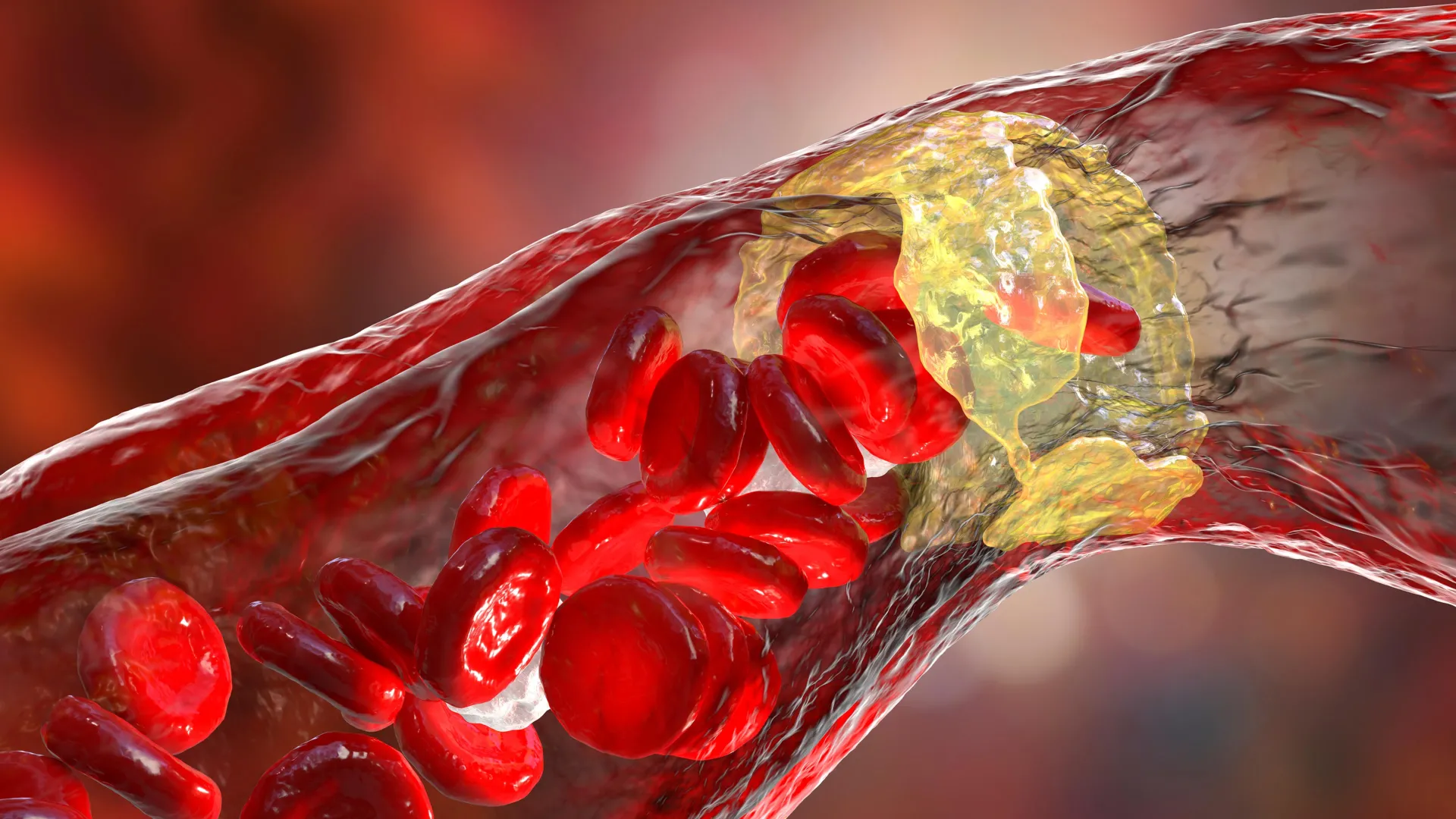
A research team at the University of California, Riverside has found that routine exposure to microplastics — tiny pieces released from packaging, fabrics, and common consumer plastics — may speed up the formation of atherosclerosis, the artery-narrowing condition associated with heart attacks and strokes. The effect appeared only in male mice, offering new insight into how microplastics may influence cardiovascular health in people.
“Our findings fit into a broader pattern seen in cardiovascular research, where males and females often respond differently,” said lead researcher Changcheng Zhou, a professor of biomedical sciences in the UCR School of Medicine. “Although the precise mechanism isn’t yet known, factors like sex chromosomes and hormones, particularly the protective effects of estrogen, may play a role.”
Microplastics Found Throughout the Environment and the Body
Microplastics are found widely in the modern environment, including in food, drinking water, and the air. They have also been detected inside the human body. Recent clinical studies have identified microplastics in atherosclerotic plaques and associated higher concentrations with elevated cardiovascular risk, although it was not clear whether these particles directly cause arterial injury.
“It’s nearly impossible to avoid microplastics completely,” Zhou said. “Still, the best strategy is to reduce exposure by limiting plastic use in food and water containers, reducing single-use plastics, and avoiding highly-processed foods. There are currently no effective ways to remove microplastics from the body, so minimizing exposure and maintaining overall cardiovascular health — through diet, exercise, and managing risk factors — remains essential.”
Study Design Using a Heart Disease Mouse Model
In their paper published in Environment International, Zhou and colleagues describe their use of LDLR-deficient mice, a common model for examining atherosclerosis. Both male and female mice were placed on a low-fat, low-cholesterol diet similar to what a lean and healthy person might eat.
The team then administered microplastics daily (10 milligrams per kilogram of body weight) for nine weeks. This amount reflects levels that could realistically be encountered through contaminated food and water.
Microplastics Intensify Plaque Formation in Male Mice
The results showed a sharp increase in atherosclerosis, but only in males. Male mice exposed to microplastics developed 63% more plaque in the aortic root, the segment of the aorta connected to the heart, and 624% more plaque in the brachiocephalic artery, a major vessel branching from the aorta in the upper chest. Female mice exposed to the same conditions did not show significant plaque progression.
The researchers confirmed that microplastics did not cause weight gain or increased cholesterol in either sex. The mice stayed lean, and their lipid profiles remained unchanged, indicating that traditional risk factors such as obesity or high cholesterol did not explain the heightened arterial damage.
Disruption of Artery-Lining Cells
The study also showed that microplastics interfered with the function and makeup of cells lining the arteries. Using single-cell RNA sequencing, which identifies gene activity in individual cells, the researchers observed that microplastics altered several cell types involved in atherosclerosis. Endothelial cells — the cells that form the inner lining of blood vessels and help regulate inflammation and circulation — were affected the most.
“We found endothelial cells were the most affected by microplastic exposure,” Zhou said. “Since endothelial cells are the first to encounter circulating microplastics, their dysfunction can initiate inflammation and plaque formation.”
Microplastics Enter Arterial Plaques and Alter Gene Activity
Fluorescent microplastics used in the study were found inside plaques and concentrated within the endothelial layer, consistent with reports from human samples that have revealed microplastics in arterial lesions.
Another key observation was that microplastics activated harmful gene pathways in endothelial cells from both mice and humans. This included genes associated with pro-atherogenic (plaque-promoting) activity, suggesting that microplastics trigger similar biological responses across species.
“Our study provides some of the strongest evidence so far that microplastics may directly contribute to cardiovascular disease, not just correlate with it,” Zhou said. “The surprising sex-specific effect — harming males but not females — could help researchers uncover protective factors or mechanisms that differ between men and women.”
Future Research on Sex Differences and Microplastic Types
Zhou and his team emphasize that more work is needed to determine why males appear more susceptible. The group plans to investigate whether humans show similar patterns.
“We would like to investigate how different types or sizes of microplastics affect vascular cells,” Zhou said. “We will also look into the molecular mechanisms behind endothelial dysfunction and explore how microplastics affect male and female arteries differently. As microplastic pollution continues to rise worldwide, understanding its impacts on human health — including heart disease — is becoming more urgent than ever.”
Zhou conducted the study with collaborators from UCR, Boston Children’s Hospital and Harvard Medical School in Massachusetts, and the University of New Mexico Health Sciences.
The work received partial support from the National Institutes of Health.
The title of the paper is “Microplastic exposure elicits sex-specific atherosclerosis development in lean low-density lipoprotein receptor-deficient mice.”
The Princess of Wales tells business leaders about the importance of supporting families with young children.
The Foreign Office list now includes India, Morocco and Nepal following a “global increase” in reported cases.
