A gene from 100-year-olds could help kids who age too fast

Scientists have uncovered a breakthrough in the fight against a rare genetic condition that causes children to age much faster than normal. The discovery involves “longevity genes” found in people who live exceptionally long lives, often beyond 100 years. Researchers from the University of Bristol and IRCCS MultiMedica found that these genes, which help maintain the health of the heart and blood vessels during aging, could reverse some of the damage caused by this devastating disease.

The study, published in Signal Transduction and Targeted Therapy, is the first to show that a gene from long-lived individuals can slow down heart aging in a model of Progeria. Known scientifically as Hutchinson-Gilford Progeria Syndrome (HGPS), this rare and fatal disorder causes children to exhibit signs of “accelerated aging.”

Progeria stems from a mutation in the LMNA gene, which leads to the creation of a harmful protein called progerin. This protein disrupts normal cell function, particularly in the heart and blood vessels. Most affected children die in their teenage years from heart complications, though some, like Sammy Basso — the oldest known person with Progeria — live longer. Sammy passed away on October 24, 2024, at the age of 28.

Progerin harms cells by destabilizing their nucleus, the “control center” that manages cell activity. This damage accelerates aging, especially in the cardiovascular system.

At present, the only drug approved by the United States Food and Drug Administration (FDA) is lonafarnib, which reduces the accumulation of progerin. Researchers are now testing a combination of lonafarnib with another experimental drug, Progerinin, to determine whether the two work better together.

Testing Longevity Genes from Supercentenarians

To explore new treatments, Dr. Yan Qiu and Professor Paolo Madeddu of the Bristol Heart Institute collaborated with Professor Annibale Puca’s team at IRCCS MultiMedica in Italy. Their goal was to determine if genes from people who live to extreme old age — supercentenarians — could protect against the cellular damage caused by Progeria.

The scientists focused on one particular gene, LAV-BPIFB4, which previous research has shown supports healthy heart and blood vessel function during aging.

Using genetically engineered mice that develop Progeria, the researchers observed early heart problems similar to those found in children with the disease. After a single injection of the LAV-BPIFB4 longevity gene, the mice showed improved heart function, particularly in the way the heart relaxes and fills with blood (a process known as diastolic function).

The gene treatment also reduced heart tissue damage, known as fibrosis, and lowered the number of “aged” cells in the heart. In addition, it promoted the growth of new small blood vessels, potentially improving heart health and resilience.

The team then tested the gene on human cells derived from Progeria patients. These experiments revealed that introducing the longevity gene reduced cellular aging and fibrosis without altering progerin levels directly. This suggests that the gene helps cells withstand the toxic effects of progerin rather than eliminating it. The approach strengthens the body’s natural defenses instead of attacking the defective protein itself.

A New Approach to Treating Progeria and Heart Aging

Dr. Yan Qiu, Honorary Research Fellow in the Bristol Heart Institute at the University of Bristol, said: “Our research has identified a protective effect of a “supercentenarian longevity gene” against progeria heart dysfunction in both animal and cell models.

“The results offer hope to a new type of therapy for Progeria; one based on the natural biology of healthy aging rather than blocking the faulty protein. This approach, in time, could also help fight normal age-related heart disease.

“Our research brings new hope in the fight against Progeria and suggests the genetics of supercentenarians could lead to new treatments for premature or accelerated cardiac aging, which might help us all live longer, healthier lives.”

Looking Ahead: Toward New Anti-Aging Therapies

Professor Annibale Puca, Research Group Leader at IRCCS MultiMedica and Dean of the Faculty of Medicine at the University of Salerno, added: “This is the first study to indicate that a longevity-associated gene can counteract the cardiovascular damage caused by progeria.

“The results pave the way for new treatment strategies for this rare disease, which urgently requires innovative cardiovascular drugs capable of improving both long-term survival and patient quality of life. Looking ahead, the administration of the LAV-BPIFB4 gene through gene therapy could be replaced and/or complemented by new protein- or RNA-based delivery methods.

“We are currently conducting numerous studies to investigate the potential of LAV-BPIFB4 in counteracting the deterioration of the cardiovascular and immune systems in various pathological conditions, with the goal of translating these experimental findings into a new biologic drug.”

Share Button

Private baby scan clinics ‘putting expectant mothers at risk’

Some high-street clinics are risking lives by letting untrained staff do baby scans, warn experts.

Share Button

A prehistoric battle just rewrote T. rex’s story

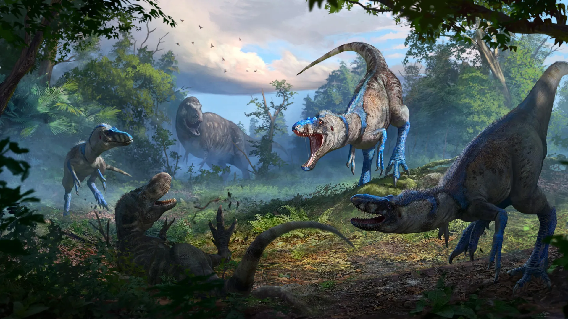
Could everything we thought we knew about T. rex growth be wrong? A remarkably complete tyrannosaur skeleton has brought new clarity to one of paleontology’s longest debates: whether Nanotyrannus was its own species or merely a young Tyrannosaurus rex.

The fossil comes from the famous “Dueling Dinosaurs” discovery in Montana, which preserved two creatures locked in ancient combat — a Triceratops and a smaller-bodied tyrannosaur. After extensive analysis, scientists have confirmed that the smaller predator was not a juvenile T. rex, but an adult Nanotyrannus lancensis.

“This fossil doesn’t just settle the debate. It flips decades of T. rex research on its head,” says Lindsay Zanno, associate research professor at North Carolina State University and head of paleontology at the North Carolina Museum of Natural Sciences. Zanno co-authored the study recently published in Nature.

Evidence Points to a Fully Mature Predator

By studying bone growth rings, spinal fusion, and developmental anatomy, researchers determined that the animal was roughly 20 years old when it died — well into adulthood. Distinct traits, including longer arms, a greater number of teeth, fewer tail vertebrae, and unique skull nerve structures, all appeared early in development and are biologically inconsistent with T. rex.

“For Nanotyrannus to be a juvenile T. rex, it would need to defy everything we know about vertebrate growth,” explains James Napoli, an anatomist at Stony Brook University and co-author of the research. “It’s not just unlikely — it’s impossible.”

The findings carry major implications. For years, scientists relied on Nanotyrannus fossils to understand T. rex growth, behavior, and ecology. This new study reveals that such comparisons were mistaken — those bones belonged to two separate species. It also indicates that several kinds of tyrannosaurs may have lived side by side during the final million years before the asteroid impact that ended the age of dinosaurs.

A Hidden Species Revealed

Zanno and Napoli reviewed more than 200 tyrannosaur fossils during their research. One specimen, long thought to be a teenage T. rex, turned out to differ slightly from Nanotyrannus lancensis. The team designated it a new species, Nanotyrannus lethaeus, referencing the River Lethe from Greek mythology — a fitting tribute to a species “forgotten” for decades.

Recognizing Nanotyrannus as a valid genus reshapes our picture of the late Cretaceous ecosystem. Predator diversity during that time appears to have been far greater than scientists once assumed, suggesting that other small dinosaur species might also have been misclassified.

“This discovery paints a richer, more competitive picture of the last days of the dinosaurs,” says Zanno. “With enormous size, a powerful bite force and stereoscopic vision, T. rex was a formidable predator, but it did not reign uncontested. Darting alongside was Nanotyrannus — a leaner, swifter and more agile hunter.”

The research, published in Nature, was supported by the State of North Carolina, NC State University, the Friends of the North Carolina Museum of Natural Sciences, and the Dueling Dinosaurs Capital Campaign.

Share Button

After 25 years, scientists solve the bird-eating bat mystery

After nearly a quarter century of investigation, scientists have solved a remarkable mystery. Europe’s largest bat doesn’t merely snack on small birds — it hunts and captures them more than a kilometer above the ground and consumes them while still in flight.

An international research team has uncovered how this massive bat hunts and eats its prey. Their findings, published in Science, reveal an astonishing story of night-time aerial chases, precision attacks, and predation in total darkness.

Each year, billions of songbirds migrate between breeding and wintering grounds. Many travel at night and at high altitudes to avoid daytime predators. Yet flying in darkness comes with its own dangers, as bats rule the night skies.

Riding on the Bats’ Backs

To understand these elusive hunters, scientists effectively “rode along” with Europe’s largest bat — the greater noctule (Nyctalus lasiopterus) — by fitting individuals with tiny “backpacks” containing biologgers developed at Aarhus University. These lightweight devices measured the bats’ altitude, acceleration, movement, and sounds (including their echolocation calls), providing an unprecedented look at their nocturnal hunting strategies more than a kilometer above ground.

The data revealed that the bats soar high into the night sky to find and ambush unsuspecting birds. Unlike insects, birds cannot detect the bats’ ultrasonic calls and only realize the danger moments before being caught.

Their success depends on powerful, low-frequency echolocation calls that can detect birds at long distances. When they close in on a target, the bats unleash rapid bursts of short calls, signaling the final stage of attack.

Daring Dives

Information from the biologgers showed that the bats plunge toward their prey in steep, high-speed dives reminiscent of fighter jets in combat.

In two documented chases, the bats dove for 30 and 176 seconds respectively, flapping harder, tripling their acceleration, and continuously emitting attack calls.

The first bat eventually abandoned its pursuit — birds are agile aerialists too — but the second succeeded after a nearly three-minute chase, capturing a robin near the ground.

Microphones recorded 21 distress calls from the robin, followed by 23 minutes of chewing as the bat flew low, feeding on the wing.

Combined with X-ray and DNA analysis of bird wings found beneath hunting areas, these results confirm what happens next: the bat kills the bird with a bite, removes its wings (likely to reduce drag), and then uses the membrane between its hind legs as a pouch to hold and eat the prey while still airborne.

Wild Maneuvers

“We know that songbirds perform wild evasive maneuvers such as loops and spirals to escape predators like hawks during the day — and they seem to use the same tactics against bats at night. It’s fascinating that bats are not only able to catch them, but also to kill and eat them while flying. A bird like that weighs about half as much as the bat itself — it would be like me catching and eating a 35-kilo animal while jogging,” explains Assistant Professor Laura Stidsholt from the Department of Biology at Aarhus University.

Stidsholt, a lead author of the study, has spent years perfecting biologger technology in bat research, resulting in numerous discoveries. When she completed the data collection and analysis for this project, she was a Postdoc at the Leibniz Institute for Zoo and Wildlife Research (Leibniz-IZW) in Berlin.

A 25-Year Hypothesis Confirmed

For decades, scientists suspected that some large bat species prey on small birds during flight. Much of that work originated from Spanish bat expert Carlos Ibáñez and colleagues at the Doñana Biological Station (CSIC) in Seville.

Nearly 25 years ago, Ibáñez found bird feathers in greater noctule droppings and spent years gathering evidence that these bats were indeed bird predators.

His team has closely monitored this elusive forest-dwelling species using “smart” roosts equipped with antennas to detect implanted microchips in the bats. The system tracks movements, stores data, and sends alerts to researchers’ phones in real time.

Despite the evidence, the idea that bats could catch birds midair was met with skepticism, as birds can weigh nearly half as much as the bats themselves.

Filming these hunts proved impossible in the dark. Over the years, researchers experimented with roost cameras, military radar, hot-air balloons with ultrasound recorders, and GPS trackers — struggling to create tools light enough for the bats to carry.

Finally, with new miniature biologgers from Aarhus University — and just as Ibáñez neared retirement — the team succeeded in recording a greater noctule hunting and eating a bird in flight.

Essential for Bat Conservation

For co-author Elena Tena, hearing the recording was both thrilling and sobering:

“While it evokes empathy for the prey, it is part of nature. We knew we had documented something extraordinary. For the team, it confirmed what we had been seeking for so long. I had to listen to it several times to fully grasp what we had recorded.”

Fortunately, these bats pose no threat to songbird populations. The greater noctule is extremely rare and endangered in many regions due to the loss of forest habitats.

Understanding its behavior and ecology is now vital for developing conservation and management strategies that can help protect one of Europe’s most extraordinary nocturnal predators.

Share Button

Thousands of poorest Scots to receive free weight-loss jabs in trial

Up to 5,000 people in Scotland will take the injections as part of a multi-million pound study being led by Glasgow University.

Share Button

Resetting the body’s rhythm could protect the brain from Alzheimer’s

Disrupting communication between the body’s internal clock and the brain could help limit neurodegeneration in Alzheimer’s disease, according to new research from Washington University School of Medicine in St. Louis (WashU Medicine). The study, published in Nature Aging, explored how changes in the circadian system affect brain health and memory in mouse models of Alzheimer’s disease.

Led by Erik Musiek, MD, PhD, the Charlotte & Paul Hagemann Professor of Neurology at WashU Medicine, and first author Jiyeon Lee, PhD, the research team investigated whether blocking a specific circadian clock protein might slow the progression of neurodegeneration. They found that inhibiting the activity of this protein lowered levels of tau, a toxic protein linked to Alzheimer’s pathology, and reduced damage to brain tissue.

REV-ERBα, NAD+, and Brain Aging

The circadian protein under investigation, called REV-ERBα, helps regulate the body’s daily rhythms of metabolism and inflammation. While its role in the brain has been less understood, earlier studies in other tissues showed that REV-ERBα influences levels of nicotinamide adenine dinucleotide (NAD+), a molecule vital for metabolism, energy production, and DNA repair. Declining NAD+ levels are closely associated with brain aging and neurodegenerative conditions. Many over-the-counter supplements aim to raise NAD+ as a strategy to slow aging and promote cellular health.

To test REV-ERBα’s role, the team genetically deleted the protein in two groups of mice: one in which the deletion occurred throughout the body, and another where it was removed only in astrocytes (supportive glial cells that form a major part of the central nervous system). In both cases, NAD+ levels rose significantly. The results suggest that eliminating REV-ERBα in astrocytes directly boosts NAD+ in the brain, pointing to a potential path for future treatments targeting neurodegeneration.

Drug Treatment Protects Against Tau Pathology

In a further experiment, the researchers blocked REV-ERBα using both genetic methods and a new drug that has also shown promise in studies of amyloid-β and Parkinson’s disease. This approach increased NAD+ levels and shielded the mice from tau-related brain damage. Tau aggregates are known to disrupt brain function and drive neurodegenerative diseases such as Alzheimer’s.

The findings suggest that manipulating the body’s internal clock — specifically by inhibiting REV-ERBα — could represent a new way to protect the brain, prevent tau buildup, and potentially slow or halt the progression of Alzheimer’s disease.

Share Button

Scientists just shattered a major exercise myth

Forget the old idea that physical activity drains your supply of heartbeats. New research from Australia shows that people who are physically fit actually use fewer heartbeats each day, which may help them live longer.

According to the study, athletes had an average heart rate of 68 beats per minute (bpm), while non-athletes averaged 76 bpm. Over a 24-hour period, this equals roughly 97,920 beats for athletes compared to 109,440 for non-athletes — about 10 percent fewer.

“That’s an incredible saving of about 11,500 beats a day,” says Professor La Gerche, head of the HEART Laboratory supported by the St Vincent’s Institute of Medical Research (SVI) and the Victor Chang Cardiac Research Institute (VCCRI).

He adds, “Even though athletes’ hearts work harder during exercise, their lower resting rates more than make up for it.”

Exercise Efficiency and Heart Health

The study, published in JACC: Advances, found that the most physically fit individuals had resting heart rates as low as 40 bpm, compared to the typical 70-80 bpm range. This means that, despite higher peaks during training, athletes still end up with fewer total beats across the day than those who are inactive.

These results challenge a long-held belief, once repeated by US President Donald Trump, that the human body is like a battery with a limited energy supply and that exercise only depletes it.

“The fitter you are, the more metabolically efficient your body becomes,” Professor La Gerche explains. “Even if you’re training hard for an hour a day, your heart beats more slowly for the other 23 hours. The net effect is fewer beats used overall.”

A slower resting heart rate is not only a sign of good physical condition but also a strong indicator of long-term health. Increasing physical activity, when done safely, can enhance heart performance and reduce the risk of cardiovascular disease.

“Exercise is strongly linked with improved mental health, longer lifespan and lower rates of heart disease,” Professor La Gerche says.

Moderate Exercise Delivers the Biggest Benefits

Although extreme endurance events such as the Tour de France can cause temporary increases in daily heartbeats, Professor La Gerche notes that the health benefits of consistent, moderate exercise far outweigh any risks.

“The biggest bang for your health buck is going from unfit to moderately fit. Just a few hours of purposeful exercise each week can transform your heart’s efficiency and help make every beat count. It may even extend your life by years,” he says.

About the HEART Lab

Professor La Gerche leads the Heart, Exercise & Research Trials (HEART) Lab, which investigates how exercise influences heart health, in partnership with SVI and VCCRI. By studying elite athletes, the team uncovers findings that reach well beyond sports performance, offering valuable insights for people living with serious heart conditions.

Share Button

Too much screen time may be hurting kids’ hearts

  • More time using electronic devices or watching TV among children and young adults was linked with higher cardiometabolic disease risk, including high blood pressure, high cholesterol and insulin resistance, based on data from more than 1,000 participants in Denmark.
  • The association between screen time and cardiometabolic risks was strongest in youth who slept fewer hours, suggesting that screen use may harm health by “stealing” time from sleep, researchers said.
  • Researchers said the findings underscore the importance of addressing screen habits among young people as a potential way to protect long-term heart and metabolic health.

Screen time tied to early heart and metabolic risks

Children and teens who spend many hours on TVs, phones, tablets, computers or gaming systems appear to face higher chances of cardiometabolic problems, such as elevated blood pressure, unfavorable cholesterol levels and insulin resistance. The findings are reported in the Journal of the American Heart Association, an open-access, peer-reviewed journal of the American Heart Association.

A 2023 scientific statement from the American Heart Association reported that “cardiometabolic risk is accruing at younger and younger ages,” and that only 29% of U.S. youth ages 2 to 19 had favorable cardiometabolic health in 2013-2018 National Health and Nutrition Examination Survey data.

Danish cohorts show a consistent pattern

An evaluation of more than 1,000 participants from two Danish studies found a clear connection: more recreational screen time was significantly associated with greater cardiovascular and overall cardiometabolic risk among children and adolescents.

“Limiting discretionary screen time in childhood and adolescence may protect long-term heart and metabolic health,” said study lead author David Horner, M.D., PhD., a researcher at the Copenhagen Prospective Studies on Asthma in Childhood (COPSAC) at the University of Copenhagen in Denmark. “Our study provides evidence that this connection starts early and highlights the importance of having balanced daily routines.”

What researchers measured

The team analyzed two COPSAC groups: one of 10-year-olds followed in 2010 and one of 18-year-olds followed in 2000. They examined how leisure screen use related to cardiometabolic risk factors. Screen time included watching TV and movies, gaming and time on phones, tablets or computers for fun.

To capture overall risk, researchers created a composite cardiometabolic score based on multiple components of metabolic syndrome, including waist size, blood pressure, high-density lipoprotein or HDL “good” cholesterol, triglycerides and blood sugar levels. They adjusted for sex and age. The score reflects each participant’s risk relative to the study average (in standard deviations): 0 indicates average risk, and 1 indicates one standard deviation above average.

Each hour adds up

The analysis showed that every additional hour of recreational screen time was linked with an increase of about 0.08 standard deviations in the cardiometabolic score for the 10-year-olds and 0.13 standard deviations for the 18-year-olds. “This means a child with three extra hours of screen time a day would have roughly a quarter to half a standard-deviation higher risk than their peers,” Horner said.

“It’s a small change per hour, but when screen time accumulates to three, five or even six hours a day, as we saw in many adolescents, that adds up,” he said. “Multiply that across a whole population of children, and you’re looking at a meaningful shift in early cardiometabolic risk that could carry into adulthood.”

Sleep appears to intensify the risk

Short sleep and later bedtimes strengthened the relationship between screen time and cardiometabolic risk. Youth who slept less showed notably higher risk linked to the same amount of screen exposure.

“In childhood, sleep duration not only moderated this relationship but also partially explained it: about 12% of the association between screen time and cardiometabolic risk was mediated through shorter sleep duration,” Horner said. “These findings suggest that insufficient sleep may not only magnify the impact of screen time but could be a key pathway linking screen habits to early metabolic changes.”

Metabolic “fingerprint” linked to screen use

In a machine learning analysis, investigators identified a distinctive pattern of blood metabolites that appeared to correlate with screen time.

“We were able to detect a set of blood-metabolite changes, a ‘screen-time fingerprint,’ validating the potential biological impact of the screen time behavior,” he said. “Using the same metabolomics data, we also assessed whether screen time was linked to predicted cardiovascular risk in adulthood, finding a positive trend in childhood and a significant association in adolescence. This suggests that screen-related metabolic changes may carry early signals of long-term heart health risk.

“Recognizing and discussing screen habits during pediatric appointments could become part of broader lifestyle counseling, much like diet or physical activity,” he said. “These results also open the door to using metabolomic signatures as early objective markers of lifestyle risk.”

Practical guidance from experts

Amanda Marma Perak, M.D., M.S.CI., FAHA, chair of the American Heart Association’s Young Hearts Cardiovascular Disease Prevention Committee, who was not involved in this research, said focusing on sleep is a great starting point to change screen time patterns.

“If cutting back on screen time feels difficult, start by moving screentime earlier and focusing on getting into bed earlier and for longer,” said Perak, an assistant professor of pediatrics and preventive medicine at Northwestern University Feinberg School of Medicine in Chicago.

Adults can also set an example, she said. “All of us use screens, so it’s important to guide kids, teens and young adults to healthy screen use in a way that grows with them. As a parent, you can model healthy screen use — when to put it away, how to use it, how to avoid multitasking. And as kids get a little older, be more explicit, narrating why you put away your devices during dinner or other times together.

“Make sure they know how to entertain and soothe themselves without a screen and can handle being bored! Boredom breeds brilliance and creativity, so don’t be bothered when your kids complain they’re bored. Loneliness and discomfort will happen throughout life, so those are opportunities to support and mentor your kids in healthy ways to respond that don’t involve scrolling.”

Important caveats and next questions

Because this work is observational, it reveals associations rather than direct cause and effect. In addition, screen use for the 10-year-olds and 18-year-olds was reported by parents through questionnaires, which may not perfectly reflect actual time spent on screens.

Horner noted that future studies could test whether reducing screen exposure in the hours before bedtime, when screen light may disrupt circadian rhythms and delay sleep onset, helps lower cardiometabolic risk.

Study details, background and design

  • The two prospective research groups at COPSAC in Denmark consisted of mother-child pairs, with analysis of data collected at planned clinical visits and study assessments from the birth of the children through age 10 in the 2010 study group and age 18 in the 2000 study group.
  • Through questionnaires, parents of children in the 10-year-old group and 18-year-olds detailed the number of hours the young participants spent watching TV or movies, gaming on a console/TV and using phones, tablets or computers for leisure.
  • For the 2010 group, the number of hours of screen time was available for 657 children at age 6 and 630 children at age 10. Average screen time was two hours per day at age 6, and 3.2 hours per day at age 10, representing a significant increase over time.
  • For the 2000 group of 18-year-olds, screen time was available for 364 individuals. Screen time at 18 years was significantly higher at an average of 6.1 hours per day.
  • Sleep was measured by sensors over a 14-day period.
Share Button

2 million-year-old teeth reveal secrets from the dawn of humanity

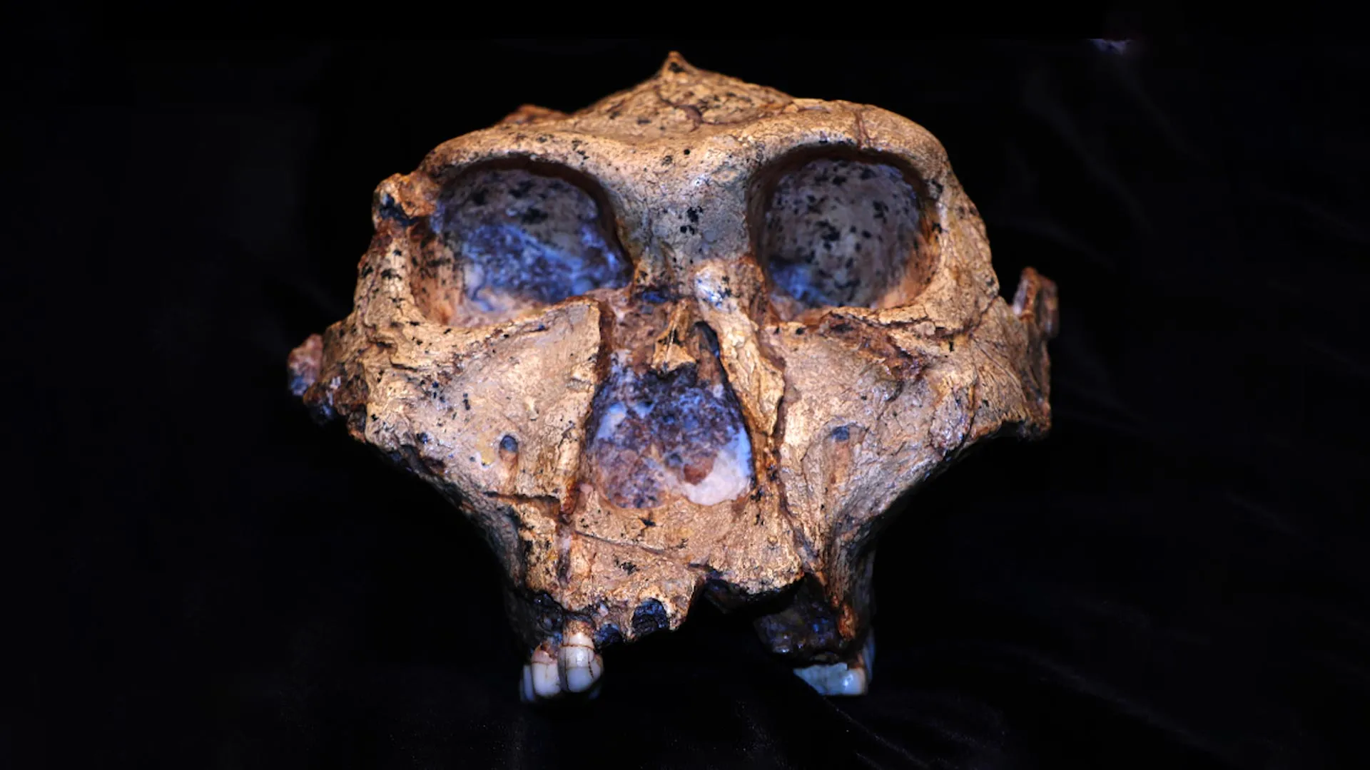
For nearly 100 years, scientists have been intrigued by the fossils of Paranthropus robustus, a sturdy, distant cousin of early humans. This upright-walking hominin was built for chewing tough foods, equipped with powerful jaws and large teeth coated in thick enamel. Researchers believe it lived in southern Africa between 2.25 million and 1.7 million years ago.

South Africa’s fossil record preserves an extraordinary range of early human relatives. These include Australopithecus prometheus, A. africanus (best known for the Taung child), A. sediba, and P. robustus. Later species such as Homo habilis, Homo erectus/ergaster, Homo naledi, and ultimately Homo sapiens (modern humans) also appear in the record.

These fossils reveal key milestones in our evolutionary journey — from walking on two legs to crafting tools and developing larger brains. Over time, this lineage led to the emergence of Homo sapiens in South Africa roughly 153,000 years ago.

Decades of Questions

When P. robustus fossils were first unearthed in 1938, they raised questions that persisted for generations. How much variation existed within this species? Were size differences linked to biological sex or evidence of multiple species? What genetic traits set P. robustus apart from other early hominins and the first members of the Homo genus?

Researchers have long sought genetic clues to answer these questions, but ancient DNA does not survive well in Africa’s warm climate. To overcome this limitation, a team of African and European scientists turned to a different method: paleoproteomics, the study of ancient proteins.

They successfully extracted proteins from the enamel of four P. robustus teeth found at Swartkrans Cave, part of South Africa’s Cradle of Humankind. Unlike DNA, proteins can survive for millions of years because they bond tightly to bone and enamel, making them more resistant to heat and decay.

One of these ancient proteins revealed the sex of the fossils, showing that two of the individuals were male and two were female.

The Oldest Genetic Clues from Africa

This work marks a major step in human origins research. It provides some of the oldest molecular data ever recovered from Africa, allowing scientists to reexamine how early human relatives varied and how they might have been related to one another — or even represented multiple species.

The protein sequences also revealed intriguing genetic differences. One gene, responsible for producing enamelin (a key enamel-forming protein), varied among the specimens. Two fossils shared an amino acid sequence found in humans, chimpanzees, and gorillas, while the others had a version so far unique to Paranthropus.

Even more remarkably, one fossil carried both variants of the amino acid. This provided the first-ever evidence of heterozygosity — two versions of a gene — preserved in proteins that are 2 million years old.

A More Complex Family Tree

Mutations in protein sequences can signal evolutionary divergence. What appeared at first to be a mutation exclusive to P. robustus turned out to vary among individuals of the same group. This suggests that P. robustus may not have been a single uniform species, but a mix of populations with different ancestries.

By combining molecular data with physical anatomy (morphology), scientists can now create a more detailed picture of early human relationships. Future work will involve analyzing enamel proteins from additional P. robustus fossils found at other South African sites to test these findings.

Protecting Africa’s Fossil Legacy

The research team took great care to preserve these irreplaceable fossils, following strict South African regulations and minimizing the amount of material sampled. Local laboratories and African researchers played central roles throughout the project, ensuring that both the science and the benefits remained closely tied to the continent’s heritage.

Conducting advanced molecular research in Africa on African fossils represents an important move toward transforming and decolonizing the field of paleontology. It strengthens local expertise, fosters equitable collaboration, and ensures discoveries continue to enrich the regions where they originate.

A New Blueprint for Human Origins Research

By merging molecular and morphological data, this study offers a new model for exploring ancient diversity among early hominins. The findings suggest our ancient family tree was more complex than previously thought — and potentially far richer.

As techniques in paleoproteomics continue to advance, researchers expect more revelations about the distant ancestors who shaped the human story. For now, the Paranthropus robustus mystery has grown deeper, more intricate, and infinitely more fascinating.

(Jesper V. Olsen, Rebecca R. Ackermann and Enrico Cappellini were also the principal investigators on this project.)

Written by Palesa P. Madupe, Claire Koenig, and Ioannis Patramanis from the University of Copenhagen.

Share Button

At 21, I was crushed by a stranger’s joke about going bald. Then the way I looked at myself changed

There are multiple ways for men to combat hair loss now, but should they have to?

Share Button