Dolphins may be getting Alzheimer’s from toxic ocean blooms

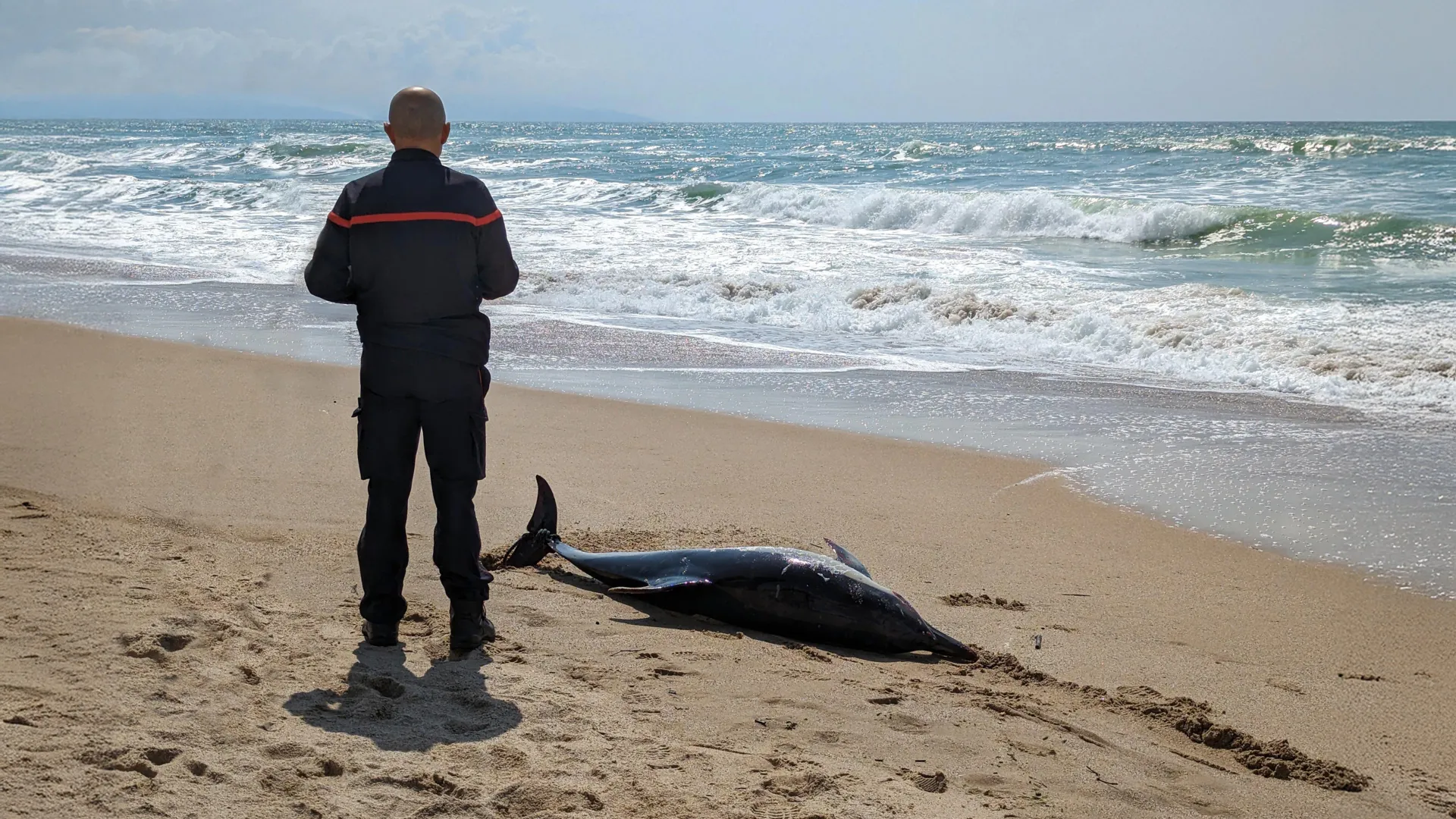
For many nature enthusiasts, few scenes are as distressing as finding a stranded whale or dolphin lying helpless on the beach. When these animals are still alive, marine biologists and volunteers rush to assist, shielding them from the sun and preventing their skin from drying out by pouring seawater over them or draping them with wet towels. Others work tirelessly to guide the animal back to deeper waters once the tide returns.

Tragically, not every rescue attempt comes in time. Some dolphins and whales are discovered already dead, leaving behind a haunting mystery that has puzzled scientists for decades: what drives these intelligent creatures to beach themselves in the first place?

A team of researchers from Florida to Wyoming believes they may have uncovered a surprising explanation. They suggest that, much like humans with dementia who sometimes wander away from familiar places, dolphins might also become disoriented due to a form of Alzheimer’s-like disease. Their findings point to a connection between this disorientation and long-term exposure to harmful compounds produced by cyanobacteria — microscopic organisms that thrive in warm, nutrient-rich waters.

Research involving residents of Guam has shown that people who regularly consume foods containing cyanobacterial toxins are more likely to develop the same brain abnormalities seen in Alzheimer’s disease, including misfolded tau proteins and amyloid plaques. One of the most concerning of these toxins is β-N-methylamino-L-alanine (BMAA), along with its chemical relatives 2,4-Diaminobutyric acid (2,4-DAB) and N-2-aminoethylglycine (AEG). These compounds are known to be highly toxic to nerve cells. Experiments in animals demonstrate that BMAA exposure can lead to Alzheimer’s-like brain damage and cognitive decline. Once released into marine ecosystems, these toxins can build up in the food chain, eventually reaching top predators such as dolphins.

When researchers examined the brains of twenty bottlenose dolphins that had stranded along Florida’s Indian River Lagoon, they found significant levels of BMAA and its related toxins, especially 2,4-DAB. Dolphins that washed ashore during peak cyanobacterial bloom periods contained up to 2,900 times more 2,4-DAB than those stranded at other times of the year. Their brains showed many of the same pathological features seen in humans with Alzheimer’s disease, including β-amyloid plaques and hyperphosphorylated tau proteins. The scientists also identified TDP-43 protein inclusions, a marker associated with more aggressive forms of Alzheimer’s, along with 536 genes expressed in patterns consistent with the disease.

The duration of cyanobacterial blooms is increasing with climate warming and nutrient inputs associated with agricultural runoff and sewage discharges. Cyanobacterial-laden waters have often been released down the St. Lucie River from Lake Okeechobee into the Indian River Lagoon. “Since dolphins are considered environmental sentinels for toxic exposures in marine environments,” Dr. David Davis at the Miller School of Medicine explains, “there are concerns about human health issues associated with cyanobacterial blooms.”

In 2024, Miami Dade County had the highest prevalence of Alzheimer’s disease in the United States. “Although there are likely many paths to Alzheimer’s disease, cyanobacterial exposures increasingly appear to be a risk factor,” adds Dr. Davis.

“Among Guam villagers, exposure to cyanobacterial toxins appeared to trigger neurological disease,” said Dr. Paul Alan Cox of the Brain Chemistry Labs in Jackson Hole.

This research was published in the current Nature journal Communication Biology by researchers at Hubbs-SeaWorld Research Institute in Melbourne Beach, Florida, The Blue World Research Institute in Cocoa, Florida, The University of Miami Miller School of Medicine, Miami, Florida, Brain Chemistry Labs, Jackson Hole, Wyoming, Rosenstiel School of Marine, Atmospheric, and Earth Science, University of Miami, Miami, Florida.

Share Button

‘We are inventing captivity medicine’: Hospital prepares for freed Israeli hostages

Several of the hostages will be brought from Gaza to the Rabin Medical Center in Petah Tikva.

Share Button

Scientists unearth a 112-million-year-old time capsule filled with ancient insects

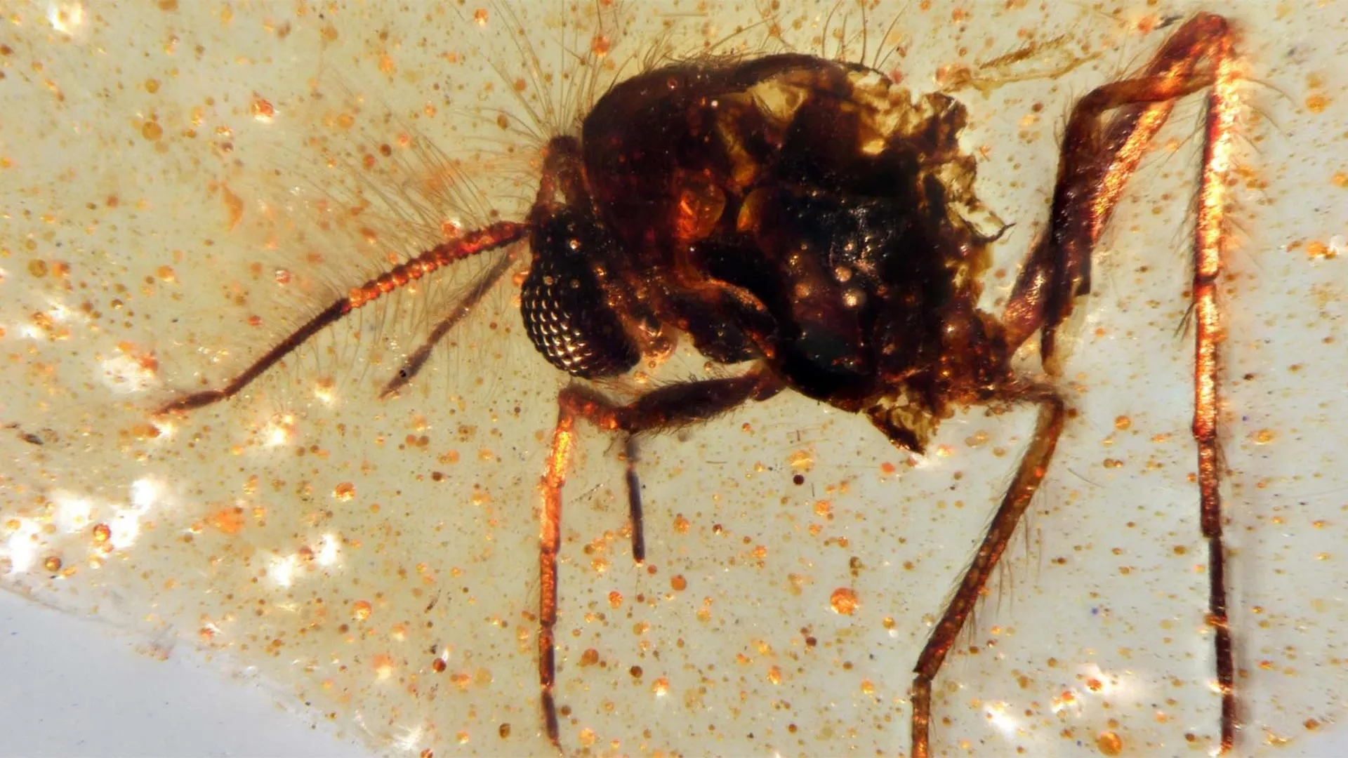
Scientists have uncovered the first South American amber deposits containing preserved insects in a quarry in Ecuador, according to a study published in Communications Earth & Environment. The discovery captures a vivid picture of a 112-million-year-old forest that once thrived on the ancient supercontinent Gondwana and opens new doors for exploring a long-overlooked prehistoric ecosystem.

Amber (fossilized tree resin) has been found in samples dating back as far as 320 million years, but it became far more common between 120 million and 70 million years ago, during the Cretaceous era (143.1 million to 66 million years ago). These ancient resins sometimes contain “bio-inclusions” — trapped remains of plants or animals — that offer rare, detailed glimpses of life forms such as insects and flowers that are not usually preserved as fossils. Until now, nearly all known major amber deposits were located in the Northern Hemisphere, leaving scientists with limited insight into what Southern Hemisphere ecosystems looked like during the time when the continents were beginning to separate from Gondwana.

To investigate, Xavier Delclòs and his research team examined amber and surrounding rock samples collected from the Genoveva quarry in Ecuador. The amber, dated to roughly 112 million years ago, belongs to the Hollín Formation, a sedimentary layer that stretches across Ecuador’s Oriente Basin. The team identified two distinct kinds of amber: one formed underground near the roots of resin-producing plants, and another that developed in the open air. Among 60 samples of the latter, the researchers found 21 bio-inclusions representing five insect orders, including Diptera (flies), Coleoptera (beetles), and Hymenoptera (a group that includes ants and wasps), as well as a fragment of spider web. In addition, the rock surrounding the amber contained numerous plant fossils such as spores, pollen, and other botanical traces.

According to the researchers, the characteristics of the fossils indicate that the amber originated in a warm, humid forest filled with dense vegetation and resin-producing trees in southern Gondwana. They emphasize that this rare find provides a crucial new resource for understanding life and biodiversity during this key period in Earth’s history.

Share Button

Report shows ‘stark’ gender gap in HIV prevention

A meeting will discuss how to tackle what organisers say is a growing inequality in HIV prevention.

Share Button

MIT’s “stealth” immune cells could change cancer treatment forever

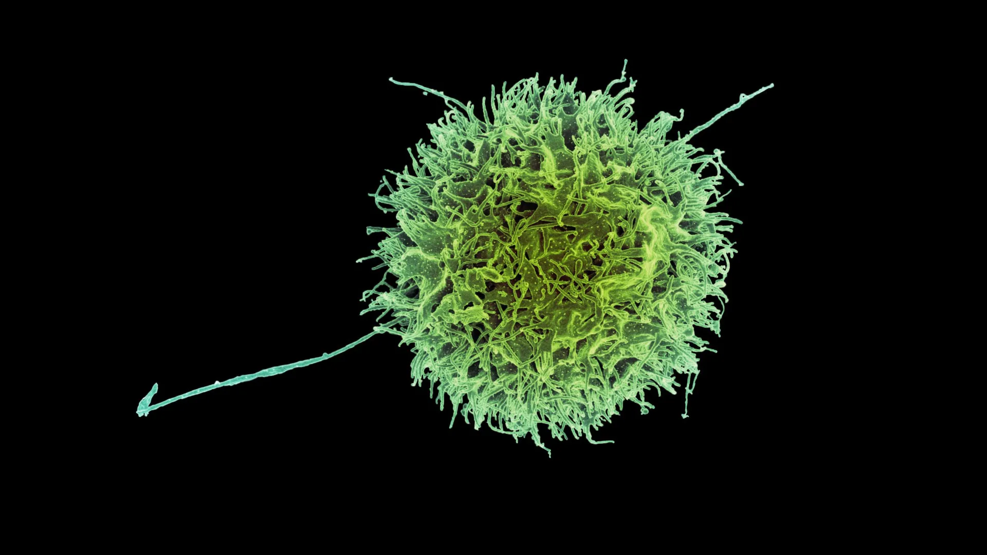
Scientists have created a new and more advanced form of immune-based cancer therapy using engineered cells known as CAR-NK (natural killer) cells. Like CAR-T cells, these modified immune cells can be programmed to recognize and attack cancer, but they rely on a different type of immune cell that naturally targets abnormal or infected cells.

A team from MIT and Harvard Medical School has now developed a more effective way to engineer CAR-NK cells that dramatically reduces the chance of the body’s immune system rejecting them. Immune rejection has been one of the biggest limitations of cell-based therapies, often weakening their effectiveness.

This innovation could also make it possible to produce “off-the-shelf” CAR-NK treatments that are available immediately after diagnosis, rather than waiting weeks for custom-engineered cells. Traditional CAR-NK and CAR-T manufacturing methods typically require several weeks to complete before patients can begin treatment.

“This enables us to do one-step engineering of CAR-NK cells that can avoid rejection by host T cells and other immune cells. And, they kill cancer cells better and they’re safer,” says Jianzhu Chen, an MIT professor of biology, a member of the Koch Institute for Integrative Cancer Research, and one of the senior authors of the study. 

In tests using mice with humanized immune systems, the newly engineered cells successfully destroyed most cancer cells while avoiding attack from the host’s own immune defenses.

Rizwan Romee, an associate professor of medicine at Harvard Medical School and Dana-Farber Cancer Institute, is also a senior author of the paper, which was published in Nature Communications. The study’s lead author is Fuguo Liu, a postdoctoral researcher at the Koch Institute and a research fellow at Dana-Farber.

Evading the immune system

Natural killer (NK) cells are a vital part of the body’s built-in immune defense, responsible for identifying and destroying cancerous and virus-infected cells. They eliminate these threats through a process called degranulation, which releases a protein known as perforin. This protein punctures the membrane of target cells, leading to their death.

To produce CAR-NK cells for treatment, doctors typically collect a blood sample from the patient. NK cells are then extracted and engineered to express a specialized protein called a chimeric antigen receptor (CAR), which is designed to target specific markers found on cancer cells.

Once modified, the cells must multiply in the lab for several weeks before there are enough to be infused back into the patient. The same general process is used for CAR-T cell therapies, some of which have already been approved to treat blood cancers like lymphoma and leukemia. CAR-NK therapies, however, are still being tested in clinical trials.

Because growing enough personalized CAR-NK cells takes time and the patient’s cells may not always be healthy enough for reliable use, scientists have been exploring an alternative: creating NK cells from healthy donors. These donor-derived cells could be mass-produced and stored for rapid use. The challenge, however, is that the recipient’s immune system often identifies donor cells as foreign and destroys them before they can attack the cancer.

In their latest research, the MIT team aimed to solve this problem by helping NK cells “hide” from immune detection. Their experiments showed that removing surface proteins known as HLA class 1 molecules allowed NK cells to avoid attack from T cells in the host’s immune system. These proteins normally act as identity markers that tell the immune system whether a cell belongs to the body.

To make use of this insight, the researchers added a sequence of siRNA (short interfering RNA) that silences the genes responsible for producing HLA class 1 proteins. Along with this genetic tweak, they introduced the CAR gene itself and another gene that encodes either PD-L1 or single-chain HLA-E (SCE), both of which help strengthen the NK cells’ cancer-fighting abilities.

All of these genetic components were combined into a single DNA construct, which allowed the team to efficiently convert donor NK cells into immune-evasive CAR-NK cells. Using this method, they engineered cells that target CD-19, a protein commonly found on malignant B cells in lymphoma patients.

NK cells unleashed

The researchers tested these CAR-NK cells in mice with a human-like immune system. These mice were also injected with lymphoma cells.

Mice that received CAR-NK cells with the new construct maintained the NK cell population for at least three weeks, and the NK cells were able to nearly eliminate cancer in those mice. In mice that received either NK cells with no genetic modifications or NK cells with only the CAR gene, the host immune cells attacked the donor NK cells. In these mice, the NK cells died out within two weeks, and the cancer spread unchecked.

The researchers also found that these engineered CAR-NK cells were much less likely to induce cytokine release syndrome — a common side effect of immunotherapy treatments, which can cause life-threatening complications.

Because of CAR-NK cells’ potentially better safety profile, Chen anticipates that they could eventually be used in place of CAR-T cells. For any CAR-NK cells that are now in development to target lymphoma or other types of cancer, it should be possible to adapt them by adding the construct developed in this study, he says.

The researchers now hope to run a clinical trial of this approach, working with colleagues at Dana-Farber. They are also working with a local biotech company to test CAR-NK cells to treat lupus, an autoimmune disorder that causes the immune system to attack healthy tissues and organs.

The research was funded, in part, by Skyline Therapeutics, the Koch Institute Frontier Research Program through the Kathy and Curt Marble Cancer Research Fund and the Elisa Rah Memorial Fund, the Claudia Adams Barr Foundation, and the Koch Institute Support (core) Grant from the National Cancer Institute.

Share Button

12,000-year-old rock art found in Arabia reveals a lost civilization

Recent research has revealed the crucial role of early human groups who settled in northern Arabia soon after the intensely dry period known as the Last Glacial Maximum (LGM). These communities followed the return of seasonal water sources and left behind an extraordinary record of their presence through monumental rock art.

An international team of archaeologists, coordinated by the Heritage Commission of the Saudi Ministry of Culture, uncovered the findings through the Green Arabia Project. The team included experts from the Max Planck Institute of Geoanthropology, KAUST (King Abdullah University of Science and Technology), University College London, Griffith University, and several other institutions.

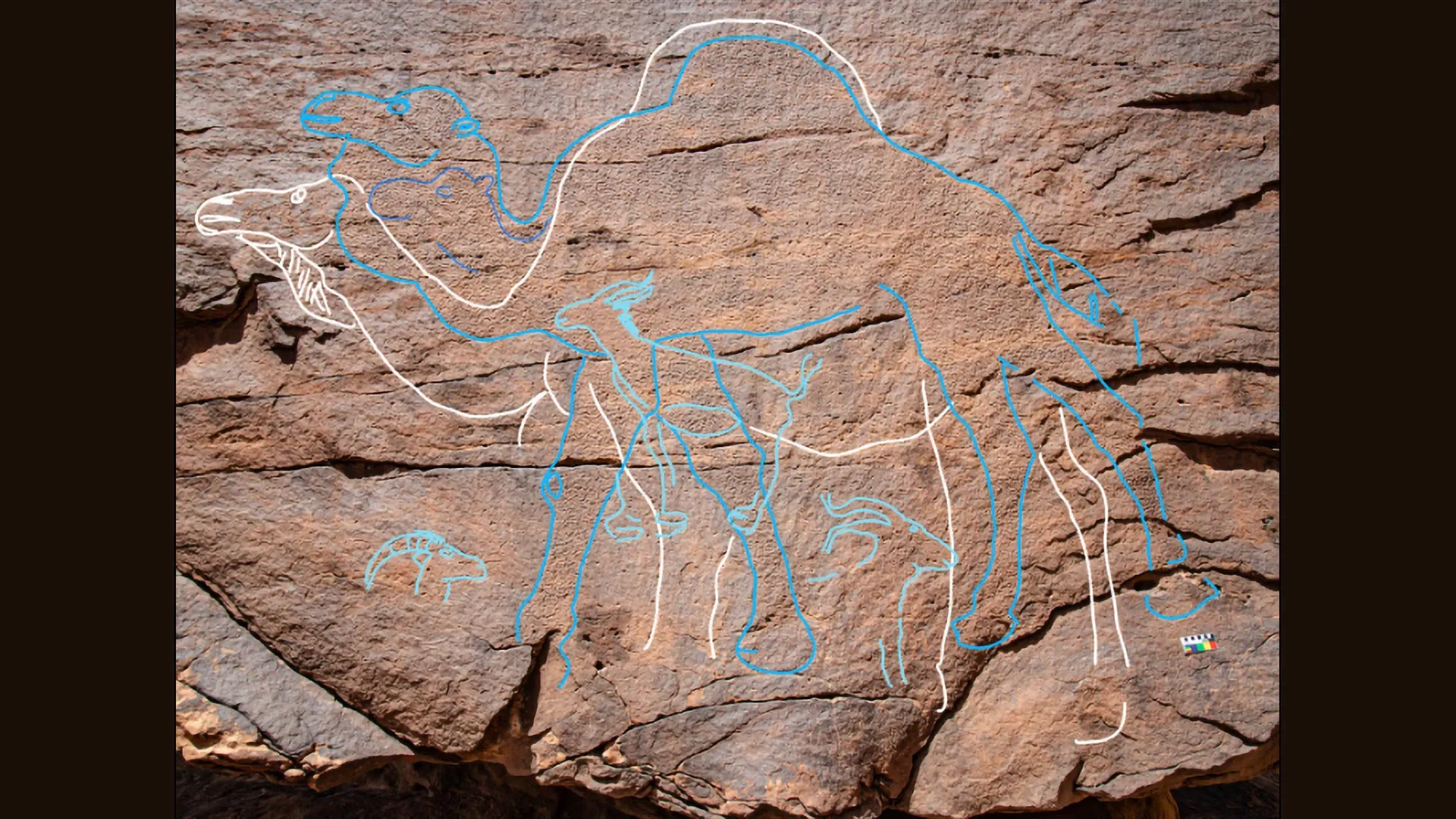
Researchers documented over 60 rock art panels featuring 176 individual engravings across three previously unexplored locations: Jebel Arnaan, Jebel Mleiha, and Jebel Misma, situated along the southern edge of the Nefud Desert in northern Saudi Arabia.

The engravings, which depict animals such as camels, ibex, equids, gazelles, and aurochs, include 130 highly detailed and life-sized figures, some reaching up to 3 meters long and more than 2 meters tall.

Dating to between 12,800 and 11,400 years ago, the engravings correspond to a time when temporary lakes and rivers reappeared after centuries of extreme aridity.

Sediment analysis confirmed the existence of these ancient water sources, which would have provided essential support for human groups venturing into the desert interior and allowed them to survive in this challenging environment.

“These large engravings are not just rock art – they were probably statements of presence, access, and cultural identity,” said lead author, Dr. Maria Guagnin from Max Planck Institute of Geoanthropology.

Dr. Ceri Shipton, co-lead author from the Institute of Archaeology, University College London, said: “The rock art marks water sources and movement routes, possibly signifying territorial rights and intergenerational memory.”

Unlike previously known sites where engravings were hidden in crevices, the Jebel Mleiha and the Jebel Arnaan panels were etched onto towering cliff faces, some up to 39 meters high, in visually commanding locations.

One panel would have required ancient artists to climb and work precariously on narrow ledges, underscoring the sheer effort and significance of the imagery.

Artefacts including Levantine-style El Khiam and Helwan stone points, green pigment, and dentalium beads suggest long-distance connections to Pre-Pottery Neolithic (PPN) populations in the Levant region.

However, the scale, content, and placement of the Arabian engravings set them apart.

“This unique form of symbolic expression belongs to a distinct cultural identity adapted to life in a challenging, arid environment,” said Dr. Faisal Al-Jibreen, from the Heritage Commmission, Saudi Ministry of Culture.

“The project’s interdisciplinary approach has begun to fill a critical gap in the archaeological record of northern Arabia between the LGM and the Holocene, shedding light on the resilience and innovation of early desert communities,” said Michael Petraglia, lead of the Green Arabia project.

The study ‘Monumental rock art illustrates that humans thrived in the Arabian Desert during the Pleistocene-Holocene transition’ has been published in Nature Communications.

Share Button

For the first time, scientists pinpoint brain cells linked to depression

Scientists at McGill University and the Douglas Institute have discovered that two distinct types of brain cells show changes in people experiencing depression.

Published in Nature Genetics, the research provides new clues that could guide the creation of treatments focused on these specific cells. It also enhances scientific understanding of depression, a condition that affects more than 264 million people globally and is one of the leading causes of disability.

“This is the first time we’ve been able to identify what specific brain cell types are affected in depression by mapping gene activity together with mechanisms that regulate the DNA code,” said senior author Dr. Gustavo Turecki, a professor at McGill, clinician-scientist at the Douglas Institute and Canada Research Chair in Major Depressive Disorder and Suicide. “It gives us a much clearer picture of where disruptions are happening, and which cells are involved.”

Rare brain bank enables breakthrough

The team conducted their work using post-mortem brain tissue from the Douglas-Bell Canada Brain Bank, one of the few collections worldwide that includes donations from people with psychiatric conditions.

Through advanced single-cell genomic analysis, the researchers examined RNA and DNA from thousands of individual brain cells to determine which ones behaved differently in people with depression and which DNA sequences might explain these variations. The study analyzed tissue from 59 individuals who had depression and 41 who did not.

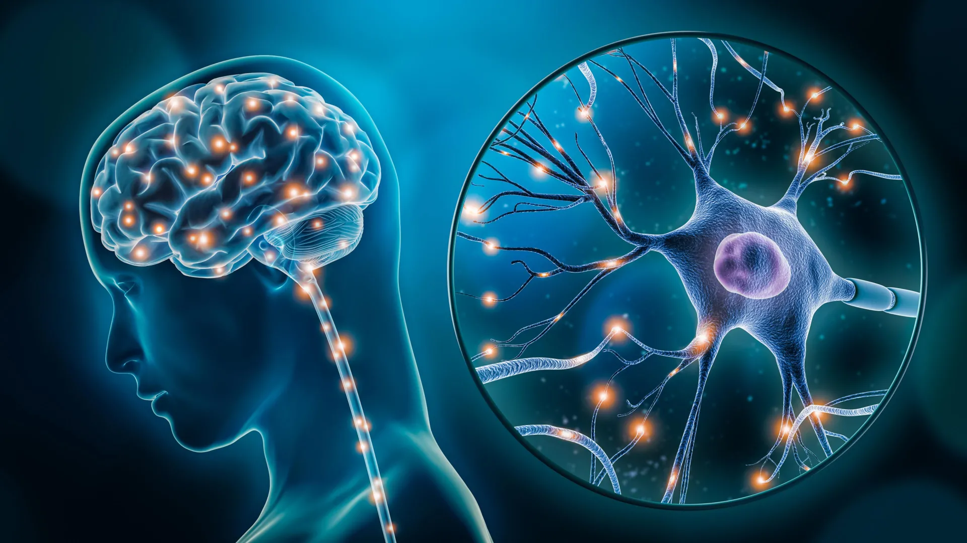
They discovered that gene activity was altered in two types of brain cells: a class of excitatory neurons responsible for mood and stress regulation, and a subtype of microglia, the immune cells that manage inflammation in the brain. In both cell types, many genes were expressed differently in people with depression, pointing to possible disruptions in vital neural systems.

By identifying the specific cells affected, the research deepens understanding of the biological foundation of depression and helps dispel outdated views of the condition.

“This research reinforces what neuroscience has been telling us for years,” Turecki said. “Depression isn’t just emotional, it reflects real, measurable changes in the brain.”

Looking ahead, the scientists intend to explore how these cellular changes influence brain function and whether targeting them could lead to more effective treatments.

About the study

“Single-nucleus chromatin accessibility profiling identifies cell types and functional variants contributing to major depression” by Anjali Chawla and Gustavo Turecki et al., was published in Nature Genetics.

The study was funded by Canadian Institutes of Health Research, Brain Canada Foundation, Fonds de recherche du Québec – Santé and Healthy Brains, Healthy Lives initiative at McGill University.

Share Button

A facelift at 28? Why young people are turning to plastic surgery

Gone are the days when facelifts were for the ageing wealthy. Now younger people are going under the knife.

Share Button

William visibly moved as widow tells of husband’s suicide

Prince William’s foundation is giving £1m to set up a suicide prevention network.

Share Button

‘Marginalised and menopausal’ women given spotlight

Women from ethnic minority communities typically experience perimenopause symptoms earlier and for longer.

Share Button