Complaint about advert offering to replace traditional contraception with natural methods is upheld.


Complaint about advert offering to replace traditional contraception with natural methods is upheld.
Stereotypes around gender and worries about looks contribute to children’s unhappiness, a report says.
Signs of stiffening arteries show up by that age and can lead to heart problems in later life.
A team of researchers at the University of Minnesota have, for the first time, fully 3D printed an array of light receptors on a hemispherical surface. This discovery marks a significant step toward creating a “bionic eye” that could someday help blind people see or sighted people see better.
The research is published today in Advanced Materials, a peer-reviewed scientific journal covering materials science. The author also holds the patent for 3D-printed semiconducting devices.
“Bionic eyes are usually thought of as science fiction, but now we are closer than ever using a multimaterial 3D printer,” said Michael McAlpine, a co-author of the study and University of Minnesota Benjamin Mayhugh Associate Professor of Mechanical Engineering.
Researchers started with a hemispherical glass dome to show how they could overcome the challenge of printing electronics on a curved surface. Using their custom-built 3D printer, they started with a base ink of silver particles. The dispensed ink stayed in place and dried uniformly instead of running down the curved surface. The researchers then used semiconducting polymer materials to print photodiodes, which convert light into electricity. The entire process takes about an hour.
McAlpine said the most surprising part of the process was the 25 percent efficiency in converting the light into electricity they achieved with the fully 3D-printed semiconductors.
“We have a long way to go to routinely print active electronics reliably, but our 3D-printed semiconductors are now starting to show that they could potentially rival the efficiency of semiconducting devices fabricated in microfabrication facilities,” McAlpine said. “Plus, we can easily print a semiconducting device on a curved surface, and they can’t.”
McAlpine and his team are known for integrating 3D printing, electronics, and biology on a single platform. They received international attention a few years ago for printing a “bionic ear.” Since then, they have 3D printed life-like artificial organs for surgical practice, electronic fabric that could serve as “bionic skin,” electronics directly on a moving hand, and cells and scaffolds that could help people living with spinal cord injuries regain some function.
McAlpine’s drive to create a bionic eye is a little more personal.
“My mother is blind in one eye, and whenever I talk about my work, she says, ‘When are you going to print me a bionic eye?'” McAlpine said.
McAlpine says the next steps are to create a prototype with more light receptors that are even more efficient. They’d also like to find a way to print on a soft hemispherical material that can be implanted into a real eye.
McAlpine’s research team includes University of Minnesota mechanical engineering graduate student Ruitao Su, postdoctoral researchers Sung Hyun Park, Shuang-Zhuang Guo, Kaiyan Qiu, Daeha Joung, Fanben Meng, and undergraduate student Jaewoo Jeong.
The research was funded by the National Institute of Biomedical Imaging and Bioengineering of the National Institutes of Health (Award No. 1DP2EB020537), The Boeing Company, and the Minnesota Discovery, Research, and InnoVation Economy (MnDRIVE) Initiative through the State of Minnesota.
Related video: https://www.youtube.com/watch?v=U2_zhpXZkS0
Story Source:
Materials provided by University of Minnesota. Note: Content may be edited for style and length.

Clinicians should consider how the way we think can make us vulnerable to obesity, and how obesity is genetically intertwined with brain structure and mental performance, according to new research.
The study, led by researchers at the Montreal Neurological Institute and Hospital (The Neuro) and published in the Proceedings of the National Academy of Sciences on Aug. 28, 2018, was an examination of magnetic resonance imaging (MRI) and cognitive test data from 1,200 individuals, supplied as part of the Human Connectome Project.
Researchers found that people with higher a body mass index (BMI) showed reduced cognitive flexibility, ability to delay gratification, visuospatial ability and verbal memory. They also found that people with increased BMI tended to have a thicker left prefrontal cortex and a thinner right prefrontal cortex. Previous studies have shown damage to the right prefrontal cortex can lead to increased eating.
Subjects with higher BMI also had increased volume in the left amygdala, which is believed to play a role in response to food cues. They also had decreased volume in the entorhinal-parahippocampal structures, which are associated with episodic memory and context mediation. This suggests a model where people prone to obesity are more sensitive to visual food cues, and less able to resist them by considering the negative context of eating, like weight gain.
Many of the subjects were siblings, including fraternal and identical twins. This allowed researchers to determine the heritability of the traits as well as obesity, measured by BMI. Using statistical methods, researchers found that many of the cognitive and neurological traits have genetic links with obesity. This suggests the role genetics play in obesity is manifested at least partially through brain anatomy and cognitive functions.
“This research will be useful in developing interventions to help people with obesity,” says study’s lead author, Uku Vainik, a researcher at The Neuro and the Institute of Psychology at University of Tartu, Estonia. “Modifying neurobehavioural factors with cognitive training, to improve people’s ability to resist food, for example, could hold promise. Interventions shouldn’t just focus on diet, but also acknowledge the neurobehavioural profile that obesity is genetically intertwined with. Such interventions might help people to stay lean despite their genetic signature.”
“This work adds support to the theory that body weight in humans is partly under control of higher-level brain systems involved in cognition, decision-making and motivation,” says Dr. Alain Dagher, the paper’s senior author. “Furthermore, individual differences in these brain systems that regulate food intake appear to be moderately heritable.”
Story Source:
Materials provided by McGill University. Note: Content may be edited for style and length.

In a finding that caps years of exploration into the tiny particle known as the Higgs boson, researchers have traced the fifth and most prominent way that the particle decays into other particles. The discovery gives researchers a new pathway by which to study the physical laws that govern the universe.
Physicists at Princeton University led one of the two main teams that today announced the detection of the Higgs particle via its decay into two particles called bottom quarks. This pathway is the last to be detected of the five main signature pathways that can identify the Higgs particle.
“We found it exactly where we expected to find it and now we can use this new pathway to study the Higgs’ properties,” said James Olsen, professor of physics and leader of the team at Princeton. “This has been a truly collaborative effort from the beginning and it is exciting to see the amplification of effort that comes from people working together.”
Long-sought because it confirms theories about the nature of matter, the Higgs particle exists only fleetingly before transforming into other, so-called “daughter” particles. Because the boson lasts only for about one septillionth of a second, researchers use the particle’s offspring as evidence of its existence.
These daughter particles are scattered among the shower of particles created from the collision of two protons at the Large Hadron Collider at the European Organization for Nuclear Research (CERN). The Higgs particle was observed for the first time in 2012 through three of the other modes of decay.
Of these lineages or decay patterns, the decay into two bottom quarks occurs most often, making up about 60 percent of the decay events from the Higgs, according to Olsen.
But at the LHC, the bottom-quark pattern is the hardest to trace back definitively to the Higgs because many other particles can also give off bottom quarks.
Quarks are tiny constituents of protons, which themselves are some of the building blocks of atoms. The bottom quark is one of the six types of quarks that make up the menagerie of particles in the “standard model” that explains matter and their interactions.
Discerning which bottom quarks came from the Higgs versus other particles has been the main challenge facing the LHC’s two Higgs detectors, the Compact Muon Solenoid (CMS) with which Olsen works, and its companion, ATLAS. The detectors operate independently and are run by separate teams of scientists.
Once produced, these bottom quarks split into jets of particles, making them hard to trace back to the original parent particles. Because of this background noise, the researchers required more data than was needed for the other pathways to be sure of their finding.
Both detectors are adept at spotting particles such as electrons, photons and muons, but are more challenged by quarks. The quarks due to their nature are not observed as free particles. Instead they are bound and appear as other particles such as mesons and baryons or decay quickly.
“It is a messy business because you have to collect all of those jets and measure their properties to calculate the mass of the object that decayed into the jets,” Olsen said.
The two detectors are massive, complex structures that sit at the end of the LHC tunnel where protons are accelerated and smashed together at high energy levels. The structures contain layers of smaller detectors arranged like layers of an onion.
The devices detect particles at each layer of the onion to reconstruct their paths. This allows researchers to trace the path of a particle back to its source in a manner analogous to following the trail of a firework’s light back to the place where the first burst occurred. By following many of these paths, the researchers can identify where and when the Higgs first formed in the proton-proton collision.
“The Higgs-to-bottom-quark decay is important because it is the most frequent decay, so a precise measurement of its rate tells us a lot about the nature of this particle,” said Christopher Palmer, an associate research scholar at Princeton, who describes the work in this video: https://www.youtube.com/watch?v=NfCgbGtVZCo
Additional Princeton researchers on the team included physics graduate student Stephane Cooperstein and undergraduate Jan Offerman, Class of 2018. The team also included collaborators at the University of Florida and Fermilab, as well as groups from Italy, Switzerland and Germany.
The main challenge in detecting the bottom-quark decay mode was the amount of background bottom quarks produced by non-Higgs events. In addition to gathering more data from collisions, the researchers searched for a distinct way that the Higgs gave rise to the bottom quarks.
Olsen began working on this challenge 10 years ago, before the LHC had switched on and when physicists were running simulations on computers. Around that time, Olsen learned about a theoretical study showing that it is possible to find bottom quarks created from the Higgs recoiling off another particle, such as a Z boson or a W boson.
“It was an idea that nobody had before, to search for it in that channel, and the only question was whether it was possible experimentally and whether it really would pay off,” Olsen said.
Olsen said it is exciting to see the work come to fruition. “This is a very satisfying moment.”
Princeton University’s research at the LHC is funded by the U.S. Department of Energy and the National Science Foundation.

Researchers have discovered a link between three degenerative eye diseases and Alzheimer’s disease.
They say their findings could lead to new ways to identify people at high risk for Alzheimer’s.
“We don’t mean people with these eye conditions will get Alzheimer’s disease,” said lead researcher Cecilia Lee, an assistant professor of ophthalmology at the University of Washington School of Medicine.
Alzheimer’s expected to increase
“The main message from this study is that ophthalmologists should be more aware of the risks of developing dementia for people with these eye conditions and primary care doctors seeing patients with these eye conditions might be more careful on checking on possible dementia or memory loss,” Lee explained in a university news release.
The study involved 3 877 randomly selected patients, aged 65 and older. They were tracked over the course of five years, during which time 792 were diagnosed with Alzheimer’s disease.
Patients with age-related macular degeneration, diabetic retinopathy or glaucoma had a 40% to 50% greater risk of Alzheimer’s disease than those without the eye conditions, the researchers said.
“What we found was not subtle,” said Paul Crane, a professor of medicine at the university. “This study solidifies that there are mechanistic things we can learn from the brain by looking at the eye.”
The study was published August eight in Alzheimer’s & Dementia: The Journal of the Alzheimer’s Association.
Alzheimer’s is the most common form of dementia, which affects more than 46 million people worldwide. That number is expected to rise to 131.5 million by 2050, the researchers estimate.
According to South Africa’s 2011 census, there are approximately 2.2 million people in South Africa with some form of dementia.


For many people, hearing aids symbolise old age and decades of deteriorating hearing, but new technology is allowing these stereotypes to be broken.
In the past, hearing aids were highly visible because they were big and cumbersome, but technology has improved the size, functionality and appearance of hearing aids.
In fact, there are now hearing aids which fit completely into your ear and are, for the most part, invisible as the devices are only about the size of a large bead.

Paul Crusius, co-founder of Audibene, points out that only about 20% of people with hearing problems actually wear hearing aids.
It is an established fact that many older people need hearing aids because their hearing has deteriorated over the years, but not everyone knows that there are many younger people who need these devices as well.
Modern technology has progressed to such a degree that people are able to control their hearing aids with an app on their smartphones, and some even allow users to listen to music through their devices.
Image credit: iStock


The quality of life of many South Africans of different ages is affected by hearing loss. Although hearing aids offer a solution for many, they’re not always equally effective. Wouldn’t it be simpler if one could fix hearing problems with surgery?
Only a small percentage eligible
The unfortunate reality is, however, that only a small percentage of people suffering from hearing loss are eligible for surgery. “It depends on the type of hearing loss,” according to a Healthy Hearing article.
There are many reasons for hearing loss. One of the most common causes of deafness is sensorineural hearing loss, which involves the nerves. It is caused by old age, consistent exposure to loud noise, infections, trauma to the head or ears, tumours and even medication. This type of hearing loss is permanent.
Other reasons for hearing loss include disorders of the auditory process, where the brain has trouble processing sounds, conductive hearing loss where the middle or outer structure of the ear has problems transferring sounds to the inner ear, or mixed hearing loss where there is a combination of causes.
Types of hearing surgery
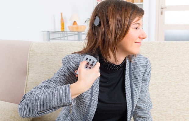
1. Cochlear implants
What it is: Cochlear implants are often performed in adults and children with profound sensorineural hearing loss. The implants can restore partial hearing and, unlike a hearing aid, the implant bypasses the damaged part of nerve instead of merely amplifying sound.
How it’s performed: A small electronic device is surgically placed under the skin behind your ear. This device is connected to electrodes which are inserted into the cochlea.
When it is considered: According to the Ear Institute of South Africa, cochlear implants are considered when no hearing aid can restore your sense of hearing. A comprehensive evaluation will be done by a team of experts before the procedure is considered, as it is highly invasive and costly.
2. Hearing aid implants
What it is: External hearing aids have developed significantly over the past few decades, but for extended wear an internal implant is often considered.
How it’s performed: One of the most commonly performed hearing aid implants is a bone-anchored implant (BAHA), which entails the stimulation of the cochlea by transmitting sounds through the bone of the skull.
When it is considered: The BAHA is often reserved for patients with chronic middle ear or outer ear problems or congenital defects of the ear and who can’t benefit from hearing aids. The prerequisite is that the patient should have one ear with moderate hearing.
3. Pressure equalising (PE) tubes
What it is: This procedure entails the insertion of tubes into the ear to equalise pressure and allow air through in cases where the Eustachian tube (the tube that connects the middle of the ear to the back of the nose) becomes blocked with fluid.
How it’s performed: An incision is made in the eardrums and any fluid drained out. Tiny, hollow metal or plastic tubes are then inserted to allow air through to the middle of the ear, and as the eardrum heals, these get pushed out again. Sometimes long-term tubes are needed, which are surgically removed.
When it is considered: This procedure is often considered for young children suffering from chronic ear infections, but in many cases the surgery is also performed on adults to correct hearing loss linked to malformed eardrums or problematic Eustachian tubes.

4. Stapedectomy
What it is: This surgical procedure is often used to treat hearing loss caused by otosclerosis, a condition that causes abnormal bone growth inside the middle ear, which prohibits vibrations to travel through the ear.
How it’s performed: An incision is made behind the ear and muscle or tissue is removed. Excess or abnormal bone (usually referred to as the “hearing bone” or the stapes bone) is fixed. A prosthesis is then inserted and connected to the second hearing bone (the incus) to allow sound waves to travel correctly.
When it’s considered: A stapedectomy will be performed when there is a deformity of the stapes bone and a hearing aid is not providing sufficient correction of hearing loss.
So, surgery or not?
It is likely that medical technology will come up with more non-invasive options to restore hearing loss in the future. But since hearing surgery can currently only restore specific cases of hearing loss when hearing aids are not an option, hearing aids still remain the most effective non-invasive method to help correct hearing.
Image credit: iStock


The Spice Girl says she’s been drinking “to numb my pain”.
