Snake fungal disease alters skin microbiome in eastern Massasaugas

In the first study of its kind, researchers characterized the skin microbiome of a population of free-ranging snakes to begin to understand how the animals’ environmental microbial community may promote disease resistance as well as how it may be disrupted by infection.

The study, which was recently published in Scientific Reports, a Nature research journal, focused on eastern massasaugas in Illinois. This species of endangered rattlesnake is highly susceptible to the fungal pathogen Ophidiomyces ophiodiicola, which causes snake fungal disease (SFD). SFD results in disfiguring sores on snake skin, has a high mortality rate, and poses a significant threat to snake populations in North America and Europe. The mechanism by which the pathogen causes disease is unknown.

“Globally, fungal pathogens are increasingly associated with wildlife epidemics, such as white-nose syndrome in bats and chytridiomycosis in amphibians,” said Dr. Matt Allender, a faculty member at the University of Illinois College of Veterinary Medicine and an affiliate of the Illinois Natural History Survey (INHS), part of the university’s Prairie Research Institute. “Snake fungal disease has been identified in a number of snake species, but very little is known about contributing factors for infection.”

Dr. Allender, who heads the Wildlife Epidemiology Laboratory, has been investigating SFD for more than 8 years. In 2014 he introduced a quantitative polymerase chain reaction (qPCR) test to quickly identify the fungus from a swabbed sample.

“In a 20-year collaborative study led by INHS researchers, we have been the primary investigator of numerous studies documenting disease trends in the eastern massasauga including overall health, but none of these health parameters seemed to explain the emergence of SFD. This study was undertaken in light of recent promising findings about the importance of environmental microbial communities in animal and human health.”

Based on their analysis of 144 skin swabs collected from 44 snakes in 2015 and 52 snakes in 2016, all near Carlyle Lake, Ill., researchers determined that infection with SFD altered the bacterial and fungal diversity of the snakes studied. On the infected snakes, Ophidiomyces was present even at locations on the snakes’ bodies distant to the open sores, indicating that the skin’s entire microbiome is altered by the infection.

No Ophidiomyces spores were detected on SFD-negative snakes, as would have been expected had those snakes’ microbiome proven protective against the pathogen.

Findings related to the specific bacteria and fungi found in greater or lesser abundance depending on the disease status of the snake are detailed in the study.

The researchers believe their findings will have broad relevance to other snake species and habitats and will provide insight into mechanisms of pathogen emergence, fluctuations in wellness of individuals, and development of therapeutic interventions.

Story Source:

Materials provided by University of Illinois at Urbana-Champaign. Note: Content may be edited for style and length.

Share Button

Early opaque universe linked to galaxy scarcity

A team of astronomers led by George Becker at the University of California, Riverside, has made a surprising discovery: 12.5 billion years ago, the most opaque place in the universe contained relatively little matter.

It has long been known that the universe is filled with a web-like network of dark matter and gas. This “cosmic web” accounts for most of the matter in the universe, whereas galaxies like our own Milky Way make up only a small fraction. Today, the gas between galaxies is almost totally transparent because it is kept ionized — electrons detached from their atoms — by an energetic bath of ultraviolet radiation.

Over a decade ago, astronomers noticed that in the very distant past — roughly 12.5 billion years ago, or about 1 billion years after the Big Bang — the gas in deep space was not only highly opaque to ultraviolet light, but its transparency varied widely from place to place, obscuring much of the light emitted by distant galaxies.

Then a few years ago, a team led by Becker, then at the University of Cambridge, found that these differences in opacity were so large that either the amount of gas itself, or more likely the radiation in which it is immersed, must vary substantially from place to place.

“Today, we live in a fairly homogeneous universe,” said Becker, an expert on the intergalactic medium, which includes dark matter and the gas that permeates the space between galaxies. “If you look in any direction you find, on average, roughly the same number of galaxies and similar properties for the gas between galaxies, the so-called intergalactic gas. At that early time, however, the gas in deep space looked very different from one region of the universe to another.”

To find out what created these differences, the team of University of California astronomers from the Riverside, Santa Barbara, and Los Angeles campuses turned to one of the largest telescopes in the world: the Subaru telescope on the summit of Mauna Kea in Hawaii. Using its powerful camera, the team looked for galaxies in a vast region, roughly 300 million light years in size, where they knew the intergalactic gas was extremely opaque.

For the cosmic web more opacity normally means more gas, and hence more galaxies. But the team found the opposite: this region contained far fewer galaxies than average. Because the gas in deep space is kept transparent by the ultraviolet light from galaxies, fewer galaxies nearby might make it murkier.

“Normally it doesn’t matter how many galaxies are nearby; the ultraviolet light that keeps the gas in deep space transparent often comes from galaxies that are extremely far away. That’s true for most of cosmic history, anyway,” said Becker, an assistant professor in the Department of Physics and Astronomy. “At this very early time, it looks like the UV light can’t travel very far, and so a patch of the universe with few galaxies in it will look much darker than one with plenty of galaxies around.”

This discovery, reported in the August 2018 issue of the Astrophysical Journal, may eventually shed light on another phase in cosmic history. In the first billion years after the Big Bang, ultraviolet light from the first galaxies filled the universe and permanently transformed the gas in deep space. Astronomers believe that this occurred earlier in regions with more galaxies, meaning the large fluctuations in intergalactic radiation inferred by Becker and his team may be a relic of this patchy process, and could offer clues to how and when it occurred.

“There is still a lot we don’t know about when the first galaxies formed and how they altered their surroundings,” Becker said.

By studying both galaxies and the gas in deep space, astronomers hope to get closer to understanding how this intergalactic ecosystem took shape in the early universe.

Story Source:

Materials provided by University of California – Riverside. Original written by Iqbal Pittalwala. Note: Content may be edited for style and length.

Share Button

Health24.com | SEE: Alzheimers disease – are you at risk?

The information on Health24 is for educational purposes only, and is not intended as medical advice, diagnosis or treatment. If you are experiencing symptoms or need health advice, please consult a healthcare professional. See additional information.

Share Button

Babies in prams ‘exposed to more pollution’

Infants in prams can be exposed to up to 60% more pollution than adults, a study suggests.

Share Button

Health24.com | Contact lens stuck inside woman’s eyelid for 28 years

When a 14-year-old Scottish girl was hit in the face by a shuttlecock while playing badminton, she thought the contact lens in her left eye had simply fallen out during the impact. 

The doctors who examined her came to the same conclusion. 

But almost 30 years later, medical personnel were shocked to discover the lens had actually lodged itself in her eye, and had been stuck there for the past 28 years. 

The case of the woman, who is now 42 years old and prefers to remain anonymous, was recently reported in BMJ Case Reports.

contact lens, contact lens stuck

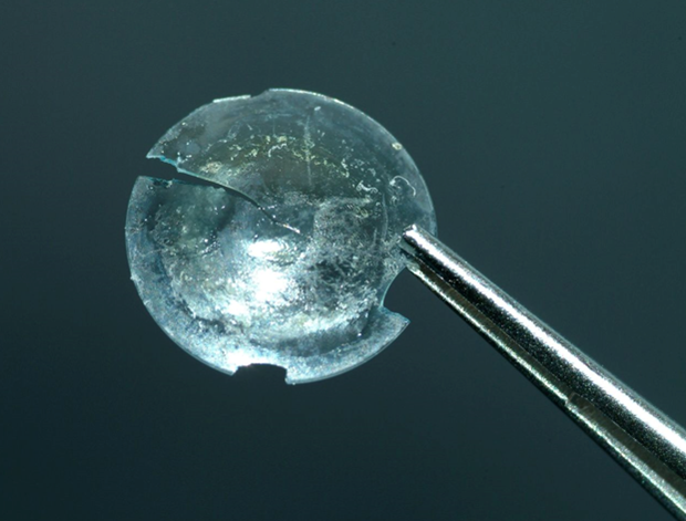
                                                        The retrieved rigid gas permeable lens within the cyst

According to the Daily Mail, the lens came to light after the patient complained to her GP of swelling in her left upper eyelid.

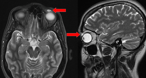
“The MRI reported a cyst with proteinaceous content,” the researchers said. “Proteinaceous” refers to the presence of a protein substance. 

“On surgical excision of the cyst, a rigid gas permeable (RGP) contact lens was found. The RGP lens was encapsulated within the upper eyelid soft tissue.”

contact lens, contact lens stuck

                 MRI of the head: Red arrow: high-intensity signal nodular lesion in the left upper eyelid

The surgeons noted that there have been four other reported cases of contact lens migration because of trauma to the eye.

“This case report exhibits the longest time between traumatic RGP lens migration into the eyelid and presentation of eyelid swelling,” they wrote. 

Doctors speculate that this must be the lens that was the residue of the childhood accident. “It can be inferred that the lens migrated into the eyelid and resided there asymptomatically for 28 years.”

The patient has since then been released from hospital and has made a full recovery.

Images credit: BMJ Case Studies

NEXT ON HEALTH24X

Share Button

Health24.com | SEE: Can doctors make you younger?

The information on Health24 is for educational purposes only, and is not intended as medical advice, diagnosis or treatment. If you are experiencing symptoms or need health advice, please consult a healthcare professional. See additional information.

Share Button

The radio ‘saved my little girl’s life’

A large trial in Burkina Faso suggests listening to the radio could improve child health.

Share Button

Vaping ‘can damage vital immune system cells’

E-cigarette vapour disables important immune cells in the lung and boosts inflammation.

Share Button

Adult-child conversations strengthen language regions of developing brain

Society for Neuroscience. “Adult-child conversations strengthen language regions of developing brain: Study suggests talking with children from early age could promote language skills regardless of socioeconomic status.” ScienceDaily. ScienceDaily, 13 August 2018. <www.sciencedaily.com/releases/2018/08/180813133422.htm>.

Society for Neuroscience. (2018, August 13). Adult-child conversations strengthen language regions of developing brain: Study suggests talking with children from early age could promote language skills regardless of socioeconomic status. ScienceDaily. Retrieved August 13, 2018 from www.sciencedaily.com/releases/2018/08/180813133422.htm

Society for Neuroscience. “Adult-child conversations strengthen language regions of developing brain: Study suggests talking with children from early age could promote language skills regardless of socioeconomic status.” ScienceDaily. www.sciencedaily.com/releases/2018/08/180813133422.htm (accessed August 13, 2018).

Share Button

Potential biomarker for autism in infancy

A study of young children with autism spectrum disorder published in JNeurosci reveals altered brain waves compared to typically developing children during a motor control task. The non-invasive neuroimaging technique used in this study could be employed to detect autism symptoms as early as infancy.

People with autism show reduced inhibitory neural activity, reflected in gamma brain waves, and movement abnormalities. Mitsuru Kikuchi and colleagues investigated these two aspects of the disorder by recording the brain activity of five- to seven-year-old children diagnosed with autism as they played a video game that required them to press a button to feed a puppy.

Compared to a matched group of typically developing children, the children with autism exhibited longer reaction times and reduced gamma oscillations that were associated with the severity of their symptoms.

The researchers demonstrate that these features alone can distinguish the two groups, which may represent an objective approach toward diagnosing autism in young children.

Story Source:

Materials provided by Society for Neuroscience. Note: Content may be edited for style and length.

Share Button