NASA Curiosity rover finds mysterious life linked molecules on mars

NASA’s Curiosity rover has identified a wide range of organic molecules on Mars, including compounds that scientists consider key ingredients for the origin of life on Earth.

The discovery comes from a chemical experiment carried out on another planet for the first time. Results show that the Martian surface is capable of preserving molecules that could act as potential signs of ancient life. However, the experiment cannot determine whether these organic compounds came from past life on Mars, natural geological processes, or meteorites that struck the planet.

To confirm any true evidence of past life, scientists would need to bring Martian rock samples back to Earth for detailed study.

New Experiment Reveals Preserved Ancient Chemistry

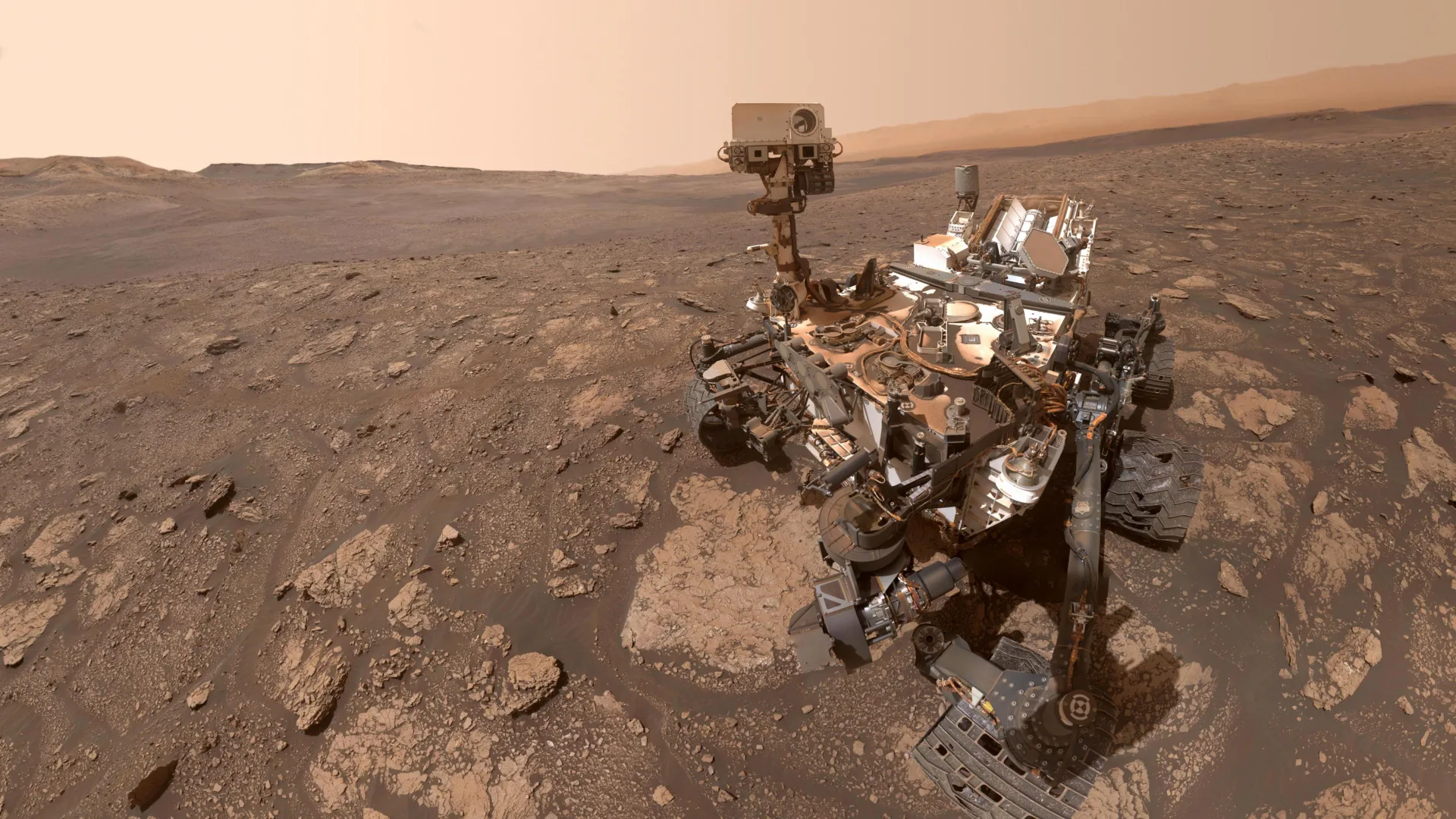
The research was led by Amy Williams, Ph.D., a geological sciences professor at the University of Florida and a member of both the Curiosity and Perseverance rover science teams. Curiosity arrived on Mars in 2012 to investigate whether the planet once had conditions suitable for microbial life. Perseverance, which landed in 2021, is focused on searching for direct signs of ancient life.

“We think we’re looking at organic matter that’s been preserved on Mars for 3.5 billion years,” said Williams, who helped develop the experiment. “It’s really useful to have evidence that ancient organic matter is preserved, because that is a way to assess the habitability of an environment. And if we want to search for evidence of life in the form of preserved organic carbon, this demonstrates it’s possible.”

Williams and an international team published the findings April 21 in the journal Nature Communications.

DNA-Like Molecule Among Key Discoveries

The experiment identified more than 20 different chemicals. Among them was a nitrogen-containing molecule with a structure similar to compounds involved in building DNA, something never before detected on Mars. The rover also found benzothiophene, a large sulfur-containing molecule with two connected rings, which is commonly delivered to planets by meteorites.

“The same stuff that rained down on Mars from meteorites is what rained down on Earth, and it probably provided the building blocks for life as we know it on our planet,” Williams said.

Gale Crater and Clay Minerals Preserve Organics

Curiosity, operated by NASA’s Jet Propulsion Laboratory, landed in Gale crater in August 2012. This site was once a lake bed. The experiment took place in 2020 in the Glen Torridon region, an area rich in clay minerals that formed in the presence of water. These clays are especially good at trapping and preserving organic material, making them ideal locations for this type of investigation.

SAM Instrument and TMAH Chemical Analysis

The analysis was carried out using the Sample Analysis at Mars instrument suite, known as SAM. Jennifer Eigenbrode, Ph.D., an astrobiologist at NASA’s Goddard Space Flight Center and co-author of the study, helps lead the instrument team. SAM has contributed many of the mission’s key discoveries about Mars’ chemistry, atmosphere, and potential habitability.

In this experiment, scientists used a chemical called TMAH to break down larger organic molecules into smaller fragments. These fragments could then be examined by SAM’s onboard instruments. Because Curiosity only carries about two cups of TMAH, researchers had to carefully plan the experiment and select the best possible sampling site.

Implications for Future Mars and Titan Missions

The success of this method is shaping future exploration plans. Upcoming missions, including the Rosalind Franklin rover on Mars and the Dragonfly mission to Saturn’s moon Titan, are expected to carry similar TMAH-based experiments to search for organic compounds.

“We now know that there are big complex organics preserved in the shallow subsurface of Mars, and that holds a lot of promise for preserving large complex organics that might be diagnostic of life,” Williams said.

Share Button

Scientists catch antimatter “atom” acting like a wave for the first time

One of the defining breakthroughs that set quantum physics apart from classical physics was the realization that matter behaves very differently at extremely small scales. Among the most important discoveries was wave-particle duality, the idea that particles can also act like waves.

This concept became widely known through the double-slit experiment. When electrons were fired through two narrow openings, they produced a pattern of alternating light and dark bands on a detector. This pattern revealed that each electron behaved like a wave, with its quantum wave-function passing through both slits at once and interfering with itself. Scientists later confirmed this effect with neutrons, helium atoms, and even larger molecules, establishing matter-wave diffraction as a key principle of quantum mechanics. However, despite these advances, this phenomenon had not been directly observed in positronium. Positronium is a short-lived, two-body system made up of an electron and a positron bound together and orbiting a shared center of mass. Because both components have equal mass, researchers have long sought to understand how such a system would behave when forming a beam and undergoing diffraction.

First Observation of Positronium Wave Behavior

A research team from Tokyo University of Science, Japan, led by Professor Yasuyuki Nagashima and joined by Associate Professor Yugo Nagata and Dr. Riki Mikami, has now achieved that goal. They successfully demonstrated matter-wave diffraction in a beam of positronium. The beam used in their experiment had the necessary energy range and coherence to produce clear interference effects. Their results, published in Nature Communications, provide strong new evidence of wave-particle duality in an unusual system.

“Positronium is the simplest atom composed of equal-mass constituents, and until it self-annihilates, it behaves as a neutral atom in a vacuum. Now, for the first time, we have observed quantum interference of a positronium beam, which can pave the way for new research in fundamental physics using positronium,” says Prof. Nagashima.

Creating a High-Quality Positronium Beam

The breakthrough relied on producing a highly controlled positronium beam. To do this, the researchers first generated negatively charged positronium ions. They then used a precisely timed laser pulse to remove an extra electron, resulting in a fast-moving, neutral, and coherent stream of positronium atoms.

This beam was directed toward a sheet of graphene. The spacing between atoms in the graphene closely matched the de Broglie wavelength of the positronium at the energies used in the experiment. As the positronium atoms passed through the two-to-three-layer graphene sheet, some of them made it through and were detected. The resulting measurements revealed a distinct diffraction pattern, confirming wave-like behavior.

Clear Diffraction Patterns and Quantum Behavior

Compared with earlier techniques, this method produces positronium beams with higher energies, reaching up to 3.3 keV. It also provides a narrower spread of energies and a more tightly directed beam. Conducting the experiment in an ultra-high vacuum kept the graphene surface clean, allowing the diffraction pattern to be observed more clearly.

The results showed that even though positronium consists of two particles, it behaves as a single quantum object. The electron and positron do not diffract separately but instead act together as one wave.

“This groundbreaking experimental milestone marks a major advance in fundamental physics. It not only demonstrates positronium’s wave nature as a bound lepton-antilepton system (a system that behaves like a tiny atom) but also opens pathways for precision measurements involving positronium,” says Dr. Nagata.

The team also investigated whether positronium would produce interference in the same way as a single particle like an electron. Their findings confirmed that it does, reinforcing the idea that it functions as a unified quantum entity.

Future Applications in Materials Science and Antimatter Research

In addition to confirming its quantum properties, positronium diffraction could lead to practical applications. Because positronium carries no electric charge, it may be useful for analyzing material surfaces without causing damage. This makes it especially valuable for studying insulators or magnetic materials that can interfere with charged particle beams.

Looking ahead, experiments involving positronium interference could also make it possible to test how antimatter responds to gravity. This remains an open question, as direct measurements have not yet been achieved, even for electrons.

About Professor Yasuyuki Nagashima from Tokyo University of Science

Dr. Yasuyuki Nagashima is a Professor in the Department of Physics at Tokyo University of Science, Japan, specializing in positron and positronium physics. His research focuses on the properties of negative ions of positronium and the positronium beam. He also studies positron annihilation-induced ion desorption from solid surfaces. In 2020, he received the Hiroshi Takuma Memorial Prize from the Matsuo Foundation. His laboratory conducts fundamental research on exotic particle-matter interactions while developing new positron-based experimental techniques for applied physics.

About Associate Professor Yugo Nagata from Tokyo University of Science

Dr. Yugo Nagata is an Associate Professor in the Department of Physics at Tokyo University of Science, Japan, specializing in positronium and atomic physics. In 2023, he received the Young Scientist Award of the Japanese Positron Science Society.

This work was supported by JSPS KAKENHI (Grants Nos. JP25H00620, JP21H04457, and JP17H01074).

Share Button

MIT scientists turn chaotic laser light into powerful brain imaging tool

Researchers at MIT have identified an unexpected effect in optical physics that could lead to a faster and more detailed way to image living tissue. Under specific conditions, what normally looks like a scattered and disordered laser signal can reorganize itself into a narrow, highly focused “pencil beam.”

With this self-formed beam, the team produced 3D images of the human blood-brain barrier at speeds about 25 times faster than the current gold-standard approach, while preserving similar image quality. The method also makes it possible to watch individual cells absorb drugs in real time. This could help scientists evaluate whether treatments for conditions such as Alzheimer’s or ALS are actually reaching their intended targets in the brain.

“The common belief in the field is that if you crank up the power in this type of laser, the light will inevitably become chaotic. But we proved that this is not the case. We followed the evidence, embraced the uncertainty, and found a way to let the light organize itself into a novel solution for bioimaging,” says Sixian You, assistant professor in the MIT Department of Electrical Engineering and Computer Science (EECS), a member of the Research Laboratory for Electronics, and senior author of a paper on this imaging technique.

She is joined on the paper by lead author Honghao Cao, an EECS graduate student; EECS graduate students Li-Yu Yu and Kunzan Liu; postdocs Sarah Spitz, Francesca Michela Pramotton, and Federico Presutti; Zhengyu Zhang PhD ’24; Subhash Kulkarni, an assistant professor at Harvard University and the Beth Israel Deaconess Medical Center; and Roger Kamm, the Cecil and Ida Green Distinguished Professor of Biological and Mechanical Engineering at MIT. The paper appears today in Nature Methods.

A Surprising Laser Behavior Emerges

The finding began with an observation that did not fit expectations.

The researchers had previously built a precise fiber shaper, a device that allows careful control of laser light traveling through a multimode optical fiber, which is capable of carrying high levels of power.

Cao gradually increased the laser power to test the limits of the fiber.

Normally, increasing power causes the light to scatter more due to imperfections inside the fiber. Instead, as the power approached the threshold where the fiber might be damaged, the light suddenly concentrated into a single, extremely sharp beam.

“Disorder is intrinsic to these fibers. The light engineering you typically need to do to overcome that disorder, especially at high power, is a longstanding hassle. But with this self-organization, you can get a stable, ultrafast pencil beam without the need for custom beam-shaping components,” You says.

Conditions That Enable Self-Organizing Light

To reproduce this effect, the team identified two key requirements.

First, the laser must enter the fiber at a perfectly aligned, zero-degree angle, which is stricter than standard practice. Second, the power must be increased until the light begins interacting directly with the glass material of the fiber.

“At this critical power, the nonlinearity can counter the intrinsic disorder, creating a balance that transforms the input beam into a self-organized pencil beam,” Cao explains.

Such conditions are rarely explored because researchers typically avoid high power levels to prevent damaging the fiber. Precise alignment is also not usually necessary since multimode fibers can already carry large amounts of energy.

When combined, however, these factors allow the system to produce a stable beam without complex optical engineering.

“That is the charm of this method — you could do this with a normal, optical setup and without much domain expertise,” You says.

Sharper Imaging With Fewer Artifacts

Tests showed that this pencil beam is both stable and highly detailed compared to similar beams. Many conventional beams produce “sidelobes” — blurred halos that reduce image clarity.

In contrast, this beam remains clean and tightly focused.

The researchers then applied the technique to image the human blood-brain barrier, a dense layer of cells that shields the brain from harmful substances but also blocks many drugs.

Faster 3D Imaging of the Blood-Brain Barrier

Scientists often need to observe how drugs move through the blood vessels in this barrier and whether they successfully reach brain tissue. Traditional optical methods typically capture one 2D slice at a time, requiring repeated scans to build a complete 3D image.

Using the new pencil beam approach, the team generated rapid, high-precision images while also tracking how cells absorb proteins in real time.

“The pharmaceutical industry is especially interested in using human-based models to screen for drugs that effectively cross the barrier, as animal models often fail to predict what happens in humans. That this new method doesn’t require the cells to have a fluorescent tag is a game-changer. For the first time, we can now visualize the time-dependent entry of drugs into the brain and even identify the rate at which specific cell types internalize the drug,” says Kamm.

“Importantly, however, this approach is not limited to the blood-brain barrier but enables time-resolved tracking of diverse compounds and molecular targets across engineered tissue models, providing a powerful tool for biological engineering,” Spitz adds.

The system produced cellular-level 3D images with improved quality and did so roughly 25 times faster than existing methods.

“Usually, you have a tradeoff between image resolution and depth of focus — you can only probe so far at a time. But with our method, we can overcome this tradeoff by creating a pencil-beam with both high resolution and a large depth of focus,” You says.

Future Applications and Next Steps

Looking ahead, the researchers aim to better understand the physics behind this self-organizing beam and the mechanisms that allow it to form. They also plan to extend the method to other applications, including imaging neurons, and to explore ways to bring the technology into practical use.

This work was funded, in part, by MIT startup funds, the National Science Foundation (NSF), the Silicon Valley Community Foundation, Diacomp Foundation, the Harvard Digestive Disease Core, a MathWorks Fellowship, and the Claude E. Shannon Award.

Share Button

Scientists finally solve mystery of strange “golden orb” found 2 miles deep

A strange object known as the “golden orb” puzzled scientists and captured public attention after it was collected during a 2023 NOAA expedition. More than two years later, researchers have finally identified what it is.

The unusual golden mass, discovered at a depth of 3,250 meters (over 2 miles) in the Gulf of Alaska, turned out to be the remains of dead tissue from a giant deep-sea anemone called Relicanthus daphneae. Specifically, it was part of the anemone’s base, which anchors the animal to rocky surfaces on the seafloor.

During NOAA Ocean Exploration missions aboard NOAA Ship Okeanos Explorer, encountering unfamiliar organisms is not unusual. In many cases, scientists can quickly identify these finds by sharing knowledge and collaborating. However, some discoveries resist easy answers, and the “golden orb” became one of those rare, lingering mysteries.

[See link to video below article.]

Discovery in the Gulf of Alaska

In 2023, the remotely operated vehicle Deep Discoverer (launched from Okeanos Explorer) was exploring more than 2 miles below the surface in the Gulf of Alaska when it spotted something unusual. Resting on a rock was a rounded, golden object with a small opening, unlike anything the team had seen before.

The discovery raised immediate questions. Could it be an egg case, a sponge, or something entirely new? Some even wondered whether a creature had entered or exited through the opening. The unusual appearance sparked widespread curiosity and speculation.

To investigate further, the team carefully collected the object using a suction sampler and sent it to the Smithsonian National Museum of Natural History (NMNH) for detailed study.

A Complex Investigation Using DNA and Microscopy

Solving the mystery of the “golden orb” took years of careful analysis. Unlike more straightforward identifications, this case required multiple scientific approaches and specialized expertise.

“We work on hundreds of different samples and I suspected that our routine processes would clarify the mystery,” explains Allen Collins, Ph.D, zoologist and director of NOAA Fisheries’ National Systematics Laboratory, which is physically located within the Smithsonian National Museum of Natural History. “But this turned into a special case that required focused efforts and expertise of several different individuals. This was a complex mystery that required morphological, genetic, deep-sea and bioinformatics expertise to solve.”

Researchers from NOAA Fisheries and the Smithsonian used an integrative taxonomic approach, combining physical examination with genetic testing. Early analysis showed that the object did not have typical animal features. Instead, it consisted of fibrous layers packed with cnidocytes (stinging cells), indicating it likely belonged to a cnidarian, the group that includes corals and anemones.

Further study by National Systematics Lab scientist Abigail Reft identified the cells as spirocysts, which are unique to the Hexacorallia subgroup of cnidarians. Scientists also compared the specimen to a similar object collected in 2021 during an expedition aboard Schmidt Ocean Institute’s Research Vessel Falkor, finding matching cellular structures.

Genetic Evidence Confirms the Answer

Initial DNA barcoding attempts did not provide clear results, possibly because the sample contained genetic material from other microscopic organisms. To get a more definitive answer, the team turned to whole-genome sequencing.

This deeper analysis confirmed the presence of animal DNA and revealed a strong genetic match to the giant deep-sea anemone Relicanthus daphneae. Sequencing of mitochondrial genomes from both specimens showed they were nearly identical to a known reference genome for this species.

What the Golden Orb Really Was

With all the evidence combined, scientists concluded that the “golden orb” was not an egg, sponge, or unknown organism. It was a leftover structure from a deep-sea anemone, specifically the base that once attached the animal to the seafloor.

Although this discovery answers the question of the orb’s identity, it also highlights how much remains unknown about life in the deep ocean.

The Deep Ocean Still Holds Many Mysteries

“So often in deep ocean exploration, we find these captivating mysteries, like the ‘golden orb’. With advanced techniques like DNA sequencing, we are able to solve more and more of them,” said CAPT William Mowitt, acting director of NOAA Ocean Exploration. “This is why we keep exploring — to unlock the secrets of the deep and better understand how the ocean and its resources can drive economic growth, strengthen our national security, and sustain our planet.”

Even with this mystery solved, scientists emphasize that the deep sea continues to be one of the least understood environments on Earth, filled with discoveries still waiting to be made.

Share Button

This hidden kind of stress may be damaging your memory as you age

Stress that people turn inward may be quietly increasing the risk of memory loss in older Chinese Americans, according to new research from Rutgers Health.

The study, published in The Journal of Prevention of Alzheimer’s Disease, was conducted by researchers at the Rutgers Institute for Health, Health Care Policy and Aging Research. It explored a range of factors that could either raise or reduce the risk of cognitive decline in Chinese adults over age 60.

This group was selected in part because older Chinese Americans have often been overlooked in research on brain aging, leaving important gaps in understanding how memory loss develops in this population.

“With the number of older Asian Americans growing significantly, it’s vital to better understand the risk factors of memory decline in this understudied population,” said Michelle Chen, a core member of the Center for Healthy Aging Research at Rutgers and the study’s lead author.

Cultural Pressures and Hidden Emotional Strain

The researchers noted that cultural expectations may play a role in shaping mental health outcomes. The model minority stereotype — which portrays Asian Americans as consistently successful, educated and healthy — can create added pressure while also masking emotional struggles.

At the same time, many older immigrants face challenges such as language barriers and cultural differences, which can contribute to ongoing stress. While these issues are not unique to Asian Americans, the researchers say they may be particularly relevant in this context.

“Stress and hopelessness may go unnoticed in aging populations, yet they play a critical role in how the brain ages,” said Chen, who is also an assistant professor of neurology at Rutgers Robert Wood Johnson Medical School. “Because these feelings are modifiable, our goal is for this research to inform culturally sensitive stress-reduction interventions to mitigate these feelings in older adults.”

Large Study Tracks Memory Changes Over Time

To better understand these effects, the team analyzed data from the Population Study of ChINese Elderly (PINE), the largest community-based cohort study focused on older Chinese Americans. The dataset included interviews conducted from 2011 to 2017 with more than 1,500 participants living in the Chicago area.

The researchers examined three key sociobehavioral factors: stress internalization, neighborhood or community cohesion and external stress alleviation.

Key Finding Points to Internalized Stress

Among these factors, internalized stress stood out. This form of stress includes feelings of hopelessness and a tendency to absorb stressful experiences rather than express or resolve them. It was strongly linked to worsening memory across three waves of the PINE study.

In contrast, the other factors did not show a significant connection to changes in memory over time.

Implications for Prevention and Support

Because internalized stress can potentially be addressed, the findings suggest an opportunity to develop targeted strategies that support emotional well-being and cognitive health in older adults. The researchers emphasize the importance of culturally sensitive approaches that take into account the unique experiences of aging immigrant populations.

The study was supported by the Rutgers-NYU Resource Center for Alzheimer’s and Dementia Research in Asian and Pacific Americans, co-led by William Hu of Rutgers Institute for Health and Rutgers Robert Wood Johnson Medical School. Coauthors include Yiming Ma, Charu Verma, Stephanie Bergren and William Hu of Rutgers Institute for Health.

Share Button

Students build a “cosmic radio” to listen for dark matter

Modern cosmology is often associated with massive observatories, advanced instruments, and large international collaborations backed by significant funding. However, meaningful progress does not always require such scale. Even in the complex search for dark matter, smaller teams with creative approaches and institutional support can still make important contributions.

A recent study published in the Journal of Cosmology and Astroparticle Physics (JCAP) highlights this idea. A group of undergraduate students from the University of Hamburg designed and built a cavity detector to search for axions, which are among the leading candidates for dark matter. Despite working with limited resources, they were able to establish new experimental limits on axion properties, demonstrating that smaller experiments can still advance one of physics’ biggest unresolved problems.

Student Funding and Institutional Support

The project was funded through a student research grant from the University of Hamburg, provided by the Hub for Crossdisciplinary Learning. This program supports independent research projects led by students.

“We were kind of embedded in the research group of the MADMAX dark matter experiment,” explains Nabil Salama, one of the study’s authors and a current M.Sc. student in Physics at the University of Hamburg. “MADMAX carries out a similar experiment on a much larger and more complex scale, and we benefited from their expertise and support.”

“We are very grateful for this help,” he adds, “and also to the University of Hamburg and the Quantum Universe Cluster of Excellence, which provided funding, access to key equipment such as the magnet, and invaluable support from researchers.”

Building a Simple Detector to Search for Axions

“The benefit of working with dark matter, or axions, is that we expect it to be present everywhere in our galaxy,” says Agit Akgümüs, the study’s first author, who is pursuing an M.Sc. in Mathematical Physics at the University of Hamburg. “So essentially, no matter where you perform the experiment, you have some dark matter on your hand you can do experiments with.”

Using their funding, the team assembled a compact experimental setup centered on a resonant cavity made from highly conductive materials. They also integrated the required electronics, cabling, structural supports, and measurement tools.

“The detector we built is essentially the simplest version of a cavity detector for dark matter,” says Salama.

The students did not begin entirely from scratch. They made use of existing facilities, equipment, and guidance provided by the university and collaborating research groups. After construction, the system was carefully tested, calibrated, and operated to collect data.

“We reduced very complex experiments to their essential components,” says Salama. “The result is a less sensitive setup, limited to a small search window, but still capable of producing new scientific data.”

No Detection, but Important New Constraints

“The search for axions involves exploring a wide range of possible parameters,” adds Akgümüs. “Our experiment covers only a small region, with limited sensitivity, but it still helps narrow down the possibilities. To actually find the particle, we need either much larger experiments or many different ones, each probing a specific region.”

After completing their data collection, the team did not detect any signal that could be attributed to axions. However, this outcome still carries scientific value. It allows researchers to rule out the existence of axions with certain characteristics within the tested mass range, especially those that would interact more strongly with photons. By excluding these possibilities, the study helps refine the search and guide future experiments.

A Model for Scalable Dark Matter Experiments

“I think the point of our experiment is that things can be done on a smaller scale,” says Salama. Akgümüs adds: “Our results are naturally more limited than those of larger experiments. Performance scales with resources and complexity. However, we have shown that it is possible to reduce these setups to a much smaller scale — even to projects developed almost independently by students — while still producing real scientific data.”

During peer review, one referee made an especially notable observation, Salama recalls. The referee suggested that once the axion is discovered and its properties — especially its mass — are known, experiments like this could become much more accessible and might even be used in teaching laboratories.

“We were told that setups like ours could one day become standard student lab experiments,” says Salama. “In a way, we may have anticipated that future, showing that it is already possible to build and operate such an experiment on a small scale.”

Share Button

Care home manager struck off over ‘horrific’ restraining of disabled person

A tribunal hearing criticised Janette Donnelly’s use of force at Millport Care Centre was “unnecessary”.

Share Button

UK healthy life expectancy falls by two years in past decade

Poor housing, obesity and the effects of deprivation have been suggested as underlying cause.

Share Button

This one change to your exercise routine could add years to your life

Regularly engaging in a variety of physical activities could be one of the most effective ways to extend lifespan, according to research published in the open access journal BMJ Medicine. The findings suggest the relationship is not simply about doing more exercise. Instead, benefits appear to level off after a certain point, indicating there may be an optimal amount of activity.

The results also show that variety itself matters. People who participate in different types of physical activity tend to have a lower risk of death regardless of how much total exercise they do. Still, the researchers emphasize that staying active overall remains important.

Why Exercise Variety Matters

Physical activity has long been linked to better physical and mental health, along with a reduced risk of death. However, it has been less clear whether certain types of exercise offer unique advantages, or whether mixing activities provides additional benefits beyond total volume.

To investigate this, researchers analyzed data from two major long-term studies: the Nurses’ Health Study (121,700 female participants) and the Health Professionals Follow-Up Study (51,529 male participants). These studies tracked participants for more than 30 years, with regular updates on lifestyle, health history, and exercise habits collected every 2 years through questionnaires.

Decades of Data on Movement and Lifestyle

Participants reported a wide range of physical activities over time. Since 1986, this included walking, jogging, running, cycling (including stationary machines), lap swimming, rowing or callisthenics, tennis and squash or racquetball.

Later surveys added more detail, covering weight training or resistance exercise; lower intensity exercise, such as yoga, stretching, and toning; vigorous tasks like lawn mowing; moderate outdoor work such as maintenance and gardening; and heavy outdoor work like digging and chopping.

Participants also reported how many flights of stairs they climbed daily, based on the estimate that each flight takes 8 seconds to ascend.

The analysis included 111,467 participants for total physical activity and 111,373 participants for activity variety. To measure activity levels, researchers used MET scores, calculated by multiplying the average time spent on each activity (in hours/week) by its MET value. METs indicate how much more energy an activity uses compared to resting.

Activity Levels, Habits, and Health Profiles

Across both groups, individuals could report up to 11 or 13 different activities depending on the study. Walking was the most common form of leisure exercise, while men were more likely than women to jog or run.

People who reported higher overall activity levels were generally healthier. They were less likely to smoke or have high blood pressure or high cholesterol. They also tended to have a lower body weight (lower BMI), eat healthier diets, drink alcohol, maintain stronger social connections, and take part in a wider range of activities.

Exercise and Risk of Death Over 30 Years

During more than three decades of follow-up, 38,847 participants died, including 9901 from cardiovascular disease, 10,719 from cancer, and 3,159 from respiratory disease.

Higher levels of physical activity, along with most individual types of exercise except swimming, were linked to a lower risk of death from any cause. However, the relationship was not linear. The benefits of total activity appeared to level off after about 20 weekly MET hours, suggesting there may be a point beyond which additional activity provides less added benefit.

Which Activities Were Linked to Lower Risk

Walking showed one of the strongest associations, with those who walked the most having a 17% lower risk of death compared with those who walked the least. Climbing stairs was linked to a 10% lower risk.

Other activities were also associated with reduced risk when comparing the least active to the most active participants. Tennis, squash, or racquetball were linked to a 15% lower risk. Rowing or callisthenics showed a 14% reduction. Weight training or resistance exercises and running were each linked to a 13% lower risk. Jogging was associated with an 11% reduction, while cycling showed a smaller 4% decrease.

The Added Benefit of Exercise Variety

Engaging in a wider range of activities was linked to even greater benefits. After accounting for total exercise levels, participants who performed the most diverse set of activities had a 19% lower risk of death from all causes.

They also showed a 13-41% lower risk of death from cardiovascular disease, cancer, respiratory disease, and other causes compared with those who engaged in fewer types of activity.

Study Limitations and What It Means

This research is observational, which means it cannot prove cause and effect. The researchers also point out several limitations. Physical activity was self-reported rather than directly measured, which may affect accuracy.

In addition, MET scores were calculated under the assumption that participants were fully engaged in each activity, and the lack of detailed information on intensity could have led to some misclassification of energy use. The study population was also mostly White, which may limit how widely the findings apply.

Even so, the researchers conclude: “Overall, these data support the notion that long term engagement in multiple types of physical activity may help extend the lifespan.”

Share Button

Blood vessels found in T. rex bones are rewriting dinosaur science

Despite decades of effort, scientists have never recovered dinosaur DNA. Most paleontology research today still focuses on searching for traces of original organic material in fossils, but DNA has not survived the passage of time.

Much of what we understand about dinosaurs comes from fossilized bones and teeth. These durable remains preserve well, but they offer only limited insight into how these animals actually lived.

Soft tissues, on the other hand, can reveal far more. These rare fossilized materials include muscles and ligaments, pigments or even skin (like scales or feathers). They provide important clues about appearance, movement, and behavior.

Another type of soft tissue sometimes preserved inside bones is blood vessels. My research team and I identified preserved blood vessels in a Tyrannosaurus rex fossil, and our findings were recently published in Scientific Reports.

A Discovery That Began With Physics

As an undergraduate physics student at the University of Regina, I joined a research group that used particle accelerators to study fossils. During that time, I used advanced 3D imaging techniques to examine a T. rex bone and noticed structures that appeared to be blood vessels.

Nearly six years later, I am now pursuing a PhD, continuing to apply physics-based methods to improve how fossils are analyzed.

The Largest T. Rex Ever Found

The preserved vessels came from an extraordinary specimen known as Scotty. Housed at the Royal Saskatchewan Museum in Canada, Scotty is the largest T. rex ever discovered and one of the most complete.

Evidence suggests Scotty lived a difficult life around 66 million years ago. Many of its bones show signs of injury, possibly from combat with another dinosaur or from disease. One rib stands out, showing a large fracture that had only partially healed.

When bones are damaged, the body increases blood vessel activity in the affected area to support healing. The structures we observed in Scotty’s rib appear to be part of that process, forming a dense network of mineralized vessels that we reconstructed using 3D models.

Advanced Imaging Reveals Hidden Structures

Studying the inside of fossil bones presents two major challenges. First, researchers need to look inside without damaging the specimen. Second, fossilized bones are extremely dense because minerals have replaced the original organic material over millions of years.

We initially considered using an computed topography (CT) scan, similar to those used in medicine. While this method is non-destructive, standard CT scanners cannot penetrate the dense structure of large fossils.

Instead, we turned to synchrotron light, a powerful form of high-intensity x-rays produced at specialized particle accelerator facilities. This technique allowed us to visualize tiny internal features such as blood vessels with remarkable clarity.

Synchrotron imaging also made it possible to analyze the chemical composition of the structures. The vessels had been preserved as iron-rich mineralized casts, which is a common fossilization process. Interestingly, they appeared in two distinct layers, reflecting a complex environmental history that contributed to their preservation.

What Blood Vessels Reveal About Dinosaur Life

The partially healed fracture in Scotty’s rib offers a rare opportunity to study how a T. rex recovered from injury. By examining the preserved blood vessels, researchers can gain insight into healing processes and survival strategies in large predatory dinosaurs.

This work may also provide a basis for comparison with other dinosaur species and with modern animals such as birds, which are closely related to dinosaurs.

The findings could guide future fossil discoveries as well. Bones that show signs of injury or disease may be more likely to preserve blood vessels or other soft tissues, helping scientists target promising specimens.

With the combination of physics, paleontology, and advanced imaging technologies, researchers are beginning to uncover details about dinosaur biology that were once thought impossible to study.

Share Button