But the health secretary says he will not negotiate on pay, adding that the union has lost goodwill with their five-day walkout.


But the health secretary says he will not negotiate on pay, adding that the union has lost goodwill with their five-day walkout.
Parents should wash their children’s hands with soap and warm water before food, say health officials.
Resident doctors in England went on strike over pay for five days at the end of July 2025.

Immersing in virtual reality (VR) nature scenes helped relieve symptoms that are often seen in people living with long-term pain, with those who felt more present experiencing the strongest effects.
A new study led by the University of Exeter, published in the journal Pain, tested the impact of immersive 360-degree nature films delivered using VR compared with 2D video images in reducing experience of pain, finding VR almost twice as effective.
Long-term (chronic) pain typically lasts more than three months and is particularly difficult to treat. The researchers simulated this type of pain in healthy participants, finding that nature VR had an effect similar to that of painkillers, which endured for at least five minutes after the VR experience had ended.
Dr Sam Hughes, Senior Lecturer in Pain Neuroscience at the University of Exeter, led the study. He said: “We’ve seen a growing body of evidence show that exposure to nature can help reduce short term, everyday pain, but there has been less research into how this might work for people living with chronic or longer-term pain. Also, not everyone is able to get out for walks in nature, particularly those living with long term health conditions like chronic pain. Our study is the first to look at the effect of prolonged exposure to a virtual reality nature scene on symptoms seen during long term pain sensitivity. Our results suggest that immersive nature experiences can reduce the development of this pain sensitivity through an enhanced sense of presence and through harnessing the brains in-built pain suppression systems”
The study, which was funded by the Academy of Medical Sciences, involved 29 healthy participants who were shown two types of nature scene after having pain delivered on the forearm using electric shocks. On the first visit, they measured the changes in pain that occur over a 50-minute period following the electric shocks and showed how the healthy participants developed sensitivity to sharp pricking stimuli in the absence of any nature scenes. The results showed that the participants developed a type of sensitivity that closely resembles that seen in people living with nerve pain — which occurs due to changes in how pain signals are processed in the brain and spinal cord.
On the second visit, they immersed the same participants in a 45-minute virtual reality 360-degree experience of the waterfalls of Oregon to see how this could change how the development of pain sensitivity. The scene was specially chosen to maximize therapeutic effects.
In the second visit, they explored the same scene, but on a 2D screen.
They completed questionnaires on their experience of pain after watching the scenes in each case, and also on how present they felt in each experience, and to what extent they felt the nature scenes to be restorative[LV1] .
On a separate visit, participants underwent MRI brain scans at the University of Exeter’s Mireille Gillings Neuroimaging Centre. Researchers administered a cold gel to illicit a type of ongoing pain and then scanned participants to study how their brains respond.
The researchers found that the immersive VR experience significantly reduced the development and spread of feelings of pain sensitivity to pricking stimuli, and these pain-reducing effects were still there even at the end of the 45-minute experience.
The more present the person felt during the VR experience, the stronger this pain-relieving effect. The fMRI brain scans also revealed that people with stronger connectivity in brain regions involved in modulating pain responses experienced less pain. The results suggest that nature scenes delivered using VR can help to change how pain signals are transmitted in the brain and spinal cord during long-term pain conditions.
Dr Sonia Medina, of the University of Exeter Medical School and one of the authors on the study, said: “We think VR has a particularly strong effect on reducing experience of pain because it’s so immersive. It really created that feeling of being present in nature – and we found the pain – reducing effect was greatest in people for whom that perception was strongest. We hope our study leads to more research to investigate further how exposure to nature effects our pain responses, so we could one day see nature scenes incorporated into ways of reducing pain for people in settings like care homes or hospitals.”
The paper is titled ‘Immersion in nature through virtual reality attenuates the development and spread of mechanical secondary hyperalgesia: a role for insulo-thalamic effective connectivity’ and is published in the journal Pain.
With doctors returning to work after five-day walkout, is there an opportunity for talks to re-start?
The two-year project involves 70 patients recruited in north Staffordshire and Birminmgham.
People with experience of compulsive buying say more NHS help is needed to tackle the issue.
Tim Hull, who has a rare neurological condition, says his quality of life is “very, very low”.

A groundbreaking international study, recently published in Health Data Science, analyzed objective sleep data from 88,461 adults in the UK Biobank and found significant associations between sleep traits and 172 diseases. The research, led by teams from Peking University and Army Medical University, highlights sleep regularity — such as bedtime consistency and circadian rhythm stability — as an underrecognized but critical factor in disease risk.
Using actigraphy data over an average of 6.8 years, researchers identified that 92 diseases had over 20% of their risk attributable to poor sleep behavior. Notably, irregular bedtime (after 00:30) was linked to a 2.57-fold higher risk of liver cirrhosis, while low interdaily stability increased the risk of gangrene by 2.61 times.
Importantly, the study challenges previous claims that “long sleep” (≥9 hours) is harmful. While subjective reports have linked long sleep to stroke and heart disease, objective data revealed this association in only one disease. Misclassification may be to blame: 21.67% of “long sleepers” actually slept less than 6 hours, suggesting that time spent in bed is often confused with actual sleep time.
“Our findings underscore the overlooked importance of sleep regularity,” said Prof. Shengfeng Wang, senior author of the study. “It’s time we broaden our definition of good sleep beyond just duration.”
The team confirmed several associations in U.S. populations and identified inflammatory pathways as a possible biological link. Future research will explore causality and assess the impact of sleep interventions on chronic disease outcomes.
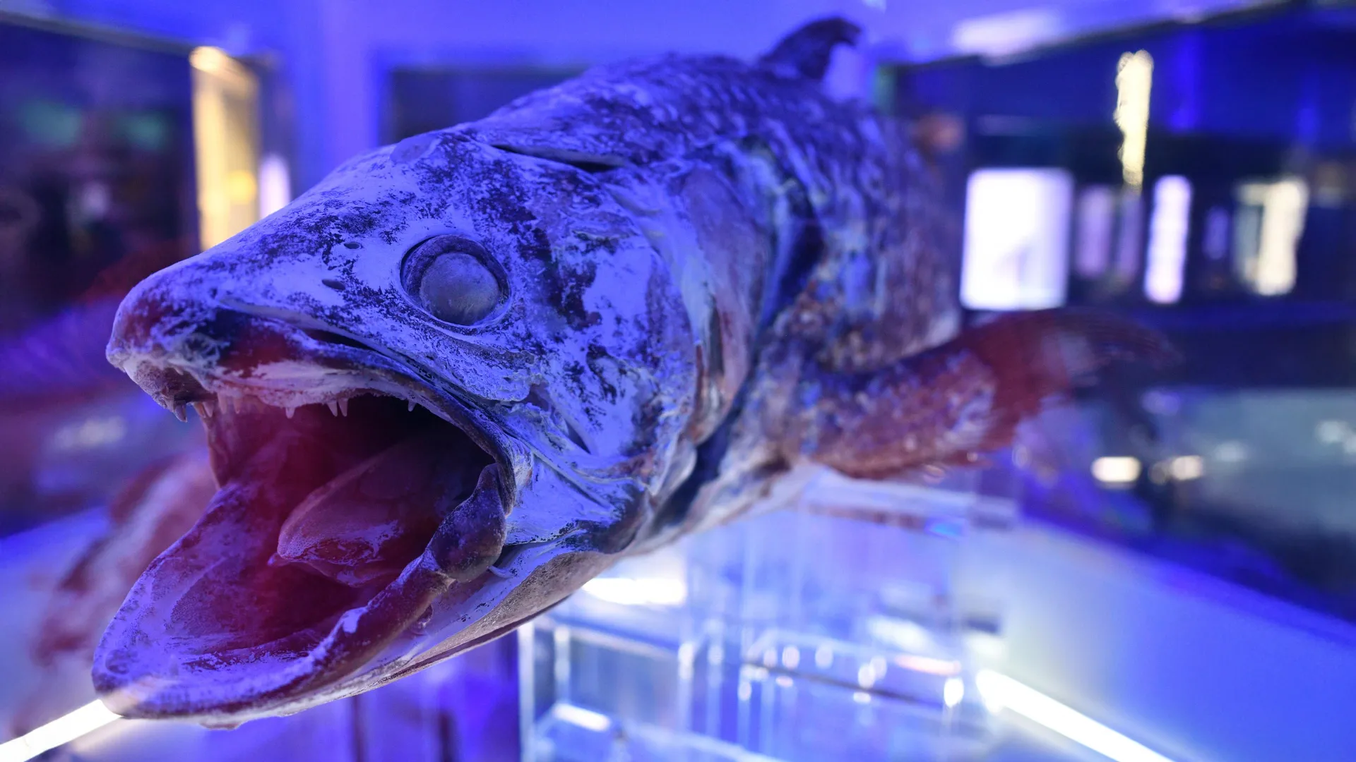
The coelacanth is known as a “living fossil” because its anatomy has changed little in the last 65 million years. Despite being one of the most studied fish in history, it continues to reveal new information that could transform our understanding of vertebrate evolution. This is revealed in a study published in the journal Science Advances by researchers from the University of São Paulo (USP) in Brazil and the Smithsonian Institution in the United States.
Upon re-examining the cranial musculature of the African coelacanth (Latimeria chalumnae), the authors discovered that only 13% of the previously identified evolutionary muscle novelties for the largest vertebrate lineages were accurate. The study also identified nine new evolutionary transformations related to innovations in feeding and respiration in these groups.
“Ultimately, it’s even more similar to cartilaginous fish [sharks, rays, and chimaeras] and tetrapods [birds, mammals, amphibians, and reptiles] than previously thought. And even more distinct from ray-finned fish, which make up about half of living vertebrates,” says Aléssio Datovo, a professor at the Museum of Zoology (MZ) at USP supported by FAPESP, who led the study.
Among the evolutionary novelties erroneously identified as present in coelacanths are muscles responsible for actively expanding the buccopharyngeal cavity, which extends from the mouth to the pharynx. This set of muscles is directly related to food capture and respiration. However, the study showed that these supposed muscles in coelacanths were actually ligaments, which are structures incapable of contraction.
Ray-finned fish (actinopterygii) and lobe-finned fish (sarcopterygii) diverged from a common ancestor approximately 420 million years ago. The sarcopterygii include fish such as coelacanths and lungfish, as well as all other tetrapods, because they evolved from an aquatic ancestor. These include mammals, birds, reptiles, and amphibians.
In ray-finned fish, such as aquarium carp, it is easy to see how the mouth moves to suck in food. This ability gave actinopterygii a significant evolutionary advantage; today, they comprise about half of all living vertebrates.
This is a fundamental difference from other fish, such as coelacanths and sharks, which primarily feed by biting their prey.
“In previous studies, it was assumed that this set of muscles that would give greater suction capacity was also present in coelacanths and, therefore, would have evolved in the common ancestor of bony vertebrates, which we now show isn’t true. This only appeared at least 30 million years later, in the common ancestor of living ray-finned fish,” points out Datovo.
Behind the scenes
Coelacanths are extremely rare fish that live about 300 meters below the surface of the water and spend their days in underwater caves.
One reason they have changed so little since the extinction of the dinosaurs is that they have few predators and live in a relatively protected environment. This has resulted in slow changes to their genome, as shown by a 2013 study published in the journal Nature.
Coelacanths were first known only from fossils from about 400 million years ago. It was not until 1938 that a living animal was discovered, much to the astonishment of scientists. In 1999, another species (Latimeria chalumnae) was discovered in Asian waters.
Due to the rarity of specimens in museums, researchers from USP and the Smithsonian Institution’s National Museum of Natural History had to persevere to find an institution willing to lend animals for dissection.
The Field Museum in Chicago and the Virginia Institute of Marine Science, both in the United States, finally agreed to lend one specimen each. According to Datovo, G. David Johnson, co-author of the article, deserves credit for obtaining the loan.
Johnson, born in 1945, was “probably the greatest fish anatomist of his time,” according to Datovo. He died in November 2024 after a domestic accident while the study was under review.
Contribution
“Contrary to what it may seem, dissecting a specimen does not mean destroying it as long as it’s done properly,” says Datovo.
The researcher, who has been conducting this type of study for over 20 years, spent six months separating all the muscles and skull bones of the coelacanth. These structures are now preserved and can be studied individually by other scientists, eliminating the need to dissect a new animal.
Seeing each muscle and nerve firsthand allowed the authors to identify what was actually in the coelacanth’s head with certainty, point out previously undescribed structures, and correct errors that had been repeated in the scientific literature for over 70 years.
“There were many contradictions in the literature. When we finally got to examine the specimens, we detected more errors than we’d imagined. For example, 11 structures described as muscles were actually ligaments or other types of connective tissue. This has a drastic consequence for the functioning of the mouth and breathing, because muscles perform movement, while ligaments only transmit it,” he explains.
Due to the position of coelacanths in the vertebrate tree of life, the discovery impacts our understanding of cranial evolution in all other large vertebrate groups.
With this information, the researcher used three-dimensional microtomography images of the skulls of other groups of fish, both extinct and living. These images are made available by other researchers who study fish anatomy when they perform 3D scans.
From images of the skull bones of other fish from completely extinct lineages, Datovo and Johnson were able to infer where the muscles found in coelacanths would fit, elucidating the evolution of these muscles in the first jawed vertebrates. In future work, Datovo intends to analyze similarities with the muscles of tetrapods, such as amphibians and reptiles.
