Florida cat sniffs out another new virus—and scientists are listening

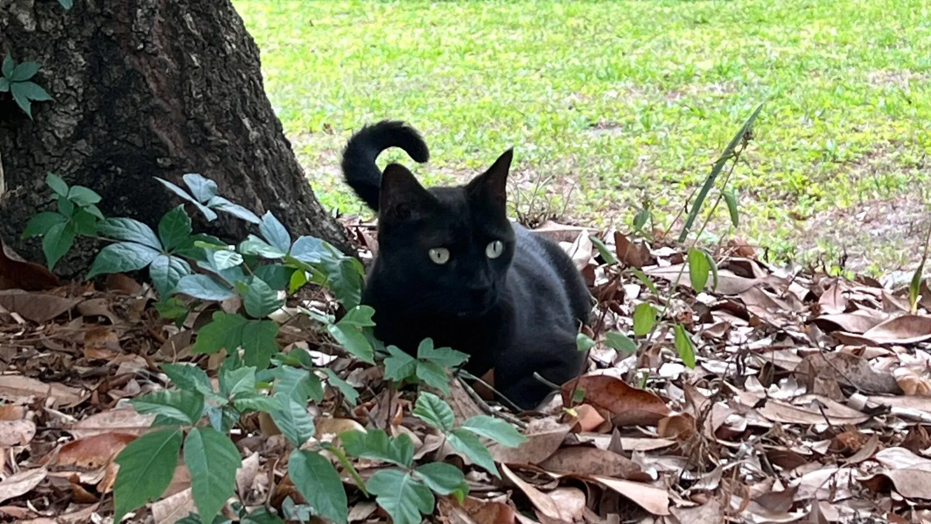
Pepper, the pet cat who made headlines last year for his role in the discovery of the first jeilongvirus found in the U.S., is at it again. This time, his hunting prowess contributed to the identification of a new strain of orthoreovirus.

John Lednicky, Ph.D., Pepper’s owner and a University of Florida College of Public Health and Health Professions virologist, took Pepper’s catch — a dead Everglades short-tailed shrew — into the lab for testing as part of his ongoing work to understand transmission of the mule deerpox virus.

Testing revealed the shrew had a previously unidentified strain of orthoreovirus. Viruses in this genus are known to infect humans, white-tailed deer, bats and other mammals. While orthoreoviruses’ effects on humans are not yet well understood, there have been rare reports of the virus being associated with cases of encephalitis, meningitis and gastroenteritis in children.

“The bottom line is we need to pay attention to orthoreoviruses, and know how to rapidly detect them,” said Lednicky, a research professor in the PHHP Department of Environmental and Global Health and a member of UF’s Emerging Pathogens Institute.

The UF team published the complete genomic coding sequences for the virus they named “Gainesville shrew mammalian orthoreovirus type 3 strain UF-1” in the journal Microbiology Resource Announcements.

“There are many different mammalian orthoreoviruses and not enough is known about this recently identified virus to be concerned,” said the paper’s lead author Emily DeRuyter, a UF Ph.D. candidate in One Health. “Mammalian orthoreoviruses were originally considered to be ‘orphan’ viruses, present in mammals including humans, but not associated with diseases. More recently, they have been implicated in respiratory, central nervous system and gastrointestinal diseases.”

The Lednicky lab’s jeilongvirus and orthoreovirus discoveries come on the heels of the team publishing their discovery of two other novel viruses found in farmed white-tailed deer. Given the propensity of viruses to constantly evolve, paired with the team’s sophisticated lab techniques, finding new viruses isn’t entirely surprising, Lednicky said.

“I’m not the first one to say this, but essentially, if you look, you’ll find, and that’s why we keep finding all these new viruses,” Lednicky said.

Like influenza virus, two different types of orthoreovirus can infect a host cell, causing the viruses’ genes to mix and match, in essence, creating a brand new virus, Lednicky said.

In 2019, Lednicky and colleagues isolated the first orthoreovirus found in a deer. That strain’s genes were nearly identical to an orthoreovirus found in farmed mink in China and a deathly ill lion in Japan. How in the world, the scientific community wondered, could the same hybrid virus appear in a farmed deer in Florida and two species of carnivores across the globe? Some experts speculated that components of the animals’ feed could have come from the same manufacturer.

With so many unanswered questions about orthoreoviruses and their modes of transmission, prevalence in human and animal hosts and just how sick they could make us, more research is needed, DeRuyter and Lednicky said.

Next steps would include serology and immunology studies to understand the threat Gainesville shrew mammalian orthoreovirus type 3 strain UF-1 may hold for humans, wildlife and pets.

For readers concerned about Pepper’s health, rest assured. He has shown no signs of illness from his outdoor adventures and will likely continue to contribute to scientific discovery through specimen collection.

“This was an opportunistic study,” Lednicky said. “If you come across a dead animal, why not test it instead of just burying it? There is a lot of information that can be gained.”

Share Button

Scientists just found 200+ hidden proteins that may drive Alzheimer’s

For decades, the story of Alzheimer’s research has been dominated by a battle between A-beta and tau amyloids, both of which can kill neurons and impact the brain’s ability to function. A new study suggests, however, that these sticky brain plaques may not be operating alone.

Johns Hopkins University researchers have identified more than 200 types of misfolded proteins in rats that could be associated with age-related cognitive decline.

The findings could lead the way to finding new therapeutic targets and treatments in humans that could provide relief for the millions of people over 65 who suffer from Alzheimer’s, dementia, or other diseases that rob them of their memories and independence as they age.

“Amyloids are the buildup of misshapen proteins. They’re big and ugly and easy to see under the microscope, so it makes sense that they catch our attention. But we’re seeing hundreds of proteins misfolding in ways that don’t clump together in an amyloid and yet still seem to impact how the brain functions,” said Stephen Fried, an assistant professor of chemistry and protein scientist who studies how molecules in the brain change during aging. “Our research is showing that amyloids are just the tip of the iceberg.”

The results were published on July 11 in Science Advances.

To understand the molecular differences between older brains that are mentally sharp and those that are experiencing decline, Fried and his team studied 17 2-year-old rats that grew up in the same colony. Seven rats performed poorly on memory and problem-solving tests and were considered cognitively impaired, while 10 performed as well as 6-month-old rats.

The researchers then measured more than 2,500 types of protein in the hippocampus, the part of the brain associated with spatial learning and memory. For the first time, scientists were able to determine for a large number of proteins whether individual proteins were misshapen or folded incorrectly, allowing the researchers to work out which proteins misfold for all the rats and are associated with aging in general versus which proteins specifically misfold in cognitively impaired rats.

More than 200 proteins were misfolded in the cognitively impaired rats yet maintained their shapes in the cognitively healthy rats. The findings suggest that some of those proteins are contributing to cognitive decline, the researchers said.

Misfolded proteins are unable to carry out tasks necessary for a cell to function properly, so cells have a natural surveillance system that identifies and destroys these misbehaving proteins. Previously, researchers thought misfolded proteins — specifically A-beta and tau proteins — were only disruptive when they clumped into amyloids.

“We think there are a lot of proteins that can be misfolded, not form amyloids, and still be problematic,” Fried said. “And that suggests these misfolded proteins have ways of escaping this surveillance system in the cell.”

But exactly how those misfolded proteins slip past a cell’s security system remains a mystery.

Next, the team plans to look at misfolded proteins under high-resolution microscopes to get a more detailed picture of what their deformities look like at the molecular level.

“A lot of us have experienced a loved one or a relative who has become less capable of doing those everyday tasks that require cognitive abilities,” Fried said. “Understanding what’s physically going on in the brain could lead to better treatments and preventive measures.”

Share Button

Inside the Maya king’s tomb that rewrites Mesoamerican history

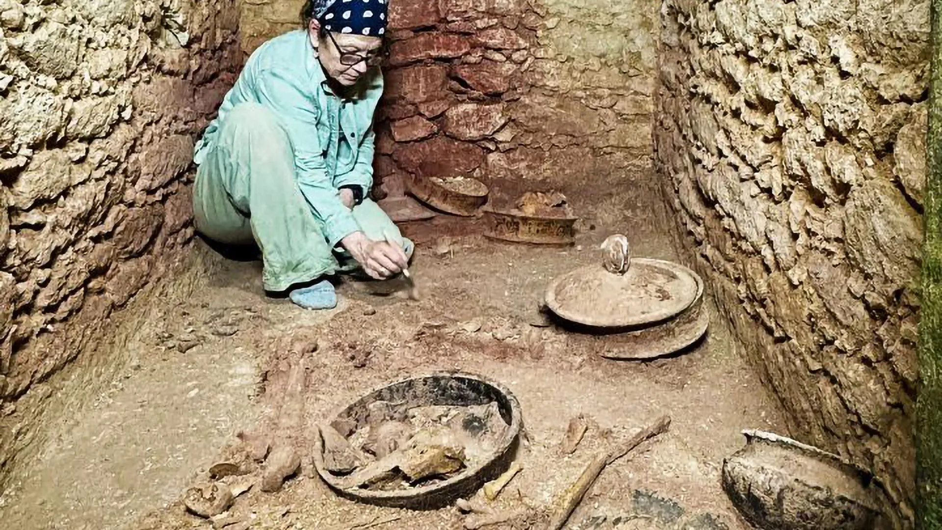
Archaeologists from the University of Houston working at Caracol in Belize, Central America have uncovered the tomb of Te K’ab Chaak, the first ruler of this ancient Maya city and the founder of its royal dynasty. Now in ruins, this metropolis was a major political player in Maya history, dominating the southern part of the Yucatan Peninsula from 560 through 680 AD before its abandonment by 900 AD.

The discovery is the first identifiable ruler’s tomb found in over four decades of work in Caracol, the largest Maya archaeological site in Belize and in the Maya lowlands.

Te K’ab Chaak, who acceded to the throne in 331 AD, was interred at the base of a royal family shrine with eleven pottery vessels, carved bone tubes, jadeite jewelry, a mosaic jadeite mask, Pacific spondylus shells, and other perishable materials. Pottery vessels in the chamber included a scene of a Maya ruler holding a spear and receiving offerings from supplicants in the form of deities. Another vessel portrays the image of Ek Chuah, Maya god of traders, surrounded by offerings. Four of the pottery vessels portray bound captives (similar vessels also appeared in two related burials). Two vessels supported lids with modeled handles of coatimundi (pisote) heads. The coatimundi, or tz’uutz’ in Maya, was used by subsequent Caracol rulers as part of their names.

A team led by husband and wife collaborators Arlen F. Chase and Diane Z. Chase of the University of Houston has been excavating at Caracol for more than 40 years; this field season was carried out in concert with Belize’s Institute of Archaeology and was supported by the Alphawood Foundation, the University of Houston, the Geraldine and Emory Ford Foundation, and the KHR Family Fund.

The Chases estimate that at the time of his burial the ruler was of advanced age and approximately 5’7″ in height. He had no remaining teeth.

Their investigations at Caracol’s Northeast Acropolis show that Te K’ab Chaak’s tomb was the first of three major burials dating to about 350 AD, a time of early contact with the central Mexican city of Teotihuacan, some 1200 kilometers distant. By 300 AD, Teotihuacan was a huge city that traded throughout Central America.

“One question that has perplexed Maya archaeologists since the 1960s is whether a new political order was introduced to the Maya area by Mexicans from Teotihuacan,” said Diane Z. Chase, archeologist and senior vice president for academic affairs and Provost at the University of Houston. “Maya carved stone monuments, hieroglyphic dates, iconography, and archaeological data all suggest that widespread pan-Mesoamerican connections occurred after an event in 378 AD referred to as the “entrada.” Whether this event represented actual Teotihuacanos in the Maya area or Maya using central Mexican symbols is still debated. The Caracol archaeological data suggests that the situation was far more complicated.”

A cremation placed in the center of Caracol’s Northeast Acropolis plaza, recovered in 2010 and placed after Te K’aab Chaak’s burial has been dated to AD 350 by radiocarbon analysis and included artifacts from central Mexico. It contained the remains of three individuals, as well as two large knives, six atlatl points, and fifteen pristine blades of green obsidian from Pachuca, Mexico (north of Teotihuacan); several pottery vessels also likely came from central Mexico. Additionally, a carved atlatl projectile tip, atypical for the Maya but typical for a Teotihuacan warrior, was included in the cremation.

The cremation itself and its placement in the center of a residential plaza are also more typical practices for a high-status Teotihuacano and do not accord with standard Maya burial practices. Based on other ceramics in this cremation, the main individual was likely a Caracol royal family member that had adopted central Mexican ritual practices. This individual may even have served as a royal Maya envoy who had lived at Teotihuacan and returned to Caracol.

A third burial – the tomb of a woman, also covered with hematite and containing four pottery vessels, a spondylus bead necklace, mirror fragments, and two Pacific spondylus shells – was recovered in the northern building of the same residential group in 2009 and is similarly dated.

The three burials interred in the Caracol Northeast Acropolis all cluster at AD 350, at least a generation before the previously recognized Teotihuacan presence in the Maya area. They demonstrate that early Maya rulers were fully enmeshed in Mesoamerican-wide contacts prior to the Teotihuacan entrada recorded on Maya monument[s].

“Both central Mexico and the Maya area were clearly aware of each other’s ritual practices, as reflected in the Caracol cremation, said Arlen F. Chase, professor and chair of Comparative Cultural Studies at the University of Houston. “The connections between the two regions were undertaken by the highest levels of society, suggesting that initial kings at various Maya cities – such as Te K’ab Chaak at Caracol – were engaged in formal diplomatic relationships with Teotihuacan.” The royal dynasty established by Te K’ab Chaak continued at Caracol for over 460 years.

The Chase’s findings also indicate that ancient peoples in the new world were travelers. A trip between Teotihuacan and Caracol today by car would take over 23 hours. The one-way walking time may be estimated to be approximately 153 days.

Research continues on the contents of the chamber with the reconstruction of the jadeite death mask and with ancient DNA and stable isotope analysis of the skeletal material. The Chases will present results of the 2025 Caracol field season at a conference on Maya-Teotihuacan interaction hosted by the Maya Working Group at the Santa Fe Institute (New Mexico) in August 2025.

About the Chases

Married since 1975, Diane and Arlen Chase are two of the most influential Maya archaeologists in the world, and the foremost experts on Caracol, one of the most significant ancient Maya cities.

Since their first dig in 1985, the Chases have become known for:

  • Providing a nuanced picture of Caracol’s ancient landscape and people, including recognition of Caracol as a city of advanced urban planning, road systems and markets and a large non-elite population upending the notion of a strict Maya social hierarchy. In 1986 they discovered an inscription that told of Caracol’s military victory over Tikal in 562 AD, overturning the myth that Tikal was the most powerful Classic Period city and illustrating that Caracol was a major military and political power.

  • Using airborne LiDAR (light detection and ranging) technology to uncover structures hidden for centuries under dense jungle, revolutionizing how archaeologists explore Maya sites. Their work from the 1980s-2000s revealed a web of interconnected roadways, proving that Caracol was a massive, urban and integrated city.

  • Overturning outdated beliefs about the Maya, while providing new insights. Their archaeological work has shown that ancient Maya society was composed of many different social gradients, contrary to more simple two-class models. From the 1980s -2000, they and their team mapped numerous causeways (or roads) and thousands of agricultural terraces spread out across Caracol as well as hundreds of reservoirs in non-elite areas, proving that water, sacred to the Maya, was not controlled by the elite, as previously thought. The Chases’ son, Adrian Chase, also an archaeologist, found that Caracol had a decentralized water system and that residential groups had their own access. He is currently studying the urban structure of Caracol and changes in governance over time.

The Chases will present detailed information on the tomb and other finds from Caracol at the Santa Fe Institute in August.

Share Button

Alzheimer’s has isolated us, says Fiona Phillips’ husband

Martin Frizell has spoken of the social isolation faced by the couple since the former GMTV presenter was diagnosed.

Share Button

‘Don’t tell me my baby wasn’t meant to be’

Siobhan Gorman was 16 weeks pregnant and home alone when she gave birth to baby Archie.

Share Button

Some GPs to offer coaching to get sick people back to work

The aim is to help people return to work quicker and reduce the length of time they need fit notes – better known as sick notes.

Share Button

Vaccine roll-outs cut deaths by 60% – study

The study looked at vaccines deployed during outbreaks of five deadly diseases.

Share Button

TV doctor and IVF pioneer quits BMA over strikes

The Labour peer says the strikes could damage public trust in doctors.

Share Button

In seconds, AI builds proteins to battle cancer and antibiotic resistance

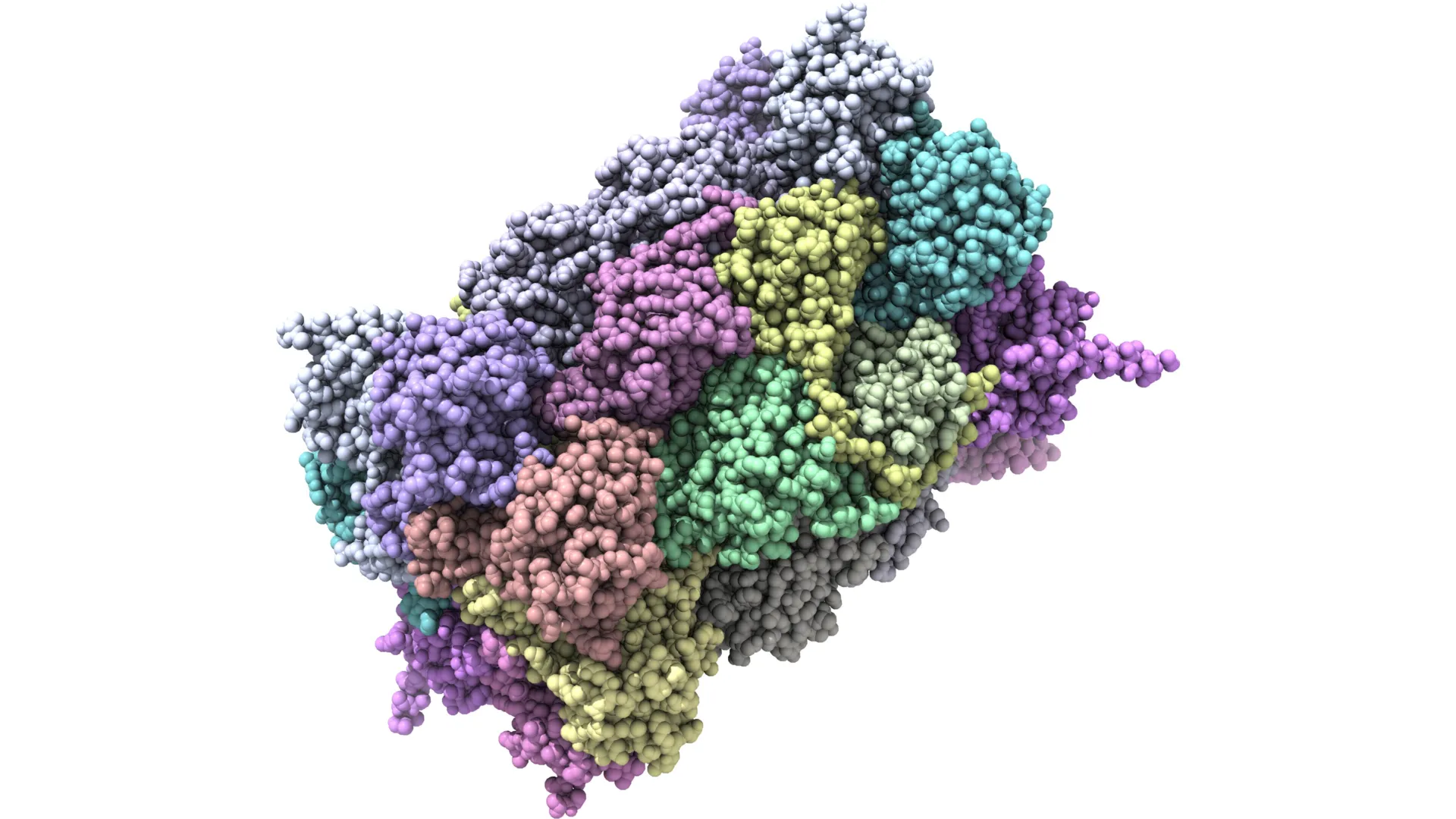
In the last year, there has been a surge in proteins developed by AI that will eventually be used in the treatment of everything from snakebites to cancer. What would normally take decades for a scientist to create — a custom-made protein for a particular disease — can now be done in seconds.

For the first time, Australian scientists have used Artificial Intelligence (AI) to generate a ready-to-use biological protein, in this case, one that can kill antibiotic resistant bacteria like E. coli.

This study, published in Nature Communications, provides a new way to combat the growing crisis caused by antibiotic resistant super bugs. By using AI in this way, Australian science has now joined countries like the US and China having developed AI platforms capable of rapidly generating thousands of ready-to-use proteins, paving the way for faster, more affordable drug development and diagnostics that could transform biomedical research and patient care.

The Nature Communications paper is co-led by Dr. Rhys Grinter and Associate Professor Gavin Knott, a Snow Medical Fellow, who lead the new AI Protein Design Program with nodes at the University of Melbourne Bio21 Institute and Monash Biomedicine Discovery Institute.

According to Dr. Grinter and A/Prof. Knott, the AI Protein Design Platform used in this work is the first in Australia that models the work done by David Baker (who won the Nobel Prize in Chemistry last year) developing an end-to-end approach that could create a wide range of proteins. “These proteins are now being developed as pharmaceuticals, vaccines, nanomaterials and tiny sensors, with many other applications yet to be tested” Associate Professor Knott said.

For this study, the AI Protein Design Platform used AI-driven protein design tools that are freely available for scientists everywhere. “It’s important to democratize protein design so that the whole world has the ability to leverage these tools,” said Daniel Fox, the PhD student who performed most of the experimental work for the study. “Using these tools and those we are developing in-house, we can engineer proteins to bind a specific target site or ligand, as inhibitors, agonists or antagonists, or engineered enzymes with improved activity and stability.”

According to Dr Grinter, currently proteins used in the treatment of diseases like cancer or infections are derived from nature and repurposed through rational design or in vitro evolution and selection. “These new methods in deep learning enable efficient de novo design of proteins with specific characteristics and functions, lowering the cost and accelerating the development of novel protein binders and engineered enzymes,” he said.

Since the work of David Baker, new tools and software are being developed, such as Bindcraft and Chai which have been incorporated into an AI Protein Design Platform co-led by Dr. Grinter and A/Prof. Knott..

Professor John Carroll, Director of the Monash Biomedicine Discovery Institute, said the new AI Protein Design Program ‘brings Australia “right up to speed in this exciting new modality for designing novel therapeutics and research tools. It is testament to the entrepreneurial spirit of two fabulous young scientists who have worked night and day to build this capability from scratch.”

“The Program, based at Monash University and the University of Melbourne, is run by a team of talented structural biologists and computer scientists who understand the design process from end-to-end. This in-depth knowledge of protein structure and machine learning makes us a highly agile program capable of regularly onboarding cutting edge tools in AI-protein design,” Associate Professor Knott said.

Share Button

How a hidden brain circuit fuels fibromyalgia, migraines, and PTSD

Pain isn’t just a physical sensation — it also carries emotional weight. That distress, anguish, and anxiety can turn a fleeting injury into long-term suffering.

Researchers at the Salk Institute have now identified a brain circuit that gives physical pain its emotional tone, revealing a new potential target for treating chronic and affective pain conditions such as fibromyalgia, migraine, and post-traumatic stress disorder (PTSD).

Published on July 9, 2025, in Proceedings of the National Academy of Sciences, the study identifies a group of neurons in a central brain area called the thalamus that appears to mediate the emotional or affective side of pain in mice. This new pathway challenges the textbook understanding of how pain is processed in the brain and body.

“For decades, the prevailing view was that the brain processes sensory and emotional aspects of pain through separate pathways,” says senior author Sung Han, associate professor and holder of the Pioneer Fund Developmental Chair at Salk. “But there’s been debate about whether the sensory pain pathway might also contribute to the emotional side of pain. Our study provides strong evidence that a branch of the sensory pain pathway directly mediates the affective experience of pain.”

The physical sensation of pain is what allows you to immediately detect it, assess its intensity, and identify its source. The affective part of pain is what makes it so unpleasant. This emotional discomfort motivates you to take action and helps you learn to associate negative feelings with the situation so you can avoid it in the future.

This is a critical distinction. Most people start to perceive pain at the same stimulus intensities, meaning we all process the sensory side of pain fairly similarly. In comparison, our ability to tolerate pain varies greatly. How much we suffer or feel threatened by pain is determined by our affective processing, and if that becomes too sensitive or lasts too long, it can result in a pain disorder. This makes it important to understand which parts of the brain control these different dimensions of pain.

Sensory pain was thought to be mediated by the spinothalamic tract, a pathway that sends pain signals from the spinal cord to the thalamus, which then relays them to sensory processing areas across the brain.

Affective pain was generally thought to be mediated by a second pathway called the spinoparabrachial tract, which sends pain information from the spinal cord into the brainstem.

However, previous studies using older research methods have suggested the circuitry of pain may be more complex. This long-standing debate inspired Han and his team to revisit the question with modern research tools.

Using advanced techniques to manipulate the activity of specific brain cells, the researchers discovered a new spinothalamic pathway in mice. In this circuit, pain signals are sent from the spinal cord into a different part of the thalamus, which has connections to the amygdala, the brain’s emotional processing center. This particular group of neurons in the thalamus can be identified by their expression of CGRP (calcitonin gene-related peptide), a neuropeptide originally discovered in Professor Ronald Evans’ lab at Salk.

When the researchers “turned off” (genetically silenced) these CGRP neurons, the mice still reacted to mild pain stimuli, such as heat or pressure, indicating their sensory processing was intact. However, they didn’t seem to associate lasting negative feelings with these situations, failing to show any learned fear or avoidance behaviors in future trials. On the other hand, when these same neurons were “turned on” (optogenetically activated), the mice showed clear signs of distress and learned to avoid that area, even when no pain stimuli had been used.

“Pain processing is not just about nerves detecting pain; it’s about the brain deciding how much that pain matters,” says first author Sukjae Kang, a senior research associate in Han’s lab. “Understanding the biology behind these two distinct processes will help us find treatments for the kinds of pain that don’t respond to traditional drugs.”

Many chronic pain conditions — such as fibromyalgia and migraine — involve long, intense, unpleasant experiences of pain, often without a clear physical source or injury. Some patients also report extreme sensitivity to ordinary stimuli like light, sound, or touch, which others would not perceive as painful.

Han says overactivation of the CGRP spinothalamic pathway may contribute to these conditions by making the brain misinterpret or overreact to sensory inputs. In fact, transcriptomic analysis of the CGRP neurons showed that they express many of the genes associated with migraine and other pain disorders.

Notably, several CGRP blockers are already being used to treat migraines. This study may help explain why these medications work and could inspire new nonaddictive treatments for affective pain disorders.

Han also sees potential relevance for psychiatric conditions that involve heightened threat perception, such as PTSD. Growing evidence from his lab suggests that the CGRP affective pain pathway acts as part of the brain’s broader alarm system, detecting and responding to not only pain but a wide range of unpleasant sensations. Quieting this pathway with CGRP blockers could offer a new approach to easing fear, avoidance, and hypervigilance in trauma-related disorders.

Importantly, the relationship between the CGRP pathway and the psychological pain associated with social experiences like grief, loneliness, and heartbreak remains unclear and requires further study.

“Our discovery of the CGRP affective pain pathway gives us a molecular and circuit-level explanation for the difference between detecting physical pain and suffering from it,” says Han. “We’re excited to continue exploring this pathway and enabling future therapies that can reduce this suffering.”

Other authors include Shijia Liu, Jong-Hyun Kim, Dong-Il Kim, Tae Gyu Oh, Jiahang Peng, Mao Ye, Kuo-Fen Lee, Ronald M. Evans, and Martyn Goulding of Salk.

The work was supported by the National Institutes of Mental Health (BRAINS grant 1R01MH116203) and the Simons Foundation (Bridge to Independence award SFARI #388708).

Share Button