Multisensory VR forest reboots your brain and lifts mood—study confirms

In Japan, Shinrin Yoku or forest bathing has already been used for therapeutic applications, for instance, to lower blood pressure and stress levels. For their study, the researchers wanted to find out whether forest bathing – consciously immersing oneself in nature – can also be effective when done virtually, and focused on whether the positive effect is stronger when several senses are addressed simultaneously.

For the project, a high-quality 360° VR video was produced in Europe’s largest Douglas fir forest, the Sonnenberg nature reserve near Parchim – complete with original sounds and the scent of essential oils from the Douglas fir. The participants experienced the virtual forest scenery either as a full sensory experience (with images, sound and scent) or in a reduced form whereby forest stimuli appealing to just a single sense – visual, auditory or olfactory – were used. In variants where only hearing or scent was activated, participants were placed in a neutral virtual environment to minimize visual stimuli and the influence of VR technology.

Significantly better effect with sensory combination

More than 130 participants were first put into an acute stress situation using stress-inducing images. Then, equipped with VR glasses, they experienced one of the four forest stimulation/ bathing variants. The results show that the combination of all three sensory stimuli led to a significantly greater improvement in mood and a stronger feeling of connection with nature compared to when individual sensory stimuli were presented. In addition to positive effects on mood, there were also limited improvements in working memory – the cognitive function that enables us to store, process and retrieve information in the short term.

However, the researchers point out that the effects are area-specific and cannot yet be considered universally valid. Further studies with larger samples are needed to confirm the results and provide a better understanding of the mechanisms behind the restorative effects of virtual nature experiences.

“We can already say that digital nature experiences can absolutely produce an emotional effect – even if they don’t replace actual nature,” reports Leonie Ascone, lead author of the study and researcher in the Neuronal Plasticity working group at the University Medical Center Hamburg-Eppendorf (UKE).

Potential for clinics, waiting rooms and urban spaces

Simone Kühn, head of the study and Director of the Center for Environmental Neuroscience at the Max Planck Institute for Human Development, adds: “Especially in places with limited access to nature – such as clinics, waiting areas or urban interiors – multisensory VR applications or targeted nature staging could support mental well-being. The images, sounds and scents of nature offer previously underestimated potential for improving mood and mental performance in everyday situations.” Kühn conducts intensive research into the effects of the environment on the human brain and, together with colleagues from universities in Vienna, Exeter and Birmingham, was recently able to prove that just from watching nature videos, patients perceive physical pain as less intense (Steininger et al., 2025).

In brief:

  • Forest bathing in Virtual Reality improves emotional well-being and increases connectedness to nature, particularly when several senses (sight, hearing, smell) are simultaneously engaged
  • The study used a 360° VR forest video complete with original sounds and the scent of Douglas fir essential oils
  • There is potential for application especially in clinical, urban and other environments with limited access to nature
Share Button

Pregnancy’s 100-million-year secret: Inside the placenta’s evolutionary power play

An international research team led by scientists from the University of Vienna has uncovered new insights into how specialized cell types and communication networks at the interface between mother and fetus evolved over millions of years. These discoveries shed light on one of nature’s most remarkable innovations – the ability to sustain a successful pregnancy. The findings have just been published in Nature Ecology & Evolution.

Pregnancy that lasts long enough to support full fetal development is a hallmark evolutionary breakthrough of placental mammals – a group that includes humans. At the center of this is the fetal-maternal interface: the site in the womb where a baby’s placenta meets the mother’s uterus, and where two genetically distinct organisms – mother and fetus – are in intimate contact and constant interaction. This interface has to strike a delicate balance: intimate enough to exchange nutrients and signals, but protected enough to prevent the maternal immune system from rejecting the genetically “foreign” fetus.

To uncover the origins and mechanisms behind this intricate structure, the team analyzed single-cell transcriptomes – snapshots of active genes in individual cells – from six mammalian species representing key branches of the mammalian evolutionary tree. These included mice and guinea pigs (rodents), macaques and humans (primates), and two more unusual mammals: the tenrec (an early placental mammal) and the opossum (a marsupial that split off from placental mammals before they evolved complex placentas).

A Cellular “Atlas of Mammal Pregnancy”

By analyzing cells at the fetal-maternal interface, the researchers were able to trace the evolutionary origin and diversification of the key cell types involved. Their focus was on two main players: placenta cells, which originate from the fetus and invade maternal tissue, and uterine stromal cells, which are of maternal origin and respond to this invasion.

Using molecular biology tools, the team identified distinct genetic signatures – patterns of gene activity unique to specific cell types and their specialized functions. Notably, they discovered a genetic signature associated with the invasive behavior of fetal placenta cells that has been conserved in mammals for over 100 million years. This finding challenges the traditional view that invasive placenta cells are unique to humans, and reveals instead that they are a deeply conserved feature of mammalian evolution. During this time, the maternal cells weren’t static, either. Placental mammals, but not marsupials, were found to have acquired new forms of hormone production, a pivotal step toward prolonged pregnancies and complex gestation, and a sign that the fetus and the mother could be driving each other’s evolution.

Cellular Dialogue: Between Cooperation and Conflict

To better understand how the fetal-maternal interface functions, the study tested two influential theories about the evolution of cellular communication between mother and fetus.

The first, the “Disambiguation Hypothesis,” predicts that over evolutionary time, hormonal signals became clearly assigned to either the fetus or the mother – a possible safeguard to ensure clarity and prevent manipulation. The results confirmed this idea: certain signals, including WNT proteins, immune modulators, and steroid hormones, could be clearly traced back to one source tissue.

The second, the “Escalation Hypothesis” (or “genomic Conflict”), suggests an evolutionary arms race between maternal and fetal genes – with, for example, the fetus boosting growth signals while the maternal side tries to dampen them. This pattern was observed in a small number of genes, notably IGF2, which regulates growth. On the whole, evidence pointed to fine-tuned cooperative signaling.

“These findings suggest that evolution may have favored more coordination between mother and fetus than previously assumed,” says Daniel J. Stadtmauer, lead author of the study and now a researcher at the Department of Evolutionary Biology, University of Vienna. “The so-called mother-fetus power struggle appears to be limited to specific genetic regions. Rather than asking whether pregnancy as a whole is conflict or cooperation, a more useful question may be: where is the conflict?”

Single-Cell Analysis: A Key to Evolutionary Discovery

The team’s discoveries were made possible by combining two powerful tools: single-cell transcriptomics – which captures the activity of genes in individual cells – and evolutionary modeling techniques that help scientists reconstruct how traits might have looked in long-extinct ancestors. By applying these methods to cell types and their gene activity, the researchers could simulate how cells communicate in different species, and even glimpse how this dialogue has evolved over millions of years.

“Our approach opens a new window into the evolution of complex biological systems – from individual cells to entire tissues,” says Silvia Basanta, co-first author and researcher at the University of Vienna. The study not only sheds light on how pregnancy evolved, but also offers a new framework for tracking evolutionary innovations at the cellular level – insights that could one day improve how we understand, diagnose, or treat pregnancy-related complications.

The research was conducted in the labs of Mihaela Pavličev at the Department of Evolutionary Biology, University of Vienna, and Günter Wagner at Yale University. Wagner is Professor Emeritus at Yale and a Senior Research Fellow at the University of Vienna. The study was supported by the John Templeton Foundation and the Austrian Science Fund (FWF).

Share Button

‘Menopause made me forget my name’

Kirsty Dixon from York runs support groups for women going through the menopause.

Share Button

Scientists thought the Arctic was sealed in ice — they were wrong

For years, scientists have debated whether a giant thick ice shelf once covered the entire Arctic Ocean during the coldest ice ages. Now a new study published in Science Advances, challenges this idea as the research team found no evidence for the presence of a massive ~1km ice shelf. Instead, the Arctic Ocean appears to have been covered by seasonal sea ice — leaving open water and life-sustaining conditions even during the harshest periods of cold periods during the last 750,000 years. This discovery gives insights crucial for our understanding of how the Arctic has responded to climate change in the past — and how it might behave in the future.

Tiny traces of life in ancient mud

Led by the European Research Council Synergy Grant project Into the Blue — i2B, the research team studied sediment cores collected from the seafloor of the central Nordic Seas and Yermak Plateau, north of Svalbard. These cores hold tiny chemical fingerprints from algae that lived in the ocean long ago. Some of these algae only grow in open water, while others thrive under seasonal sea ice that forms and melts each year.

“Our sediment cores show that marine life was active even during the coldest times,” said Jochen Knies, lead author of the study, based at UiT The Arctic University of Norway and co-lead of the Into The Blue — i2B project. “That tells us there must have been light and open water at the surface. You wouldn’t see that if the entire Arctic was locked under a kilometre-thick slab of ice.”

One of the key indicators the team looked for was a molecule called IP25, which is produced by algae that live in seasonal sea ice. Its regular appearance in the sediments shows that sea ice came and went with the seasons, rather than staying frozen solid all year round.

Simulating ancient Arctic climates

To test the findings based on the geological records, the research team used the AWI Earth System Model — a high-resolution computer model — to simulate Arctic conditions during two especially cold periods: the Last Glacial Maximum around 21,000 years ago, and a deeper freeze about 140,000 years ago when large ice sheets covered a lot of the Arctic.

“The models support what we found in the sediments,” said Knies. “Even during these extreme glaciations, warm Atlantic water still flowed into the Arctic gateway. This helped keep some parts of the ocean from freezing over completely.”

The models also showed that the ice wasn’t static. Instead, it shifted with the seasons, creating openings in the ice where light could reach the water — and where life could continue to thrive. This research not only reshapes our view of past Arctic climates but also has implications for future climate predictions. Understanding how sea ice and ocean circulation responded to past climate extremes can improve models that project future changes in a warming world.

“These reconstructions help us understand what’s possible — and what’s not — when it comes to ice cover and ocean dynamics,” said Gerrit Lohmann, co-author of this study, based at Alfred Wegener Institute, Helmholtz Centre for Polar and Marine Research (AWI) and co-lead of Into The Blue — i2B. “That matters when trying to anticipate how ice sheets and sea ice might behave in the future.”

Re-thinking the giant ice shelf theory

Some scientists have argued that features on the Arctic seafloor suggest that a huge, grounded ice shelf once covered the entire ocean. But this new study offers another explanation.

“There may have been short-lived ice shelves in some parts of the Arctic during especially severe cold phases,” said Knies. “But we don’t see any sign of a single, massive ice shelf that covered everything for thousands of years.”

One possible exception could have occurred about 650,000 years ago, when biological activity in the sediment record dropped sharply. But even then, the evidence points to a temporary event, not a long-lasting frozen lid over the Arctic.

Understanding the Arctic’s future

The study sheds new light on how the Arctic has behaved under extreme conditions in the past. This matters because the Arctic is changing rapidly today. Knowing how sea ice and ocean circulation responded to past climate shifts helps scientists understand what might lie ahead.

“These past patterns help us understand what’s possible in future scenarios,” said Knies. “We need to know how the Arctic behaves under stress — and what tipping points to watch for — as the Arctic responds to a warming world.”

The full paper, “Seasonal sea ice characterized the glacial Arctic-Atlantic gateway over the past 750,000 years,” is available in Science Advances.

This research is part of the European Research Council Synergy Grant project Into the Blue — i2B and the Research Council of Norway Centre of Excellence, iC3: Centre for ice, Cryosphere, Carbon, and Climate.

Share Button

Frozen light switches: How Arctic microbes could revolutionize neuroscience

Imagine the magnificent glaciers of Greenland, the eternal snow of the Tibetan high mountains, and the permanently ice-cold groundwater in Finland. As cold and beautiful these are, for the structural biologist Kirill Kovalev, they are more importantly home to unusual molecules that could control brain cells’ activity.

Kovalev, EIPOD Postdoctoral Fellow at EMBL Hamburg’s Schneider Group and EMBL-EBI’s Bateman Group, is a physicist passionate about solving biological problems. He is particularly hooked by rhodopsins, a group of colorful proteins that enable aquatic microorganisms to harness sunlight for energy.

“In my work, I search for unusual rhodopsins and try to understand what they do,” said Kovalev. “Such molecules could have undiscovered functions that we could benefit from.”

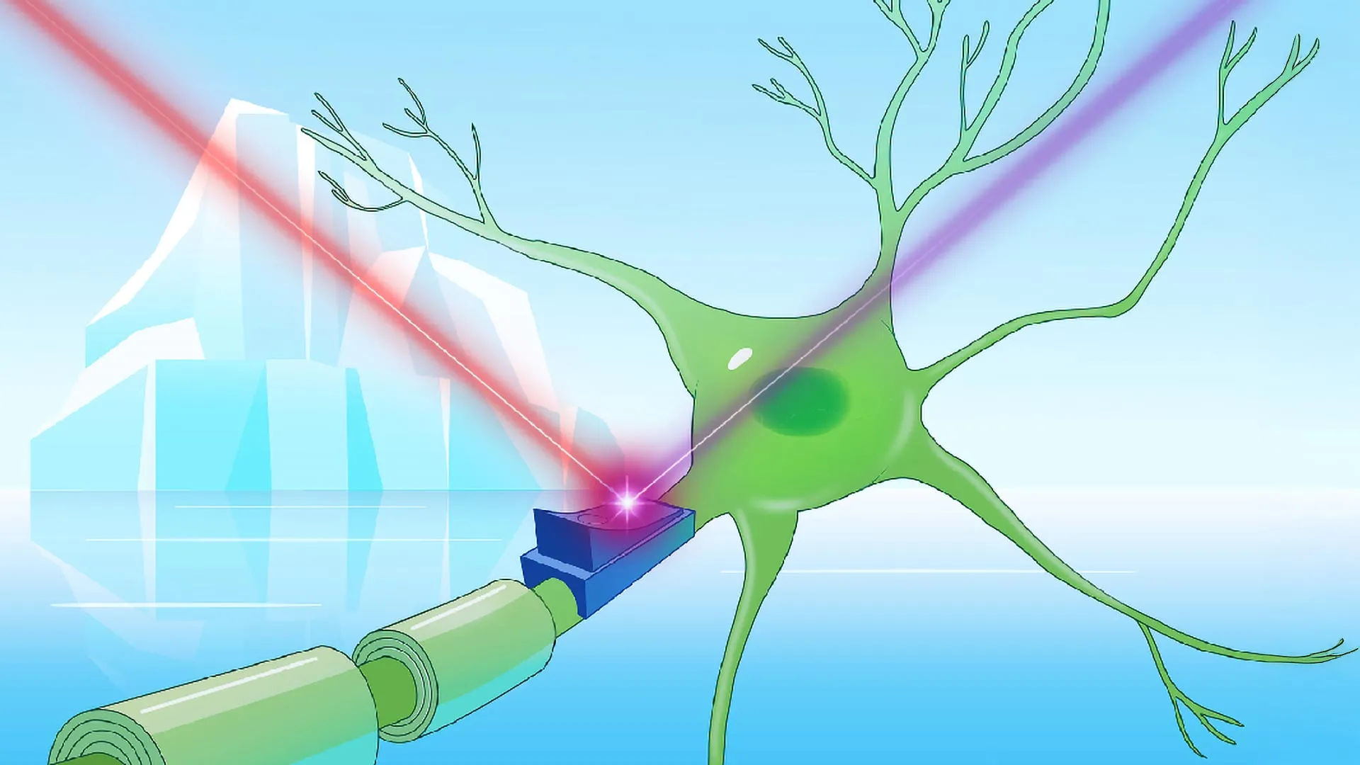
Some rhodopsins have already been modified to serve as light-operated switches for electrical activity in cells. This technique, called optogenetics, is used by neuroscientists to selectively control neuronal activity during experiments. Rhodopsins with other abilities, such as enzymatic activity, could be used to control chemical reactions with light, for example.

Having studied rhodopsins for years, Kovalev thought he knew them inside out – until he discovered a new, obscure group of rhodopsins that were unlike anything he had seen before.

As it often happens in science, it started serendipitously. While browsing online protein databases, Kovalev spotted an unusual feature common to microbial rhodopsins found exclusively in very cold environments, such as glaciers and high mountains. “That’s weird,” he thought. After all, rhodopsins are something you typically find in seas and lakes.

These cold-climate rhodopsins were almost identical to each other, even though they evolved thousands of kilometres apart. This couldn’t be a coincidence. They must be essential for surviving in the cold, concluded Kovalev, and to acknowledge this, he named them ‘cryorhodopsins’.

Rhodopsins out of the blue

Kovalev wanted to know more: what these rhodopsins look like, how they work, and, in particular, what color they are.

Color is the key feature of each rhodopsin. Most are pink-orange – they reflect pink and orange light, and absorb green and blue light, which activates them. Scientists strive to create a palette of different colored rhodopsins, so they could control neuronal activity with more precision. Blue rhodopsins have been especially sought-after because they are activated by red light, which penetrates tissues more deeply and non-invasively.

To Kovalev’s amazement, the cryorhodopsins he examined in the lab revealed an unexpected diversity of colors, and, most importantly, some were blue.

The color of each rhodopsin is determined by its molecular structure, which dictates the wavelengths of light it absorbs and reflects. Any changes in this structure can alter the color.

“I can actually tell what’s going on with cryorhodopsin simply by looking at its color,” laughed Kovalev.

Applying advanced structural biology techniques, he figured out that the secret to the blue color is the same rare structural feature that he originally spotted in the protein databases.

“Now that we understand what makes them blue, we can design synthetic blue rhodopsins tailored to different applications,” said Kovalev.

Next, Kovalev’s collaborators examined cryorhodopsins in cultured brain cells. When cells expressing cryorhodopsins were exposed to UV light, it induced electric currents inside them. Interestingly, if the researchers illuminated the cells right afterwards with green light, the cells became more excitable, whereas if they used UV/red light instead, it reduced the cells’ excitability.

“New optogenetic tools to efficiently switch the cell’s electric activity both ‘on’ and ‘off’ would be incredibly useful in research, biotechnology and medicine,” said Tobias Moser, Group Leader at the University Medical Center Göttingen who participated in the study. “For example, in my group, we develop new optical cochlear implants for patients that can optogenetically restore hearing in patients. Developing the utility of such a multi-purpose rhodopsin for future applications is an important task for the next studies.”

“Our cryorhodopsins aren’t ready to be used as tools yet, but they’re an excellent prototype. They have all the key features that, based on our findings, could be engineered to become more effective for optogenetics,” said Kovalev.

Evolution’s UV light protector

When exposed to sunlight even on a rainy winter day in Hamburg, cryorhodopsins can sense UV light, as shown using advanced spectroscopy by Kovalev’s collaborators from Goethe University Frankfurt led by Josef Wachtveitl. Wachtveitl’s team showed that cryorhodopsins are in fact the slowest among all rhodopsins in their response to light. This made the scientists suspect that those cryorhodopsins might act like photosensors letting the microbes ‘see’ UV light – a property unheard of among other cryorhodopsins.

“Can they really do that?” Kovalev kept asking himself. A typical sensor protein teams up with a messenger molecule that passes information from the cell membrane to the cell’s inside.

Kovalev grew more convinced, when together with his collaborators from Alicante, Spain, and his EIPOD co-supervisor, Alex Bateman from EMBL-EBI, they noticed that the cryorhodopsin gene is always accompanied by a gene encoding a tiny protein of unknown function – likely inherited together, and possibly functionally linked.

Kovalev wondered if this might be the missing messenger. Using the AI tool AlphaFold, the team were able to show that five copies of the small protein would form a ring and interact with the cryorhodopsin. According to their predictions, the small protein sits poised against the cryorhodopsin inside the cell. They believe that when cryorhodopsin detects UV light, the small protein could depart to carry this information into the cell.

“It was fascinating to uncover a new mechanism via which the light-sensitive signal from cryorhodopsins could be passed on to other parts of the cell. It is always a thrill to learn what the functions are for uncharacterised proteins. In fact, we find these proteins also in organisms that do not contain cryorhodopsin, perhaps hinting at a much wider range of jobs for these proteins.”

Why cryorhodopsins evolved their astonishing dual function – and why only in cold environments – remains a mystery.

“We suspect that cryorhodopsins evolved their unique features not because of the cold, but rather to let microbes sense UV light, which can be harmful to them,” said Kovalev. “In cold environments, such as the top of a mountain, bacteria face intense UV radiation. Cryorhodopsins might help them sense it, so they could protect themselves. This hypothesis aligns well with our findings.”

“Discovering extraordinary molecules like these wouldn’t be possible without scientific expeditions to often remote locations, to study the adaptations of the organisms living there,” added Kovalev. “We can learn so much from that!”

Unique approach to unique molecules

To reveal the fascinating biology of cryorhodopsins, Kovalev and his collaborators had to overcome several technical challenges.

One was that cryorhodopsins are nearly identical in structure, and even a slight change in the position of a single atom can result in different properties. Studying molecules at this level of detail requires going beyond standard experimental methods. Kovalev applied a 4D structural biology approach, combining X-ray crystallography at EMBL Hamburg beamline P14 and cryo-electron microscopy (cryo-EM) in the group of Albert Guskov in Groningen, Netherlands, with protein activation by light.

“I actually chose to do my postdoc at EMBL Hamburg, because of the unique beamline setup that made my project possible,” said Kovalev. “The whole P14 beamline team worked together to tailor the setup to my experiments – I’m very grateful for their help.”

Another challenge was that cryorhodopsins are extremely sensitive to light. For this reason, Kovalev’s collaborators had to learn to work with the samples in almost complete darkness.

Share Button

Can one vanishing particle shatter string theory — and explain dark matter?

  • Researchers from Penn and Arizona State University pinpoint a lone five-particle package (a 5-plet) that could upend string theory by detecting it at the Large Hadron Collider.
  • “Ghost” tracks that vanish mid-flight may be the smoking gun physicists are chasing.
  • Early data squeeze the search window, but the next collider runs could make — or break — the case.

In physics, there are two great pillars of thought that don’t quite fit together. The Standard Model of particle physics describes all known fundamental particles and three forces: electromagnetism, the strong nuclear force, and the weak nuclear force. Meanwhile, Einstein’s general relativity describes gravity and the fabric of spacetime.

However, these frameworks are fundamentally incompatible in many ways, says Jonathan Heckman, a theoretical physicist at the University of Pennsylvania. The Standard Model treats forces as dynamic fields of particles, while general relativity treats gravity as the smooth geometry of spacetime, so gravity “doesn’t fit into physics’ Standard Model,” he explains.

In a recent paper, Heckman; Rebecca Hicks, a Ph.D. student at Penn’s School of Arts & Sciences; and their collaborators turn that critique on its head. Instead of asking what string theory predicts, the authors ask what it definitively cannot create. Their answer points to a single exotic particle that could show up at the Large Hadron Collider (LHC). If that particle appears, the entire string-theory edifice would be, in Heckman’s words, “in enormous trouble.”

String theory: the good, the bad, the energy-hungry

For decades, physicists have sought a unified theory that can reconcile quantum mechanics,and, by extension, the behavior of subatomic particles, with gravity — which is described as a dynamic force in general relativity but is not fully understood within quantum contexts, Heckman says. A good contender for marrying gravity and quantum phenomena is string theory, which posits that all particles, including a hypothetical one representing gravity, are tiny vibrating strings and which promises a single framework encompassing all forces and matter. “But one of the drawbacks of string theory is that it operates in high-dimensional math and a vast ‘landscape’ of possible universes, making it fiendishly difficult to test experimentally,” Heckman says, pointing to how string theory necessitates more than the familiar four dimensions — x, y, z, and time — to be mathematically consistent.

“Most versions of string theory require a total of 10 or 11 spacetime dimensions, with the extra dimensions being sort of ‘curled up’ or folding in on one another to extremely small scales,” Hicks says.

To make matters even trickier, string theory’s distinctive behaviors only clearly reveal themselves at enormous energies, “those far beyond what we typically encounter or even generate in current colliders,” Heckman says.

Hicks likens it to zooming in on a distant object: at everyday, lower energies, strings look like regular point-like particles, just as a faraway rope might appear to be a single line. “But when you crank the energy way up, you start seeing the interactions as they truly are — strings vibrating and colliding,” she explains. “At lower energies, the details get lost, and we just see the familiar particles again. It’s like how from far away, you can’t make out the individual fibers in the rope. You just see a single, smooth line.”

That’s why physicists hunting for signatures of string theory must push their colliders — like the LHC — to ever-higher energies, hoping to catch glimpses of fundamental strings rather than just their lower-energy disguises as ordinary particles.

Why serve string theory a particle it likely won’t be able to return?

Testing a theory often means searching for predictions that confirm its validity. But a more powerful test, Heckman says, is finding exactly where a theory fails. If scientists discover that something a theory forbids actually exists, the theory is fundamentally incomplete or flawed. Because string theory’s predictions are vast and varied, the researchers instead asked if there’s a simple particle scenario that string theory just can’t accommodate.

They zeroed in on how string theory deals with particle “families,” groups of related particles bound together by the rules of the weak nuclear force, responsible for radioactive decay. Typically, particle families are small packages, like the electron and its neutrino sibling, that form a tidy two-member package called a doublet. String theory handles these modest particle families fairly well, without issue.

However, Heckman and Hicks identified a family that is conspicuously absent from any known string-based calculation: a five-member particle package, or a 5-plet. Heckman likens this to trying to order a Whopper meal from McDonald’s, “no matter how creatively you search the menu, it never materializes.”

“We scoured every toolbox we have, and this five-member package just never shows up,” Heckman says. But what exactly is this elusive 5-plet?

Hicks explains it as an expanded version of the doublet, “the 5-plet is its supersized cousin, packing five related particles together.” Physicists encapsulate this particle family in a concise mathematical formula known as the Lagrangian, essentially the particle-physics cookbook. The particle itself is called a Majorana fermion, meaning it acts as its own antiparticle, akin to a coin that has heads on both sides. Identifying such a particle would directly contradict what current string theory models predict is possible, making the detection of this specific particle family at the LHC a high-stakes test, one that could potentially snap string theory.

Why a 5-plet hasn’t been spotted and the vanishing-Track clue

Hicks cites two major hurdles for spotting these 5-plet structures: “production and subtlety.” In a collider, energy can literally turn into mass; Einstein’s E = mc² says that enough kinetic oomph (E) can be converted into the heft (m) of brand-new particles, so the heavier the quarry the rarer the creation event.

“The LHC has to slam protons together hard enough to conjure these hefty particles out of pure energy,” Hicks explains, citing Einstein’s E = mc², which directly links energy (E) to mass (m). “As the masses of these particles climb toward a trillion electron volts, the chance of creating them drops dramatically.”

Even if produced, detection is challenging. The charged particles in the 5-plet decay very quickly into nearly invisible products. “The heavier states decay into a soft pion and an invisible neutral particle, zero (X0),” Hicks says. “The pion is so low-energy it’s basically invisible, and X0 passes straight through. The result is a track that vanishes mid-detector, like footprints in snow suddenly stopping.”

Those signature tracks get picked up by LHC’s ATLAS (short for A Toroidal LHC ApparatuS) and CMS (Compact Muon Solenoid), house-sized “digital cameras” wrapped around the collision center. They sit at opposite collision points and operate independently, giving the physics community two sets of eyes on every big discovery. Penn physicists like Hicks are part of the ATLAS Collaboration, helping perform the searches that look for quirky signals like disappearing tracks.

Why a 5-plet matters for dark matter

Hicks says finding the 5-plet isn’t only important for testing string theory, pointing to another exciting possibility: “The neutral member of the 5-plet could explain dark matter, the mysterious mass shaping up most of our universe’s matter.”

Dark matter constitutes roughly 85 percent of all matter in the universe, yet scientists still don’t know what exactly it is. “If the 5-plet weighs around 10 TeV — about 10,000 proton masses — it neatly fits theories about dark matter’s formation after the Big Bang,” Hicks says. “Even lighter 5-plets could still play a role as part of a broader dark matter landscape.”

“If we detect a 5-plet, it’s a double win,” says Hicks. “We’d have disproven key predictions of string theory and simultaneously uncovered new clues about dark matter.”

What the LHC has already ruled out

Using existing ATLAS data from collider runs, the team searched specifically for 5-plet signals.”We reinterpreted searches originally designed for ‘charginos’ — hypothetical charged particles predicted by supersymmetry — and looked for 5-plet signatures,” Hicks says of the team’s search through the repurposed ATLAS disappearing-track data. “We found no evidence yet, which means any 5-plet particle must weigh at least 650-700 GeV, five times heavier than the Higgs boson.”

For context, Heckman says, “this early result is already a strong statement; it means lighter 5-plets don’t exist. But heavier ones are still very much on the table.”

Future searches with upgraded LHC experiments promise even sharper tests. “We’re not rooting for string theory to fail,” Hicks says. “We’re stress-testing it, applying more pressure to see if it holds up.”

“If string theory survives, fantastic,” Heckman says. “If it snaps, we’ll learn something profound about nature.”

Jonathan Heckman is a professor at the School of Arts & Sciences’ Department of Physics and Astronomy, with a secondary appointment in the Department of Mathematics.

Rebecca Hicks is a Ph.D. student in the Department of Physics and Astronomy at Penn Arts & Sciences.

Other authors include Matthew Baumgart and Panagiotis Christeas of Arizona State University.

This work received support from the Department of Energy (awards DE-SC0019470 and DE-SC0013528), the U.S.-Israel Binational Science Foundation (Grant No. 2022100), and the National Science Foundation.


Share Button

Ketamine helped me escape my negative thoughts – then it nearly killed me

Young people are taking dangerous amounts of ket because it’s cheap, easily available and helps them “disconnect”, experts say.

Share Button

No date for restarting baby deliveries at hospital

A maternity suite at the University Hospital of Hartlepool is suffering staff shortages.

Share Button

Parkinson’s reversal? One drug brings dying brain cells back to life

Putting the brakes on an enzyme might rescue neurons that are dying due to a type of Parkinson’s disease that’s caused by a single genetic mutation, according to a new Stanford Medicine-led study conducted in mice.

The genetic mutation causes an enzyme called leucine-rich repeat kinase 2, or LRRK2, to be overactive. Too much LRRK2 enzyme activity changes the structure of brain cells in a way that disrupts crucial communication between neurons that make the neurotransmitter dopamine and cells in the striatum, a region deep in the brain that is part of the dopamine system and is involved in movement, motivation and decision making.

“Findings from this study suggest that inhibiting the LRRK2 enzyme could stabilize the progression of symptoms if patients can be identified early enough,” said Suzanne Pfeffer, PhD, the Emma Pfeiffer Merner Professor in Medical Sciences and a professor of biochemistry. Researchers can mitigate overactive LRRK2 using MLi-2 LRRK2 kinase inhibitor, a molecule that attaches to the enzyme and decreases its activity.

Pfeffer added that because the genetic mutation is not the only way to end up with overactive LRRK2 enzyme, the inhibitor treatment might help with other types of Parkinson’s disease or even other neurodegenerative diseases.

Pfeffer is the senior author of the study published in Science Signaling on July 1. Ebsy Jaimon, PhD, a postdoctoral scholar in biochemistry, is the lead author. The work is part of a longstanding collaboration with Dario Alessi, PhD, at the University of Dundee in Scotland.

Cellular antennae

About 25% of Parkinson’s disease cases are caused by genetic mutations, and the single genetic mutation that makes the LRRK2 enzyme too active is one of the most common. An overactive LRRK2 enzyme causes cells to lose their primary cilia, a cellular appendage that acts like an antenna, sending and receiving chemical messages. A cell that has lost its primary cilia is like your mobile phone when the network is down — no messages come through or are sent.

In a healthy brain, many messages are sent back and forth between dopamine neurons in a region of the brain called the substantia nigra and the striatum. These cellular “conversations” are possible because dopamine neuron axons, which are tubular extensions coming off the cell body, reach all the way to the striatum to communicate with neurons and glia, cells that support neuronal function.

An important communication that is disrupted by too much LRRK2 enzyme activity occurs when dopamine neurons are stressed and release a signal in the striatum called sonic hedgehog (named after the cartoon character). In a healthy brain, it causes certain neurons and astrocytes, a type of glial support cell, in the striatum to produce proteins called neuroprotective factors. As their name suggests, these proteins help shield other cells from dying. When there is too much LRRK2 enzyme activity, many of the striatal cells lose their primary cilia — and their ability to receive the signal from dopamine neurons. This disruption in sonic hedgehog signaling means that needed neuroprotective factors are not produced.

“Many kinds of processes necessary for cells to survive are regulated through cilia sending and receiving signals. The cells in the striatum that secrete neuroprotective factors in response to hedgehog signals also need hedgehog to survive. We think that when cells have lost their cilia, they are also on the pathway to death because they need cilia to receive signals that keep them alive,” Pfeffer explained.

Restored cilia were unexpected

The goal of the study was to test if the MLi-2 LRRK2 kinase inhibitor reversed the effects of too much LRRK2 enzyme activity. Because the neurons and glia that were examined in this study were fully mature and no longer reproducing through cell division, the researchers were initially unsure whether cilia could regrow. Working with mice with the genetic mutation that causes overactive LRRK2 and symptoms consistent with early Parkinson’s disease, the scientists first tried feeding the mice the inhibitor for two weeks. There were no changes detected in brain structure, signaling or the viability of the dopamine neurons.

Recent findings on neurons involved in regulating circadian rhythms, or sleep-wake cycles, inspired the researchers to try again. The primary cilia on those cells — which were also no longer dividing — grew and shrank every 12 hours.

“The findings that other non-dividing cells grow cilia made us realize that it was theoretically possible for the inhibitor to work,” Pfeffer said.

The team decided to see what happened after mice with overactive LRRK2 enzyme consumed the inhibitor for a longer period of time; Pfeffer described the results as “astounding.”

After three months of eating the inhibitor, the percentage of striatal neurons and glia typically affected by the overactive LRRK2 enzyme that had primary cilia in mice with the genetic mutation was indistinguishable from that in mice without the genetic mutation. In the same way moving from an area with spotty cell service to one with good service restores our ability to send and receive text messages, the increase in primary cilia restored communication between dopamine neurons and the striatum.

The striatal neurons and glia were again secreting neuroprotective factors in response to hedgehog signaling from dopamine neurons in the same amounts as the brains of mice without the genetic mutation. The hedgehog signaling from dopamine neurons decreased, suggesting they were under less stress. And, indicators of the density of dopamine nerve endings within the striatum doubled, suggesting an initial recovery for neurons that had been in the process of dying.

“These findings suggest that it might be possible to improve, not just stabilize, the condition of patients with Parkinson’s disease,” Pfeffer said.

The earliest symptoms of Parkinson’s disease begin about 15 years before someone notices a tremor. Typically, these symptoms are a loss of smell, constipation and a sleep disorder in which people act out their dreams while still sleeping, according to Pfeffer. She said the hope is that people who have the LRRK2 genetic mutation can start a treatment that inhibits the enzyme as early as possible.

The next step for the research team is to test whether other forms of Parkinson’s disease that are not associated with the LRRK2 genetic mutation could benefit from this type of treatment.

“We are so excited about these findings. They suggest this approach has great promise to help patients in terms of restoring neuronal activity in this brain circuit,” Pfeffer said. “There are multiple LRRK2 inhibitor clinical trials underway, and our hope is that these findings in mice will hold true for patients in the future.”

The study was funded by The Michael J. Fox Foundation for Parkinson’s Research, the Aligning Science Across Parkinson’s initiative and the United Kingdom Medical Research Council.

Share Button

The sale of illegal cigarettes signals a deeper problem with UK high streets

Some black market cigarettes have been found to contain dead flies and asbestos. But the trade nods to a wider issue, a BBC investigation has found.

Share Button