Tiny creatures, massive impact: How zooplankton store 65 million tonnes of carbon annually

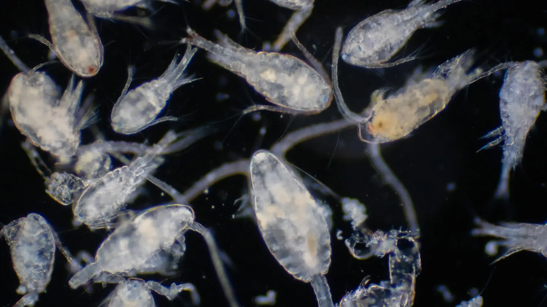
A groundbreaking study has revealed that small but mighty zooplankton — including copepods, krill, and salps — are key players in the Southern Ocean’s ability to absorb and store carbon.

Led by an international team of researchers, and published in Limnology and Oceanography, the study quantifies for the first time how these tiny creatures collectively enhance carbon sequestration through their seasonal, vertical migrations.

The Southern Ocean is a key region for carbon storage. Traditional thinking is that the carbon storage in the Southern Ocean is dominated by gravitational sinking of detritus produced by large zooplankton grazers, such as krill.

This new research concerns another more recently described process called the ‘seasonal migrant pump’. This process sees zooplankton migrate each year from surface waters to depths below 500m, storing carbon via their respiration and mortality during this deep overwintering phase.

This figure shows the traditional view of how zooplankton transport carbon to depth (left panel) by eating phytoplankton in surface waters in summer, whereby their waste material (Particulate Organic Carbon, POC) sinks passively to great depth, thereby storing the carbon for thousands of years. This new study shows that a winter process known as the ‘seasonal migrant pump’ also leads to a substantial deep carbon storage (right panel). The zooplankton migrate downwards in autumn to overwinter below 500m where their respiration and death directly inject around 65 million tonnes of carbon annually into the deep ocean.

The team first built a big database of zooplankton collected in thousands of net hauls from around the Southern Ocean, dating from the 1920s to the present day. From these they quantified the extent of the zooplankton’s annual descent to overwinter at great depths, where they respire CO2 — directly and efficiently injecting carbon into the deep ocean.

Key Findings:

  • 65 Million Tonnes of Carbon Stored Annually: The seasonal, vertical migration of zooplankton transports roughly 65 million tonnes of carbon to depths below 500 meters.
  • Copepods Dominate the ‘Seasonal Migrant Pump’: Mesozooplankton (mainly small crustaceans called copepods) account for 80% of this carbon flux, while krill and salps contribute 14% and 6%, respectively.
  • Climate Implications: The Southern Ocean is a critical carbon sink, but current Earth System Models overlook this zooplankton-driven process. As warming shifts species distributions (e.g., declining krill, increasing copepods, changing food sources), the carbon storage dynamics may change dramatically.

Why does the ‘Seasonal Migrant Pump’ matter:

The Southern Ocean absorbs approximately 40% of all human-made CO2 taken up by oceans, yet the role of zooplankton has been underestimated. Unlike sinking detritus, which removes both carbon and essential nutrients like iron, migrating zooplankton efficiently inject carbon into the deep ocean while recycling nutrients near the surface. This ‘Seasonal Migrant Pump’ could become even more important as marine ecosystems respond to climate change.

Dr Guang Yang, first author and Marine Ecologist from Institute of Oceanology, Chinese Academy of Sciences, said: “Our work shows that zooplankton are unsung heroes of carbon sequestration. Their seasonal migrations create a massive, previously unquantified carbon flux — one that models must now incorporate.”

Prof. Angus Atkinson MBE, co-author and Senior Marine Ecologist at Plymouth Marine Laboratory, added: “This study is the first to estimate the total magnitude of this carbon storage mechanism. It shows the value of large data compilations to unlock new insights and to get an overview of the relative importance of carbon storage mechanisms.”

Dr Katrin Schmidt, co-author and Marine Ecologist at the University of Plymouth, said: “The study shows the ‘seasonal migrant pump’ as an important pathway of natural carbon sequestration in polar regions. Protecting these migrants and their habitats will help to mitigate climate change.”

Dr Jen Freer, co-author and Ecological Modeller at the British Antarctic Survey (BAS), added: “Krill are famous for their role in the Antarctic food web, but we find that copepods significantly dominate carbon storage overwinter. This has big implications as the ocean warms and their habitats may shift.”

This research stresses the urgent need for updates to climate models to include zooplankton-driven carbon fluxes. It also highlights the necessity to manage and protect Southern Ocean ecosystems, where industrial fishing and warming threaten krill populations — a key species that supports both carbon export and Antarctica’s unique biodiversity.

This international study was a collaboration among scientists from China, UK, and Canada, and leverages a century’s worth of data on zooplankton biomass, distribution, respiration and mortality across the Southern Ocean.

Share Button

Parkinson’s may begin decades earlier — and your immune system might know first

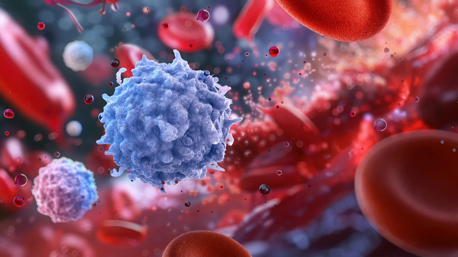
Your T cells work hard to fight disease. Unfortunately, “friendly fire” from T cells can sometimes harm the body’s healthy tissues.

For people with autoimmune disease, T cell reactivity is a big problem. Haywire T cell responses lead to autoimmune diseases such as type 1 diabetes, rheumatoid arthritis, and inflammatory bowel disease.

In recent years, scientists at La Jolla Institute for Immunology (LJI) have discovered that T cells may also contribute to the development of Parkinson’s disease. Researchers in the laboratory of LJI Professor Alessandro Sette, Dr.Biol.Sci., have found that many people with Parkinson’s disease have T cells that target key proteins, called alpha-synuclein and PINK1, on vulnerable brain cells.

Earlier this year, Sette and his colleagues published a study in npj Parkinson’s Disease that sheds light on exactly which subtypes of T cells target alpha-synuclein. Their findings offered further clues that T cell reactivity plays a role in Parkinson’s disease. Still, the scientists didn’t have a timeline to show when T cells might contribute to disease development.

“We can see these reactive T cells in people after they develop Parkinson’s, but what happens before that?” says LJI Visiting Scientist Emil Johansson, Ph.D., a researcher in the Sette Lab and co-author of the study.

Now we have answers. In a new npj Parkinson’s Disease paper, Sette and his colleagues show that potentially harmful T cell reactivity is highest during the “prodromal” period in Parkinson’s — the years before patients receive a diagnosis.

“This T cell immunity could be a marker for early Parkinson’s treatment, even before people show symptoms,” says Sette, who was senior author on the new paper. “And there’s reason to think that treating Parkinson’s in the very early stages can lead to a better outcome.”

How the study worked

The prodromal period in Parkinson’s disease can last for decades before a person develops noticeable symptoms such as tremors and cognitive impairments.

Because prodromal Parkinson’s disease is very difficult to detect, the LJI team studied T cell reactivity in research volunteers at high risk of developing Parkinson’s disease. These volunteers had genetic risk factors for Parkinson’s and some had symptoms such as disrupted REM sleep cycles and loss of sense of smell, which can be early signs of Parkinson’s disease development.

The researchers used a technique called Fluorospot to learn more about T cells found in blood samples from these study volunteers. This technique revealed which volunteers had high levels of T cells that reacted to alpha-synuclein or PINK1 — and when those T cell numbers were highest.

Sette and his colleagues found that potentially harmful T cells show up early on, well before the onset of noticeable motor symptoms, such as tremors. “You can see that T cell reactivity before diagnosis,” says Sette.

In fact, T cell reactivity to PINK1 was at an all-time high before diagnosis.

Sette warns against jumping to conclusions. Parkinson’s is a complex disease, and the new research doesn’t prove that T cells are actually driving the inflammation associated with Parkinson’s disease.

“Parkinson’s disease is associated with the destruction of nervous system cells. Does that destruction cause autoimmunity — or is the autoimmunity the cause of the disease? That’s the chicken-and-the-egg of inflammation in Parkinson’s disease,” says Sette.

“Certainly, the fact that this T cell reactivity is highest when patients are closest to a diagnosis is intriguing,” Sette adds. “The finding suggests T cells could have something to do with it.”

Next steps for helping patients

The new research may guide the development of early diagnostic tools. In the meantime, LJI scientists are looking for ways to block inflammation and protect brain cells.

As Johansson explains, some T cells actually help dial back inflammation to protect our tissues. “We want to see if there are specific T cells that are protective,” says Johansson. “Could they interfere in inflammation and maybe reduce the number of autoimmune T cells?”

Sette and his colleagues are also working to understand the role of T cells in other neurodegenerative diseases.

“We are very interested in diseases such as Alzheimer’s, for example, where a lot of progress has been made toward identifying people in very early stages of the disease progression,” says Sette.

Additional authors of the study, “T cell responses towards PINK1 and α-synuclein are elevated in prodromal Parkinson’s disease,” included first author Antoine Freuchet, Gregory P. Williams, Tanner Michealis, April Frazier, Irene Litvan, Jennifer G. Goldman, Roy N. Alcalay, David G. Standaert, Amy W. Amara, Natividad Stover, Edward A. Fon, Ronald B. Postuma, John Sidney, David Sulzer, and Cecilia S. Lindestam Arlehamn.

This study was supported by LJI & Kyowa Kirin, Inc. (KKNA- Kyowa Kirin North America), the Swedish Research Council (grant references 2024-00175), Aligning Science Across Parkinson’s (ASAP-000375), and the Michael J. Fox Foundation.

Share Button

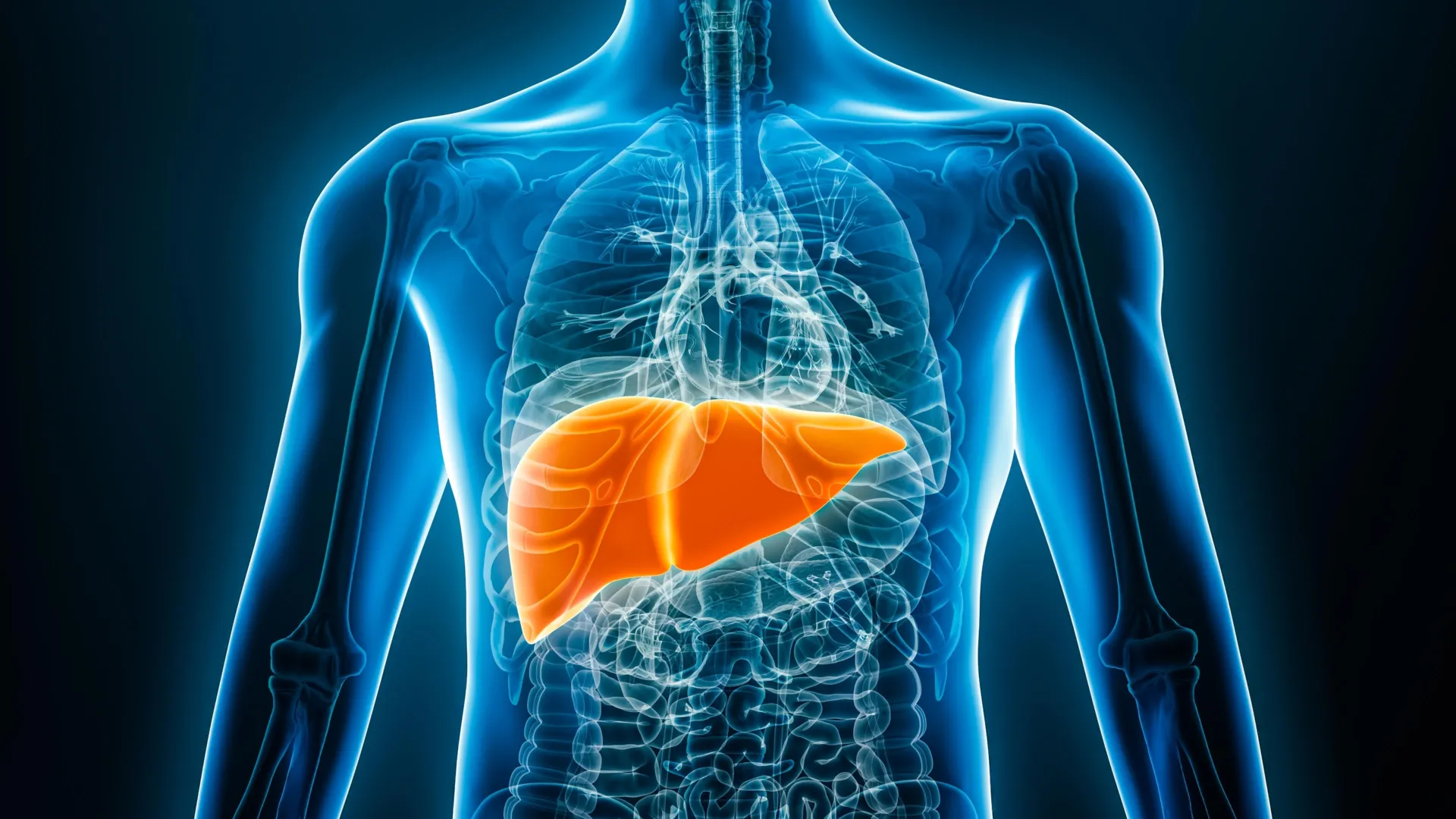
AI sees what doctors miss: Fatty liver disease hidden in chest x-rays

Fatty liver disease, caused by the accumulation of fat in the liver, is estimated to affect one in four people worldwide. If left untreated, it can lead to serious complications, such as cirrhosis and liver cancer, making it crucial to detect early and initiate treatment.

Currently, standard tests for diagnosing fatty liver disease include ultrasounds, CTs, and MRIs, which require costly specialized equipment and facilities. In contrast, chest X-rays are performed more frequently, are relatively inexpensive, and involve low radiation exposure. Although this test is primarily used to examine the condition of the lungs and heart, it also captures part of the liver, making it possible to detect signs of fatty liver disease. However, the relationship between chest X-rays and fatty liver disease has rarely been a subject of in-depth study.

Therefore, a research group led by Associate Professor Sawako Uchida-Kobayashi and Associate Professor Daiju Ueda at Osaka Metropolitan University’s Graduate School of Medicine developed an AI model that can detect the presence of fatty liver disease from chest X-ray images.

In this retrospective study, a total of 6,599 chest X-ray images containing data from 4,414 patients were used to develop an AI model utilizing controlled attenuation parameter (CAP) scores. The AI model was verified to be highly accurate, with the area under the receiver operating characteristic curve (AUC) ranging from 0.82 to 0.83.

“The development of diagnostic methods using easily obtainable and inexpensive chest X-rays has the potential to improve fatty liver detection. We hope it can be put into practical use in the future,” stated Professor Uchida-Kobayashi.

Share Button

How is your hospital doing as NHS battles to bring down waiting times?

Use our interactive tracker to see if treatment waits are getting better at your local hospital

Share Button

Drug for rare disease brings ‘joy and hope’

A teenage patient who helped discover a rare condition is the first to benefit from a new treatment.

Share Button

Why cats prefer sleeping on their left side—and how it might help them survive

Cats prefer to sleep on their left side. This is the conclusion drawn by an international research team that analyzed several hundred YouTube videos of sleeping cats. The researchers see this bias as an evolutionary advantage because it favors hunting and escape behavior after waking up. The team from the University of Bari Aldo Moro (Italy), Ruhr University Bochum, Medical School Hamburg and other partners in Germany, Canada, Switzerland and Turkey report on the study in the journal Current Biology, published online on June 23, 2025.

All animals are particularly vulnerable while sleeping. Cats sleep around 12 to 16 hours a day, preferably in elevated places where their predators can only access them from below. The research team around Dr. Sevim Isparta from the Animal Physiology and Behaviour Research Unit in Bari and Professor Onur Güntürkün from the Bochum working group Biopsychology wanted to find out whether cats prefer to sleep on one side or the other. “Asymmetries in behavior can have advantages because both hemispheres of the brain specialize in different tasks,” says Onur Güntürkün.

Perceiving dangers with the left visual field brings advantages

The group analyzed 408 publicly available YouTube videos in which a single cat was clearly visible with its entire body sleeping on one side for at least ten seconds. Only original videos were used; modified or flipped material was excluded from the study. Two thirds of the videos showed cats sleeping on their left side.

The explanation: Cats that sleep on their left side perceive their surroundings upon awakening with their left visual field, which is processed in the right hemisphere of the brain. This hemisphere is specialized in spatial awareness, the processing of threats and the coordination of rapid escape movements. If a cat sleeps on its left shoulder and wakes up, visual information about predators or prey goes directly to the right hemisphere of the brain, which is best in processing them. “Sleeping on the left side can therefore be a survival strategy,” the researchers conclude.

Cooperation partners

  • University of Bari Aldo Moro (Italy)
  • Ruhr University Bochum (Germany)
  • Medical School Hamburg (Germany)
  • Research Institute for Farm Animal Biology (Germany)
  • University of Prince Edward Island (Canada)
  • Kafkas University (Turkey)
  • Federal Food Safety and Veterinary Office (Switzerland)
Share Button

Acid-busting diet triggers 13-pound weight loss in just 16 weeks

Compared with a Mediterranean diet, dietary acid load decreased significantly on a low-fat vegan diet and was associated with weight loss, according to a randomized cross-over trial conducted by the Physicians Committee for Responsible Medicine and published in Frontiers in Nutrition.

“Eating acid-producing foods like meat, eggs, and dairy can increase the dietary acid load, or the amount of acids consumed, causing inflammation linked to weight gain,” says Hana Kahleova, MD, PhD, director of clinical research at the Physicians Committee and lead author of the study. “But replacing animal products with plant-based foods like leafy greens, berries, and legumes can help promote weight loss and create a healthy gut microbiome.”

This new research included 62 overweight adults who were randomized to a Mediterranean or a low-fat vegan diet for 16 weeks, separated by a four-week cleansing period, followed by an additional 16 weeks on the alternate diet.

Participants’ dietary records were used to calculate dietary acid load, which is commonly estimated by two scores: Potential Renal Acid Load (PRAL) and Net Endogenous Acid Production (NEAP). A higher score indicates a higher dietary acid load.

Animal products including meat, fish, eggs, and cheese cause the body to produce more acid, increasing dietary acid load, which is linked to chronic inflammation that disrupts metabolism and can lead to increased body weight. Plant-based diets, which are more alkaline, are associated with weight loss, improved insulin sensitivity, and lower blood pressure.

In the new analysis, both PRAL and NEAP scores decreased significantly on the vegan diet, with no significant change on the Mediterranean diet. The reduction in dietary acid load was associated with weight loss, and this association remained significant even after adjustment for changes in energy intake. Body weight was reduced by 13.2 pounds on the vegan diet, compared with no change on the Mediterranean diet.

The authors say that a vegan diet’s alkalizing effect, which increases the body’s pH level to make it less acidic, may also help promote weight loss. Top alkalizing foods include vegetables, particularly leafy greens, broccoli, beets, asparagus, garlic, carrots, and cabbage; fruits, such as berries, apples, cherries, apricots, or cantaloupe; legumes, for example lentils, chickpeas, peas, beans or soy; and grains, such as quinoa or millet.

Founded in 1985, the Physicians Committee for Responsible Medicine is a nonprofit organization that promotes preventive medicine, conducts clinical research, and encourages higher standards for ethics and effectiveness in education and research.

Share Button

Can these endangered lizards beat the heat? Scientists test bold relocation plan

Climate change and habitat loss are affecting animal populations around the world and reptiles such as South Australia’s own endangered pygmy bluetongue are susceptible to higher temperatures and declining long-term rainfall trends.

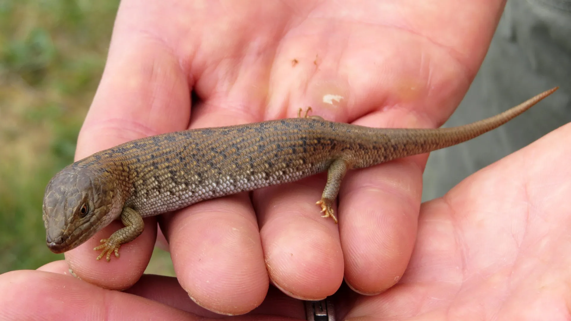
Flinders University scientists are working on securing a sustainable future for the burrow-dwelling endemic skink (Tiliqua adelaidensis) by assessing their suitability to cooler and slightly greener locations, below their usual range in the state’s drier, hotter northern regions.

While the lizards take time to acclimatize to their new homes, translocation remains one of the more important ways to conserve rare species and mitigate extinction risk to climate and habitat changes.

The latest research, outlined in a new article in Biology, compared the ability of three separate pygmy bluetongue lizard populations to withstand different microclimates in South Australia – between the northern Flinders Ranges near Jamestown, Mid North near Burra, and southern-most translocation sites near Tarlee and Kapunda.

The study, led by PhD candidate Deanne Trewartha from the College of Science and Engineering, says moving wildlife adapted to a hotter, drier location to another microclimate can mean exposure to different temperatures, water availability and humidity and needs extensive assessment.

“We need to understand how this species, which are highly dependent on body temperature, adapt to cooler and often wetter seasons in these new environments,” says Ms Trewartha, from the Flinders University Lab of Evolutionary Genetics and Sociality (LEGS) research group.

Reptiles rely on attaining certain body temperatures for basic bodily function and increasing body temperature raises dehydration risk.

She says the research so far suggests acclimatization to new sites may take longer than two years for all three populations and may vary with latitude of origin.

“Despite this acclimatization delay, our results indicate that these lizards may cope with translocation as a mitigation strategy in the longer term.

“Further monitoring of the three lineages will continue to see any behavioral variations in wet versus dry seasons and the long-term behavioral acclimatization periods for translocations.”

Australia has the highest reptile diversity in the world, and Flinders University Professor of Biodiversity and Ecology Mike Gardner says translocation may be the only way for the conservation of numerous small burrow-dwelling reptiles, other ectotherms and reptile species in future.

“With high biodiversity loss, translocation to ‘future-suitable’ sites is becoming increasingly urgent for the conservation of numerous reptile species,” says Professor Gardner, who leads an Australian Research Council Linkage project to study various pygmy bluetongue groups at different latitudes in South Australia.

“So far, these three populations are showing various responses to their new locations, but behavioral variations may not be detrimental in the long term and may potentially aid animals in acclimatizing to changed environments to optimize their chance their survival.”

A previous study published last year noted differences between the way the colonies behaved.

From spring 2020 to autumn 2021, monthly monitoring of behaviors found the translocated southern lineage lizards showed significantly less daily activity and were active at lower temperatures and higher humidity than northern lineage lizards.

Southern lineage lizards allowed a human observer to approach closer as base-of-burrow humidity increased, while northern lineage lizards were quicker to retreat into burrows, at both source and translocation sites.

This project was carried out in accordance with Flinders University ethics approval E453-17, Department of Environment and Water ‘Take from the wild’ permit 20210331 and research permit G25011. Acknowledgements: The authors acknowledge the Ngadjuri people, who are the traditional custodians of the Mid North study sites and represent the oldest human culture. We acknowledge their elders, past, present and emerging, and that sovereignty was never ceded. Thanks to the Nature Foundation, relevant private landowners, Department for Environment and Water, Renewable Energy Systems Pty Ltd, Flow Power, Nature Foundation and Adelaide Airport Limited for their support in accommodating this research.

Share Button

Weight loss jabs study begins after reports of pancreas issues

There have been hundreds of cases of acute pancreatitis in people who have also taken Mounjaro, Ozempic and Wegovy.

Share Button

Womb lining test offers miscarriage hope to women

The test can measure whether an abnormal reaction in the womb could make pregnancy loss more likely.

Share Button