Astronomers finally solve Saturn’s decades-long spin mystery

For years, Saturn appeared to be doing something impossible.

Measurements suggested the giant planet’s rotation rate was changing over time, as if Saturn were somehow speeding up or slowing down. That puzzling result left scientists searching for answers. Now, researchers using the James Webb Space Telescope (JWST) say they have finally solved the mystery.

The new findings, published in the Journal of Geophysical Research: Space Physics, reveal that Saturn’s spectacular northern lights are at the heart of the phenomenon. The study shows that the planet’s aurora drives a powerful cycle involving heat, winds, and electrical currents that can make Saturn appear to spin at different speeds depending on how it is measured.

Saturn’s Rotation Mystery

The puzzle dates back decades, but it gained renewed attention after observations from NASA’s Cassini spacecraft in 2004 suggested that Saturn’s rotation rate was gradually changing.

That result was difficult to explain because planets do not simply alter their spin rates on short timescales.

In 2021, a team led by Professor Tom Stallard of Northumbria University proposed a different explanation. Their research showed that Saturn’s rotation was not actually changing. Instead, electrical signals linked to the planet’s aurora were being affected by winds in Saturn’s upper atmosphere. Those winds generated electrical currents that altered the auroral signal scientists were using to estimate the planet’s rotation.

While that study explained the misleading measurements, one major question remained unanswered: What was driving those atmospheric winds?

James Webb Maps Saturn’s Aurora

To investigate, Stallard and colleagues from institutions across the United Kingdom and United States turned to the James Webb Space Telescope.

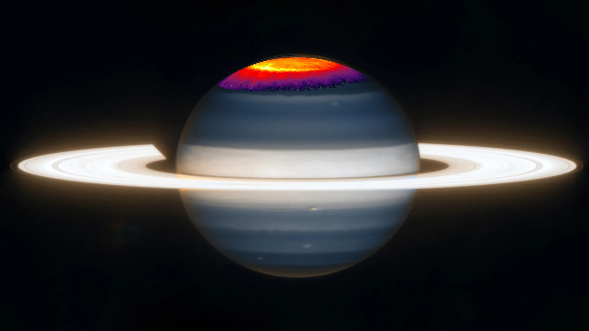
The team observed Saturn’s northern auroral region continuously for an entire Saturnian day. The observations provided a level of detail that previous instruments could not achieve.

Researchers focused on infrared light emitted by a molecule known as trihydrogen cation. This molecule forms in Saturn’s upper atmosphere and serves as a natural indicator of temperature. By analyzing its glow, the team created the most detailed maps ever produced of temperatures and charged particle densities within Saturn’s auroral region.

The improvement in accuracy was dramatic. Earlier measurements carried uncertainties of roughly 50 degrees Celsius, making it difficult to detect subtle changes. JWST’s observations were about ten times more precise, allowing scientists to identify localized patterns of heating and cooling for the first time.

A Self-Sustaining Planetary Heat Engine

The new data closely matched predictions from computer models developed more than a decade ago. However, the models only worked if the source of the atmospheric heating was located exactly where the strongest auroral particles enter Saturn’s atmosphere.

The results indicate that Saturn’s aurora is doing far more than creating a dazzling light show.

Energy deposited by the aurora heats specific regions of the atmosphere. That heating generates winds, which then create electrical currents. Those currents help power the aurora itself, which continues heating the atmosphere and sustaining the entire cycle.

Lead researcher Professor Tom Stallard said: “What we are seeing is essentially a planetary heat pump. Saturn’s aurora heats its atmosphere, the atmosphere drives winds, the winds produce currents that power the aurora, and so it goes on. The system feeds itself.

“For decades, we knew something strange was happening with Saturn’s apparent rotation rate, but we could not explain it. We then showed it was being driven by atmospheric winds, but we still did not know why those winds existed. These new observations, made possible by JWST, finally give us the evidence we needed to close that loop.”

Implications Beyond Saturn

The discovery may have significance far beyond a single planet.

Researchers found evidence that Saturn’s atmosphere and magnetosphere are closely connected. The magnetosphere is the vast region of space shaped by the planet’s magnetic field. Activity in the atmosphere appears to influence conditions in the magnetosphere, while the magnetosphere feeds energy back into the atmosphere.

This ongoing exchange could help explain why the process remains stable over long periods.

According to the researchers, similar interactions may occur on other planets as well.

Professor Stallard added: “This result changes how we think about planetary atmospheres more generally. If a planet’s atmospheric conditions can drive currents out into the surrounding space environment, then understanding what is happening in the stratospheres of other worlds may reveal interactions we have not yet even imagined.”

An International Research Effort

The James Webb Space Telescope is the world’s premier space science observatory. The telescope is designed to study objects throughout the solar system, investigate planets orbiting distant stars, and explore the origins and evolution of the universe. Webb is an international project led by NASA in partnership with ESA (European Space Agency) and CSA (Canadian Space Agency).

The study was conducted by researchers from Northumbria University together with collaborators from Boston University, the University of Leicester, Aberystwyth University, the University of Reading, Imperial College London, Lancaster University, and Johns Hopkins University Applied Physics Laboratory. Funding for the research was provided by the Science and Technology Facilities Council (STFC).

Share Button

Human organoids reveal how to reverse “irreversible” nerve damage

Scientists at the University of Cambridge have created tiny lab-grown brain and spinal cord systems that mimic how movement signals travel through the human nervous system. Using this model, the team discovered that nerve damage once believed to be permanent may actually be reversible under certain conditions.

As the human body develops from an embryo into a fetus and eventually an infant, neurons form complex communication networks between the brain and spinal cord. These signals travel through axons, the long nerve fibers that allow neurons to send messages and control muscle movement.

Over time, however, the central nervous system largely loses its ability to regrow damaged axons. As a result, injuries to the brain or spinal cord often become permanent, leading to serious disabilities such as paralysis or loss of movement. This loss of regenerative ability is also linked to neurological diseases including motor neurone disease and multiple sclerosis.

Mini Human Brain and Spinal Cord Models

In 2021, Dr. András Lakatos and his colleagues at the University of Cambridge developed miniature human brain models using stem cells taken from patients. These pea-sized “brain organoids” resembled parts of the cerebral cortex and allowed researchers to study molecular changes linked to motor neurone disease and explore ways to prevent them.

Now, in a new study published in Cell Reports, the researchers expanded on that work by building a miniature version of the connected human brain and spinal cord system.

Because the brain and spinal cord are separate but connected structures in the body, the team kept the organoids physically apart in the lab. They then observed axons from the brain tissue growing across the gap and connecting with the spinal cord tissue. The resulting neural circuit was functional enough to trigger contractions in tiny clusters of muscle cells.

Nerve Regrowth Declines During Development

The scientists maintained these miniature systems in the lab for more than a year. They discovered that until about day 150 of development, roughly corresponding to the middle stage of pregnancy, damaged axons could still regrow. After that point, the neurons showed a major decline in their ability to regenerate.

George Gibbons from the Department of Clinical Neurosciences at the University of Cambridge and first author of the study said: “Neurons taken from less mature organoids regrew long fibers after injury, but those from more mature organoids showed a sharp drop in their ability to regrow. In other words, poor regeneration is built into human neurons as they mature in the central nervous system.”

The team analyzed gene activity in neurons that connect the brain and spinal cord. Their work revealed a network of genes that acts like a biological switch, limiting axon growth as neurons mature and form synapses.

Remarkably, when researchers blocked key regulators within this network, the neurons regained the ability to grow axons again.

Existing Drug Boosted Nerve Regrowth

The researchers also searched a database of drug compounds to identify medicines that affect this newly identified gene network. One promising candidate was lynestrenol, a hormone drug currently approved for certain menstrual disorders and contraceptive use.

When the drug was tested on damaged neurons, it significantly improved axon regrowth.

The scientists noted that scar tissue and inflammation can also interfere with nerve repair after injury. However, understanding the neuron-specific biological mechanisms that limit regeneration remains critically important. Previous evidence has shown that younger neurons can grow through environments that normally block repair at injury sites.

Senior author Dr. András Lakatos, who led the study at the Department of Clinical Neurosciences, said: “When the brain and spinal cord are damaged, the nerve fibers that carry movement signals from the brain to the spinal cord rarely grow back. That’s why paralysis is usually permanent. But we didn’t know exactly when the ability of axons to regenerate becomes limited. Our model provides a good indication that this block happens during development, and it can still be reversed after this point.

“Lynestrenol itself may not be the answer to spinal cord repair, but it shows us that, in principle, it should be possible to directly target human neurons and regenerate their axons. Although we still need to show that this strategy will also help to re-establish appropriate connections between the brain and spinal cord cells, this gives us hope that one day we may be able to treat conditions previously thought untreatable.”

Why Human Organoids Matter

Organoid technology is becoming increasingly valuable for studying human biology and disease. While animal models such as mice and rats remain useful in research, important biological differences limit how accurately they reflect human nervous system function.

Human stem cell-derived organoids can more closely reproduce human biology, helping bridge the gap between animal experiments and real patient outcomes.

Dr. Lakatos added: “Much of what we know about nerve regeneration comes from rodents, whose neurons behave differently from human neurons. Our sophisticated organoid models help bridge the knowledge gap from animal models to what we see in patients. They are also an important contribution to efforts to reduce the use of animals in research.”

Researchers at the University of Cambridge are already using organoids for a wide range of medical studies, including efforts to repair damaged livers, investigate Crohn’s disease in children, and study the earliest stages of pregnancy.

The research was funded by the UK Research and Innovation Medical Research Council and Spinal Research.

Share Button

CBD may slow Alzheimer’s by calming the brain’s immune system

Cannabidiol, better known as CBD, is gaining attention from scientists studying Alzheimer’s disease. New research suggests the cannabis-derived compound may help reduce harmful inflammation in the brain, a process increasingly believed to play a major role in Alzheimer’s progression.

Alzheimer’s disease is the most common form of dementia, a condition that gradually damages memory, thinking, and behavior. For years, most Alzheimer’s research has focused on the buildup of amyloid plaques and tau tangles in the brain. These abnormal protein clumps are considered hallmark signs of the disease. However, many researchers now believe chronic inflammation in the brain may also be a key factor driving nerve cell damage.

CBD and Brain Inflammation

Inflammation is part of the body’s natural immune response. In the brain, immune cells normally help protect neurons and clear away harmful debris. But when inflammation becomes chronic, it can begin damaging healthy brain tissue instead. This ongoing immune overactivation, often called neuroinflammation, has been linked to Alzheimer’s disease and several other neurological disorders.

In a new study published in eNeuro, researchers led by Babak Baban from Augusta University investigated whether CBD could help calm this damaging inflammatory response in the brain.

The team used a well-established mouse model of Alzheimer’s disease and delivered CBD through inhalation. They then examined how the compound affected immune activity and inflammatory signaling in the central nervous system, which includes the brain and spinal cord.

Researchers Identify Changes in Key Immune Pathways

Using a variety of molecular and genetic tests, the scientists found that CBD lowered the activity of several important regulators involved in neuroinflammation. The treatment was also associated with reduced levels of proinflammatory molecules, which are substances that can worsen inflammation and contribute to tissue damage.

The researchers also identified specific immune-related pathways that appeared to interact with CBD. These findings suggest the compound may influence multiple biological systems involved in Alzheimer’s disease.

“Alzheimer’s work has long centered on plaques and tangles,” says Baban. “But our study shows that chronic autoinflammation is also a core driver of the disease. What’s exciting is that CBD not only calms this immune overactivation but, in earlier work, we’ve shown it can also help clear plaques and tangles through a different mechanism. Together, this points to a multitarget approach with real therapeutic potential.”

A Growing Interest in Multi-Target Alzheimer’s Treatments

Scientists have increasingly explored treatments that target more than one aspect of Alzheimer’s disease at the same time. Because the condition involves many overlapping biological changes, including inflammation, protein buildup, and neuron damage, researchers believe a multitarget strategy may prove more effective than focusing on a single pathway alone.

Although the findings are promising, the study was conducted in mice, not humans. More research and clinical trials will be needed before scientists know whether CBD could become a safe and effective treatment for people with Alzheimer’s disease.

Still, the results add to growing evidence that controlling brain inflammation may become an important part of future Alzheimer’s therapies.

Share Button

‘I forgot what it’s like to be outside’: Intensive care ward opens on rooftop

The outdoor ward, at King’s College Hospital in London, will monitor how much it boosts the recovery of seriously ill patients.

Share Button

DNA solves 250-year-old mystery of the Seychelles’ lost crocodiles

For more than 250 years, stories from early explorers described crocodiles as a common sight along the shores of the Seychelles. But after permanent settlers arrived in 1770, the island population disappeared rapidly. Within about 50 years, the crocodiles had been completely exterminated.

Now, scientists have finally uncovered the true identity of these vanished reptiles through a new genetic analysis. The study found that the Seychelles crocodiles were not a separate species, as some once suspected. Instead, they were the westernmost known population of the saltwater crocodile (Crocodylus porosus), the world’s largest living reptile and one of its most capable ocean travelers.

DNA Reveals the Origins of Seychelles Crocodiles

Researchers from Germany and the Seychelles investigated the evolutionary history of the saltwater crocodile by comparing DNA from modern animals with genetic material taken from historical museum specimens. The team analyzed mitochondrial genomes from preserved crocodiles belonging to the genus Crocodylus, including rare samples from the Seychelles population that vanished roughly 200 years ago.

The findings confirmed an earlier theory that had been based only on the crocodiles’ physical appearance. Genetic evidence now shows the Seychelles animals were closely connected to saltwater crocodiles living thousands of kilometers away.

Crocodiles Crossed Vast Distances Across the Indian Ocean

Among all living crocodile species, the saltwater crocodile is especially well adapted for life at sea. Specialized salt glands allow these reptiles to remove excess salt from their bodies, enabling them to survive for long periods in seawater. Over time, this ability helped the species spread across enormous stretches of coastline and remote islands.

“The founders of the Seychelles population must have drifted at least 3,000 kilometers across the Indian Ocean to reach the remote archipelago, perhaps even much further,” says reptile expert Frank Glaw of the Bavarian State Collections of Natural History (SNSB) and senior author of the study.

Scientists believe these crocodiles likely traveled with ocean currents over generations, eventually establishing a population in the isolated islands of the Seychelles.

One of the World’s Most Wide Ranging Reptiles

“The genetic patterns suggest that saltwater crocodile populations remained connected over long periods and across great distances, pointing to the high mobility of this species,” explains first author Stefanie Agne of the University of Potsdam.

Today, the saltwater crocodile remains one of the most widely distributed reptiles on Earth. Before the Seychelles population was wiped out, the species occupied an even larger range that stretched more than 12,000 kilometers from Vanuatu in the Pacific Ocean to the Seychelles in the Indian Ocean.

Share Button

What in the World

Doctors are warning that some cancers are becoming more common in people in their 20s

Share Button

Can a rooftop garden help very ill patients heal faster?

Still in her hospital bed, connected to feeding tubes and life support, Hollie is the first patient to try out the new intensive care rooftop ward at King’s College Hospital in south London.

Share Button

Prostate cancer screening only for ‘a few thousand’ high risk men

Only men with a dangerous genetic variant and a family history of cancer should be offered screening, say UK advisors.

Share Button

Why the gut is known as the second brain

Your gut doesn’t just respond to your brain – it helps shape your emotions. Professor of Biomolecular Medicine Jon Swann explains why.

Share Button

Researchers block key protein that helps Parkinson’s spread through the brain

Researchers at the Perelman School of Medicine at the University of Pennsylvania have identified a brain immune protein that may play a major role in the progression of Parkinson’s disease (PD). Their findings, published in Neuron, suggest that blocking this protein with monoclonal antibodies could eventually lead to treatments that slow the disease in its earliest stages.

The protein, known as glycoprotein nonmetastatic melanoma B (GPNMB), appears to help harmful Parkinson’s-related damage spread from one brain cell to another. Scientists say targeting it may offer a new strategy for slowing the worsening of the disease over time.

“Many patients with Parkinson’s disease are diagnosed in the early stages, when symptoms are relatively mild, but there is currently no treatment that slows the progression,” said lead author, Alice Chen-Plotkin, MD, Parker Family Professor of Neurology. “These early results are a promising step towards developing this type of treatment.”

How Parkinson’s Disease Spreads in the Brain

Parkinson’s disease affects more than one million Americans, and approximately 90,000 people in the United States are diagnosed each year. Although researchers still do not fully understand what causes the disease, scientists have known for years that it gradually spreads through the brain in stages.

A protein called alpha-synuclein is central to this process. In Parkinson’s disease, alpha-synuclein forms abnormal clumps inside neurons. These clumps damage the affected cells and can then move into nearby healthy neurons, where they continue spreading.

As more areas of the brain become affected, symptoms worsen. Patients may develop tremors, difficulty walking, balance problems, and trouble swallowing.

Current treatments, including levodopa and deep-brain stimulation, can help reduce symptoms. However, no approved therapy has been shown to slow or stop the underlying progression of Parkinson’s disease itself.

Brain Immune Cells May Help Fuel Disease Progression

In earlier research published in 2022, Chen-Plotkin and colleagues identified GPNMB as an important molecule involved in the spread of alpha-synuclein between neurons. That discovery made the protein a promising target for future therapies.

In the new study, the research team found that microglia, the brain’s immune cells, are a major source of GPNMB in Parkinson’s disease. When neurons become damaged or begin dying, nearby microglia respond by producing larger amounts of the protein.

Enzymes then cut part of GPNMB away from the cell surface, allowing it to move freely between cells in the brain.

Using preclinical laboratory experiments with cultured neurons, researchers developed antibodies designed to block GPNMB. The antibodies successfully prevented alpha-synuclein pathology from spreading from one cell to another.

“These results suggest Parkinson’s disease may be driven by a self reinforcing cycle — alpha-synuclein accumulates in neurons, damaging the neurons. The injury to the neurons initiates the release of GPNMB, which accelerates the spread of alpha-synuclein, leading to further damage,” Chen-Plotkin said. “Interrupting this cycle would hopefully slow, or even stop, the spread of alpha-synuclein through the brain and the neurodegeneration that follows.”

Human Brain Analysis Supports the Findings

To examine whether the results were relevant in people, researchers analyzed tissue samples from 1,675 brains stored in the Penn Brain Bank.

The team found that individuals carrying genetic variants linked to higher GPNMB production also showed more extensive alpha-synuclein pathology. According to the researchers, this provides strong evidence that GPNMB plays a significant role in the progression of Parkinson’s disease in humans.

Importantly, elevated GPNMB levels were not connected to markers associated with other neurodegenerative conditions, including Alzheimer’s disease.

“These results are promising for laboratory models and human brain tissue analysis, but we still have a lot of work to do before we can translate this therapy into humans,” said Chen-Plotkin. “That being said, these results are encouraging as we continue to work towards a novel treatment for PD.”

The study received support from the National Institutes of Health (R37 NS115139, P30 AG010124, U19 AG062418, P01 AG084497), SPARK-NS, the Parker Family Chair, and the Lipman Family Fund.

Share Button