Flu has come early this year with a new mutated version of the virus circulating.


Flu has come early this year with a new mutated version of the virus circulating.
Christmas is a difficult time if you suffer from a reduced tolerance to sounds, but there are ways to make it easier.
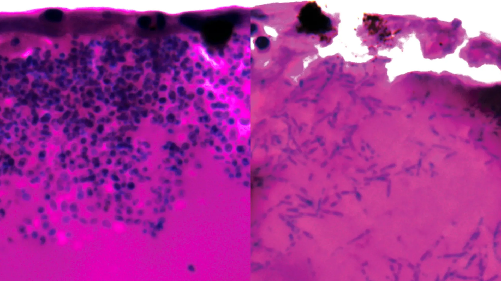
Scientists have identified a genetic mechanism that could point to new ways of treating a rare but deadly fungal infection that has forced multiple hospital intensive care units to shut down. The finding offers early hope against a pathogen that has been difficult to control and nearly impossible to treat once it spreads.
Candida auris is especially dangerous for people who are already critically ill, leaving hospitals highly exposed to outbreaks. Although the fungus can exist on the skin without causing symptoms, patients who depend on ventilators face a much greater risk. Once infection occurs, about 45 percent of patients die, and the fungus is resistant to all major types of antifungal drugs. This resistance makes treatment extremely challenging and allows the pathogen to persist in hospital wards.
A global health threat with mysterious origins
The infection was first detected in 2008, and its origin is still unknown. Since then, outbreaks have been reported in more than 40 countries, including the UK. Candida auris, also called Candidozyma auris, is now recognized as a serious global health threat and appears on the World Health Organization’s critical priority fungal pathogens list. In the UK, reported cases have continued to rise steadily.
Studying infection in a living model
Researchers at the University of Exeter have now taken a major step forward by examining how genes are activated during Candida auris infection. This marks the first time such genetic activity has been studied in a living host using an approach based on fish larvae. The study was published in the Nature portfolio journal Communications Biology and was supported by Wellcome, the Medical Research Council (MRC), and the National Center for Replacement, Reduction and Refinement (NC3Rs).
The researchers say the results could help identify a biological target for new antifungal treatments or even allow existing drugs to be reused, if the same genetic behavior is confirmed during infection in humans.
The project was co-led by NIHR Clinical Lecturer Hugh Gifford of the University of Exeter’s MRC Center for Medical Mycology (CMM). He said: “Since it emerged, Candida auris has wreaked havoc where it takes hold in hospital intensive care units. It can be deadly for vulnerable patients, and health trusts have spent millions on the difficult job of eradication. We think our research may have revealed an Achilles heel in this lethal pathogen during active infection, and we urgently need more research to explore whether we can find drugs that target and exploit this weakness.”
Why traditional research models fell short
One of the biggest obstacles in studying Candida auris has been its ability to survive high temperatures. When combined with its unusually strong tolerance for salt, this has led some researchers to suggest it may have originated in tropical oceans or marine animals. These traits also made it difficult to study using conventional laboratory models.
To overcome this, the Exeter team developed a new infection model using Arabian killifish. The eggs of this species can survive at temperatures similar to the human body, making them suitable for observing infection in conditions that closely resemble real illness.
Genetic activity reveals possible vulnerabilities
During the experiments, researchers observed that Candida auris can change its shape by forming elongated fungal structures known as filaments. These structures may help the fungus search for nutrients while infecting a host.
The team also analyzed which genes were activated or switched off during infection to identify possible weak points. Several of the genes that became active are responsible for producing nutrient pumps that capture iron-scavenging molecules and transport iron into fungal cells. Because iron is essential for survival, this process may represent a critical vulnerability.
Co-senior author Dr. Rhys Farrer of the University of Exeter’s MRC Centre for Medical Mycology said: “Until now, we’ve had no idea what genes are active during infection of a living host. We now need to find out if this also occurs during human infection. The fact that we found genes are activated to scavenge iron gives clues to where Candida auris may originate, such as an iron-poor environment in the sea. It also gives us a potential target for new and already existing drugs.”
Hope for future treatments
Dr. Gifford, who also works as a resident physician in intensive care and respiratory medicine at the Royal Devon & Exeter Hospital, emphasized the clinical importance of the findings. He said: “While there are a number of research steps to go through yet, our finding could be an exciting prospect for future treatment. We have drugs that target iron scavenging activities. We now need to explore whether they could be repurposed to stop Candida auris from killing humans and closing down hospital intensive care units.”
The Arabian killifish larvae model was developed with support from an NC3Rs project grant as an alternative to using mouse and zebrafish models, which are commonly used to study interactions between pathogens and their hosts. Dr. Katie Bates, NC3Rs Head of Research Funding, said: “This new publication demonstrates the utility of the replacement model to study Candida auris infection and enable unprecedented insights into cellular and molecular events in live infected hosts. This is a brilliant example of how innovative alternative approaches can overcome key limitations of traditional animal studies.”
The paper is titled ‘Xenosiderophore transporter gene expression and clade-specific filamentation in Candida auris killifish (Aphanius dispar) infection’ and is published in the Nature portfolio journal Communications Biology.
That Time of the Month with Naga Munchetty, Dr Nighat Arif and Dr Christine Ekechi
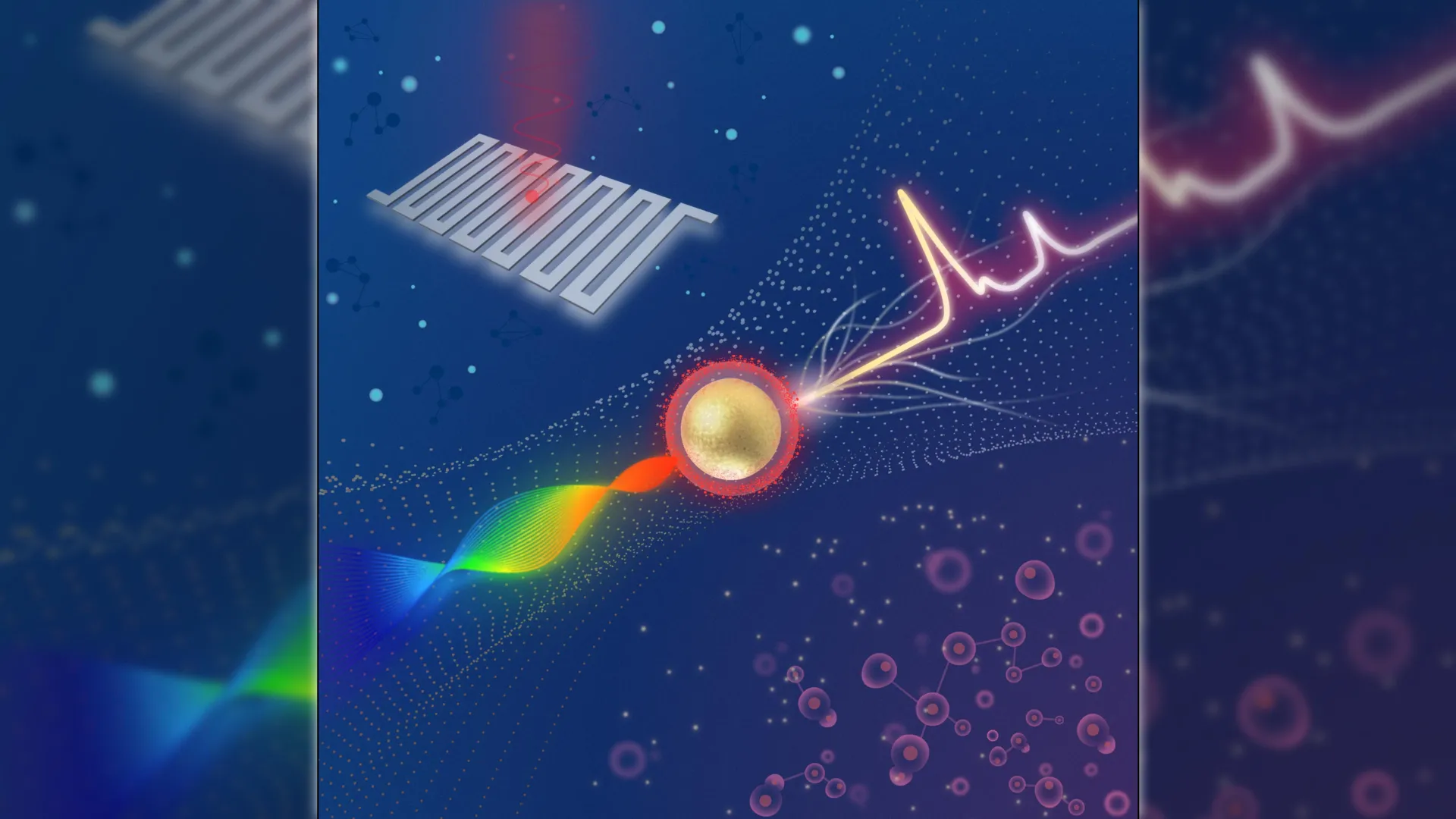
Scientists have developed a compact Raman imaging system that can reliably tell cancerous tissue apart from normal tissue. The approach could support earlier cancer detection and help move advanced molecular imaging tools beyond research laboratories and into more practical clinical settings.
The imaging system is designed to detect extremely weak signals from surface-enhanced Raman scattering (SERS) nanoparticles that are engineered to attach to tumor markers. Once these nanoparticles are applied to a sample or to the area being examined, the system reads their Raman signal and automatically highlights regions that are more likely to contain tumor tissue.
“Traditional methods for cancer-related diagnosis are time-consuming and labor-intensive because they require staining tissue samples and having a pathologist look for any abnormalities,” said research team leader Zhen Qiu from the Institute for Quantitative Health Science and Engineering (IQ), Michigan State University. “While our system would not immediately replace pathology, it could serve as a rapid screening tool to accelerate diagnosis.”
Published results show major gains in sensitivity
In Optica, Optica Publishing Group’s journal for high-impact research, Qiu and colleagues report that their system can distinguish cancerous cells from healthy ones while detecting Raman signals that are about four times weaker than those measured by a comparable commercial system. This improved sensitivity comes from combining a swept-source laser — which changes wavelength during analysis — with an ultra-sensitive detector called a superconducting nanowire single-photon detector (SNSPD).
“This technology could eventually enable portable or intraoperative devices that enable clinicians to detect cancers at earlier stages, improve the accuracy of biopsy sampling and monitor disease progression through less invasive testing,” said Qiu. “Ultimately, such advances could enhance patient outcomes and reduce diagnostic delays, accelerating the path from detection to treatment.”
Pushing detection limits with superconducting detectors
Qiu’s lab studies how SNSPDs can be used to enhance a range of imaging technologies. SNSPDs rely on a superconducting wire that can detect individual particles of light, allowing the system to capture extremely weak optical signals at high speed while keeping background noise very low.
For this project, the researchers aimed to build a platform that could measure Raman signals far fainter than those detected by existing Raman systems. Raman imaging works by mapping a sample’s chemical composition through the unique light-scattering fingerprints of its molecules. These signals can be strengthened by using SERS nanoparticles.
“Combining this advanced detector with a swept-source Raman architecture that replaces a bulky camera and collects light more efficiently resulted in a system with a detection limit well beyond that of comparable commercial systems,” said Qiu. “Also, the fiber coupling configuration and compact design facilitate system miniaturization and future clinical translation.”
Strong tumor contrast across multiple sample types
To test the system, the team used SERS nanoparticles coated with hyaluronan acid, which enables the particles to bind to CD44, a surface protein found on many tumor cells. Initial experiments with simple nanoparticle solutions showed that the system could reach femtomolar sensitivity. The researchers then applied the imaging platform to cultured breast cancer cells, mouse tumors, and healthy tissue samples.
“The SERS signals were strongly concentrated in tumor samples, with only minimal background detected in healthy tissue,” said Qiu. “This demonstrates both the system’s exceptional sensitivity and its ability to provide reliable tumor-versus-healthy contrast. Moreover, by adjusting or substituting the targeting molecule, this method could be adapted for other cancer types.”
Next steps toward clinical use
According to the researchers, additional work is needed before the system can be used in clinical settings. Future improvements will focus on increasing readout speed and expanding validation studies. The team is exploring faster laser sources, including VCSELs, and testing whether narrowing the sweep range can further improve performance. They also plan multiplexing experiments that use different nanoparticles to target multiple biomarkers at the same time.
The researchers acknowledge industry collaborator Quantum Opus, which provided the SNSPD devices used in this work.
The adverts for prescription-only drugs showed healthcare professionals impersonating the British retailer.
Bertie Melly was in hospital for 18 months after his premature birth in May 2024.
Wegovy becomes first pill of its kind to be approved, shifting weight-loss drugs beyond injections.

MIT chemists have produced verticillin A in the lab for the first time. This fungal molecule was identified more than 50 years ago and has drawn attention for its potential as an anticancer agent.
Verticillin A is notoriously hard to build because of its intricate chemical architecture. Even compared with closely related compounds, it proved far more challenging to synthesize, despite differing by only a couple of atoms.
“We have a much better appreciation for how those subtle structural changes can significantly increase the synthetic challenge,” says Mohammad Movassaghi, an MIT professor of chemistry. “Now we have the technology where we can not only access them for the first time, more than 50 years after they were isolated, but also we can make many designed variants, which can enable further detailed studies.”
In lab tests using human cancer cells, one verticillin A derivative stood out against a pediatric brain cancer known as diffuse midline glioma. The researchers emphasize that additional testing is needed to assess whether it could eventually be useful in the clinic.
Movassaghi and Jun Qi, an associate professor of medicine at Dana-Farber Cancer Institute/Boston Children’s Cancer and Blood Disorders Center and Harvard Medical School, are the senior authors of the study, published in the Journal of the American Chemical Society. Walker Knauss PhD ’24 is the paper’s lead author. Xiuqi Wang, a medicinal chemist and chemical biologist at Dana-Farber, and Mariella Filbin, research director in the Pediatric Neurology-Oncology Program at Dana-Farber/Boston Children’s Cancer and Blood Disorders Center, are also authors.
Why This Fungal Molecule Was So Hard to Make
Researchers first reported isolating verticillin A from fungi in 1970. Fungi use the compound to help defend themselves from pathogens. Verticillin A and similar fungal molecules have been explored for possible anticancer and antimicrobial activity, but their complexity has made them difficult to synthesize.
In 2009, Movassaghi’s lab reported the synthesis of (+)-11,11′-dideoxyverticillin A, a compound closely related to verticillin A. That molecule contains 10 rings and eight stereogenic centers, meaning carbon atoms that each connect to four different chemical groups. Those groups must be positioned with the correct orientation, or stereochemistry, relative to the rest of the molecule.
Even after that earlier success, verticillin A itself remained out of reach. The key difference between verticillin A and (+)-11,11′-dideoxyverticillin A is two oxygen atoms, but those additions made a major difference in how the molecule behaves during synthesis.
“Those two oxygens greatly limit the window of opportunity that you have in terms of doing chemical transformations,” Movassaghi says. “It makes the compound so much more fragile, so much more sensitive, so that even though we had had years of methodological advances, the compound continued to pose a challenge for us.”
Rethinking the Chemistry Step by Step
Both versions of the verticillin molecule are built from two identical halves that must be connected into a structure called a dimer. In the earlier synthesis of (+)-11,11′-dideoxyverticillin A, the team carried out the dimerization near the end of the process and then formed four crucial carbon-sulfur bonds.
When they tried to apply that same sequencing to verticillin A, it did not work. Adding the carbon-sulfur bonds late in the process failed to deliver the correct stereochemistry, forcing the team to redesign the entire order of steps.
“What we learned was the timing of the events is absolutely critical. We had to significantly change the order of the bond-forming events,” Movassaghi says.
The new synthesis starts from an amino acid derivative called beta-hydroxytryptophan. From there, the researchers build the structure in stages, adding chemical functional groups, including alcohols, ketones, and amides, while carefully controlling stereochemistry at each step.
To guide that control, the team introduced a group containing two carbon-sulfur bonds and a disulfide bond early in the process. Because disulfides are sensitive, they had to be “masked” by converting them into a protected pair of sulfides so the structure would not break down during later reactions. After dimerization, the disulfide-containing groups were restored.
“This particular dimerization really stands out in terms of the complexity of the substrates that we’re bringing together, which have such a dense array of functional groups and stereochemistry,” Movassaghi says.
In total, the route takes 16 steps from the beta-hydroxytryptophan starting material to reach verticillin A.
Early Tests Against Diffuse Midline Glioma
With verticillin A finally accessible, the researchers could also adjust the approach to create derivates. A Dana-Farber team tested these molecules against several types of diffuse midline glioma (DMG), a rare brain tumor with limited treatment options.
The strongest effects appeared in DMG cell lines that produce high levels of a protein called EZHIP. EZHIP influences DNA methylation and has previously been flagged as a potential drug target for DMG.
“Identifying the potential targets of these compounds will play a critical role in further understanding their mechanism of action, and more importantly, will help optimize the compounds from the Movassaghi lab to be more target specific for novel therapy development,” Qi says.
The verticillin derivatives seem to affect EZHIP in a way that increases DNA methylation, which pushes the cancer cells into programmed cell death. The most effective molecules in these experiments were N-sulfonylated (+)-11,11′-dideoxyverticillin A and N-sulfonylated verticillin A. N-sulfonylation — the addition of a functional group containing sulfur and oxygen — improves molecular stability.
“The natural product itself is not the most potent, but it’s the natural product synthesis that brought us to a point where we can make these derivatives and study them,” Movassaghi says.
Next, the Dana-Farber researchers plan to further confirm how the verticillin derivatives work, and they hope to test the compounds in animal models of pediatric brain cancers.
“Natural compounds have been valuable resources for drug discovery, and we will fully evaluate the therapeutic potential of these molecules by integrating our expertise in chemistry, chemical biology, cancer biology, and patient care. We have also profiled our lead molecules in more than 800 cancer cell lines, and will be able to understand their functions more broadly in other cancers,” Qi says.
The research was funded by the National Institute of General Medical Sciences, the Ependymoma Research Foundation, and the Curing Kids Cancer Foundation.
The question is playful and unrealistic, but it points to a serious idea: the tension between quantity and quality. New research suggests this same tradeoff has shaped evolution, especially in the rise of complex animal societies.
How ants choose numbers over toughness
A study published on December 19, 2025, in the journal Science Advances reports that some ant species organize their colonies by prioritizing numbers rather than individual strength. These ants invest less in each worker’s cuticle — the hard outer layer of the exoskeleton — which frees up valuable nutrients. Those resources can then be used to produce more workers. According to the researchers, this approach of creating many less-protected ants instead of fewer heavily armored ones proved to be evolutionarily successful. The findings help explain how individuals can change as large, complex societies develop, including those seen in humans.
“There’s this question in biology of what happens to individuals as societies they are in get more complex. For example, the individuals may themselves become simpler because tasks that a solitary organism would need to complete can be handled by a collective,” said senior author Evan Economo, chair of the Department of Entomology at the University of Maryland.
In this context, individuals can become what scientists describe as “cheaper.” That means they require fewer resources to build and can be produced in larger numbers, even if each one is less physically robust.
“That idea hasn’t been explicitly tested with large-scale analyses of social insects until now,” said Economo, who also holds the James B. Gahan and Margaret H. Gahan Professorship at UMD.
Why ants are ideal for studying social evolution
Ants offer an unusually good system for exploring how complex societies evolve. Depending on the species, ant colonies can range from just a few dozen members to many millions.
“Ants are everywhere,” said lead author Arthur Matte, a Ph.D. student in zoology at the University of Cambridge. “Yet the fundamental biological strategies which enabled their massive colonies and extraordinary diversification remain unclear.”
The research team proposed that colony size might be linked to how much ants invest in their cuticle.
The cost of building body armor
The cuticle plays several important roles. It helps protect ants from predators, drying out, and disease, and it provides structural support for their muscles. At the same time, it is expensive to produce because it requires limited nutrients such as nitrogen and various minerals. Making a thicker cuticle uses more of these resources, which could restrict how many individuals a colony can support.
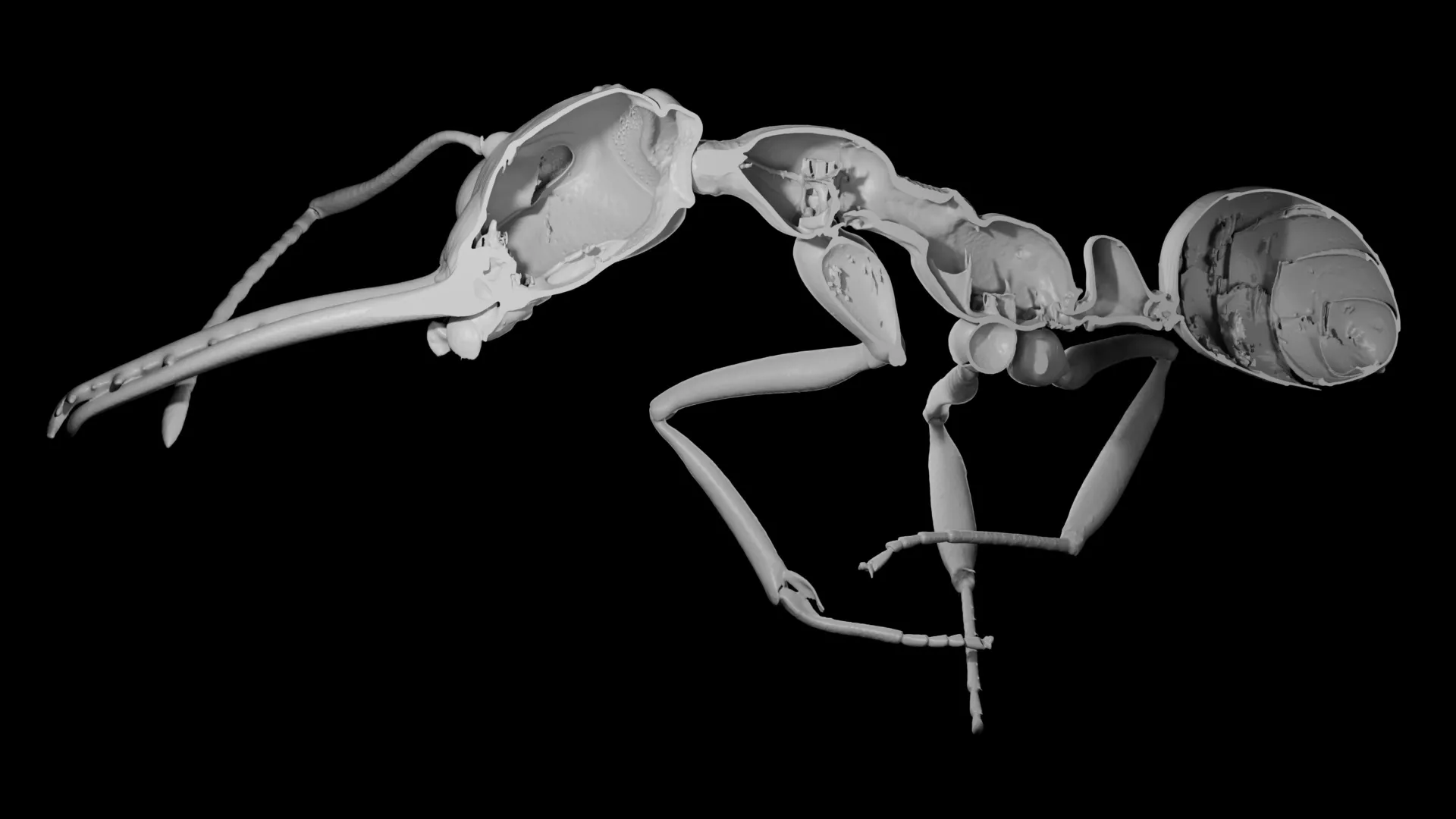
To investigate this idea, the researchers analyzed a large dataset of 3D X-ray scans from more than 500 ant species. They measured both total body volume and cuticle volume, finding that investment in the cuticle varied widely, from 6% to 35% of an ant’s body. When these measurements were fed into evolutionary models, a clear trend emerged: species that devoted less of their body to cuticle tended to form larger colonies.
Bigger colonies through collective strength
While thinner cuticles leave individual ants more vulnerable, the authors suggest this tradeoff may actually encourage the growth of large societies. Reduced armor may go hand in hand with other helpful social traits, including cooperative foraging, shared nest defense, and division of labor, all of which tend to become more pronounced as colonies grow.
“Ants reduce per-worker investment in one of the most nutritionally expensive tissues for the good of the collective,” Matte explained. “They’re shifting from self-investment toward a distributed workforce, resulting in more complex societies. It’s a pattern that echoes the evolution of multicellularity, where cooperative units can be individually simpler than a solitary cell, yet collectively capable of far greater complexity.”
The researchers also found that lower investment in the cuticle was linked to higher diversification rates. Biologists often use diversification, which reflects how frequently new species form, as a marker of evolutionary success. Economo noted that very few traits have been connected to diversification in ants, making this result especially striking.
Why less armor may lead to more species
Exactly why reduced cuticle investment promotes speciation is still unclear. One leading idea is that ants with lower nutritional demands can expand into environments where resources are limited.
“Requiring less nitrogen could make them more versatile and able to conquer new environments,” said Matte, who began the work during his master’s program while interning in Economo’s lab at the Okinawa Institute of Science and Technology in Japan.
The authors also suggest that as ant societies became more complex, group-level defenses such as collective nest protection and disease control reduced the need for heavy individual armor. This may have created a reinforcing cycle. Lower cuticle investment allows colonies to grow larger, and larger colonies further reduce the pressure for each ant to be strongly protected.
“I think of this as the evolution of squishability,” laughed Economo. “Many kids have discovered that insects aren’t all equally robust.”
Other social organisms, including termites, may have followed similar evolutionary paths, although that possibility still needs further testing.
What ant societies can teach us about humans
The findings also have implications beyond insects. The researchers draw parallels to human military history, where heavily armored knights were eventually replaced by specialized soldiers such as archers and crossbowmen. Economo also pointed to Lanchester’s Laws — mathematical equations developed during World War I that examine when large numbers of weaker fighters can overpower a smaller force of stronger ones.
“The tradeoff between quantity and quality is all around. It’s in the food you eat, the books you read, the offspring you want to raise,” Matte said. “It was fascinating to retrace how ants handled it through their long evolution. We could see lineages taking different directions, being shaped by different constraints and environments, and ultimately giving rise to the extraordinary diversity we observe today.”
The paper, “The evolution of cheaper workers facilitated larger societies and accelerated diversification in ants,” was published in the journal Science Advances on December 19, 2025.
This research was supported by the Okinawa Institute of Science and Technology, the Japan Society for the Promotion of Science KAKENHI (24K01785), the University of Cambridge and the General Research Fund 2022/2023 (17121922) from the Research Grant Council of Hong Kong. This article does not necessarily reflect the views of these organizations.
