Gut molecule shows remarkable anti-diabetes power

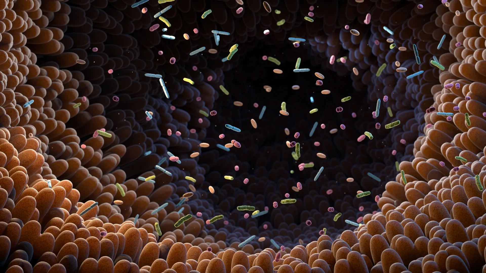
An international group of scientists led by Professor Marc-Emmanuel Dumas at Imperial College London & CNRS, along with Prof. Patrice Cani (Imperial & University of Louvain, UCLouvain), Dr. Dominique Gauguier (Imperial & INSERM, Paris) and Prof. Peter Liu (University of Ottawa Heart Institute), has identified an unexpected natural compound that helps counter insulin resistance and type 2 diabetes. The compound, trimethylamine (TMA), is a metabolite created by gut microbes from dietary choline. According to a study in Nature Metabolism, TMA can interrupt a key immune pathway and contribute to healthier blood sugar levels.

The discovery builds on work that began 20 years ago. During his postdoctoral research, Patrice Cani found that high-fat diets allow bacterial components to pass into the body, prompting the immune system to activate and ignite inflammation. This immune response was shown to play a direct role in insulin resistance among people with diabetes. Although this idea faced skepticism in 2005, it is now widely recognized and scientifically accepted.

In 2025, researchers at the University of Louvain and Imperial College London clarified how this harmful chain reaction might be counteracted. They reported that TMA, formed by gut microbes from dietary choline present in several foods, can support improved blood-sugar control.

TMA Blocks a Critical Immune Protein

The key lies in the molecule’s interaction with IRAK4, a protein that helps regulate immune activity. Under a high-fat diet, IRAK4 responds by triggering inflammation to signal that the body is experiencing dietary imbalance.

However, when the body is exposed to elevated fat intake for extended periods (as in type 2 diabetes), IRAK4 becomes overstimulated. This constant activation drives chronic inflammation, which contributes directly to insulin resistance.

Using a combination of human cell cultures, animal studies, and molecular screening tools, the research team demonstrated that TMA can attach to IRAK4 and reduce its activity. This interaction lowers inflammation caused by fatty foods and restores the body’s ability to respond to insulin. The findings suggest that TMA may help recalibrate harmful metabolic responses triggered by poor dietary habits. The molecule also showed an impressive ability to protect mice from sepsis-related death by weakening overwhelming inflammatory responses.

IRAK4 Targeting Offers New Therapeutic Possibilities

Further experiments confirmed that removing the IRAK4 gene or inhibiting it with drugs produced the same beneficial effects seen with TMA. Because IRAK4 is already a well-established target in drug development, the results point toward promising treatment strategies for diabetes.

“This flips the narrative,” said Prof. Dumas. “We’ve shown that a molecule from our gut microbes can actually protect against the harmful effects of a poor diet through a new mechanism. It’s a new way of thinking about how the microbiome influences our health.”

“This shows how nutrition and our gut microbes can work together by producing molecules that fight inflammation and improve metabolic health!” said Prof. Patrice Cani, co-senior author, University of Louvain, Belgium and visiting professor at Imperial College London.

Global Impact and Future Directions

With more than 500 million people worldwide living with diabetes, the identification of TMA as a microbial signal that shapes immune responses introduces a potential new avenue for treatment. Approaches that enhance TMA production, whether through diet or medication, could help reduce insulin resistance and improve long-term health outcomes.

“What we eat shapes our microbes and some of their molecules can protect us from diabetes. That’s nutrition in action!” said University of Louvain, Prof. Cani.

This work was supported by an extensive network of collaborators across Europe and North America, involving teams in Belgium, Canada, Australia, France, Italy, and Spain. Funding came from numerous European (ERC, FEDER) and national (MRC, Wellcome Trust, ANR, FNRS, EOS, WELRi, ARC) sources, highlighting the large-scale effort behind this breakthrough.

Share Button

New mpox strain identified in England

It’s a mix of two major types of the virus, and has been found in someone who recently returned from Asia.

Share Button

Is it a cold, ‘super flu’ or Covid? How to tell the difference

BBC Morning Live’s Dr Oscar Duke shares his advice on how to identify whether you have cold, flu or Covid and how to look after yourself.

Share Button

Low dose melanoma treatment delivers dramatically better results

A recent study reports that using reduced amounts of approved immunotherapy for malignant melanoma may lead to better control of tumors while also limiting side effects. The findings come from researchers at Karolinska Institutet and were published in the Journal of the National Cancer Institute.

“The results are highly interesting in oncology, as we show that a lower dose of an immunotherapy drug, in addition to causing significantly fewer side effects, actually gives better results against tumors and longer survival,” says last author Hildur Helgadottir, a researcher at the Department of Oncology-Pathology at Karolinska Institutet, who led the study.

Why Sweden Uses a Modified Treatment Approach

Standard treatment for malignant melanoma typically relies on the approved doses of nivolumab and ipilimumab. However, because the full-dose combination often leads to considerable side effects, Swedish clinicians have increasingly adopted a regimen that uses a reduced amount of ipilimumab. This component is both the most expensive part of the treatment and the one most strongly linked to side effects.

“In Sweden, we have greater freedom to choose doses for patients, while in many other countries, due to reimbursement policies, they are restricted by the doses approved by the drug authorities,” says Hildur Helgadottir.

Improved Responses and Survival With Reduced Ipilimumab

Nearly 400 people with advanced, inoperable malignant melanoma took part in the study, which focused on this severe form of skin cancer. According to the results, patients receiving the lower-ipilimumab regimen showed stronger responses. A total of 49 percent of these patients responded to treatment, compared to 37 percent in the group that received the traditional dose.

Progression-free survival, defined as the time a patient lives without the disease getting worse, reached a median of nine months for the lower-dose group. The traditional-dose group experienced a median of three months. Overall survival also differed significantly, with medians of 42 months and 14 months, respectively.

Reduced Side Effects May Allow Longer Treatment

Serious side effects appeared in 31 percent of people treated with the lower dose, compared to 51 percent among those given the standard regimen.

“The new immunotherapies are very valuable and effective, but at the same time they can cause serious side effects that are sometimes life-threatening or chronic. Our results suggest that this lower dosage may enable more patients to continue the treatment for a longer time, which is likely to contribute to the improved results and longer survival,” says Hildur Helgadottir.

Study Limitations and Research Collaboration

Although some differences existed between the treatment groups, the advantage of the lower-dose approach remained even after accounting for several factors, including age and tumor stage. Because this was a retrospective observational study, the research cannot conclusively prove that the lower dose directly caused the improved outcomes.

The work was carried out together with the Sahlgrenska Comprehensive Cancer Center at Sahlgrenska University Hospital and received funding from the Cancer Foundation, Region Stockholm, and the Radiumhemmet Research Fund.

Share Button

‘Life being stressful is not an illness’ – GPs on mental health over-diagnosis

Hundreds of GPs in England tell the BBC they are also worried about a lack of help for patients.

Share Button

New moonquake discovery could change NASA’s Moon plans

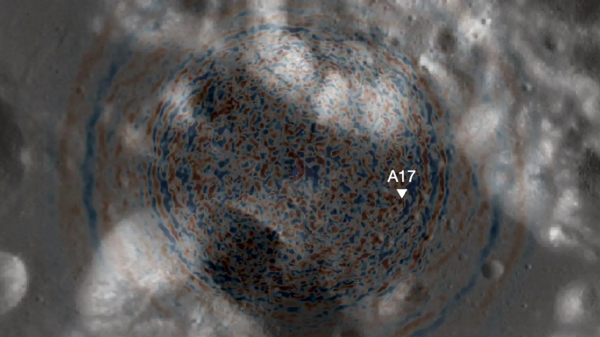
A recently published study reports that shaking from moonquakes, rather than impacts from meteoroids, was the main force behind the shifting terrain in the Taurus-Littrow valley, the site where Apollo 17 astronauts landed in 1972. The researchers also identified a likely explanation for the changing surface features and evaluated potential damage by applying updated models of lunar seismic activity — results that could influence how future missions and long-term settlements are planned on the moon.

The work, conducted by Smithsonian Senior Scientist Emeritus Thomas R. Watters and University of Maryland Associate Professor of Geology Nicholas Schmerr, appeared in the journal Science Advances.

Evidence From Apollo 17 Reveals Ancient Moonquake Activity

To investigate the region, Watters and Schmerr examined samples and observations collected during Apollo 17. Astronauts documented boulder tracks and landslides that appear to have been triggered by moonquakes. By analyzing this geological evidence, the scientists estimated how strong these past quakes were and identified the most likely fault responsible for generating them.

“We don’t have the sort of strong motion instruments that can measure seismic activity on the moon like we do on Earth, so we had to look for other ways to evaluate how much ground motion there may have been, like boulder falls and landslides that get mobilized by these seismic events,” Schmerr said.

Active Lunar Fault May Still Be Producing Quakes

According to the study, moonquakes with magnitudes near 3.0 — mild by Earth standards but significant when occurring close to their source — repeatedly shook the area over the last 90 million years. These events were linked to the Lee-Lincoln fault, a tectonic feature that cuts through the valley floor. The pattern of activity points to the possibility that this fault, one of many young thrust faults identified across the moon, has not yet gone dormant.

“The global distribution of young thrust faults like the Lee-Lincoln fault, their potential to be still active and the potential to form new thrust faults from ongoing contraction should be considered when planning the location and assessing stability of permanent outposts on the moon,” Watters said.

Assessing Daily Risk for Future Lunar Operations

Watters and Schmerr also calculated the statistical likelihood of a damaging quake near an active lunar fault. Their estimate suggests a one in 20 million chance of such an event occurring on any given day.

“It doesn’t sound like much, but everything in life is a calculated risk,” Schmerr noted. “The risk of something catastrophic happening isn’t zero, and while it’s small, it’s not something you can completely ignore while planning long-term infrastructure on the lunar surface.”

Short missions like Apollo 17 face little danger due to their limited duration. However, the researchers found that projects involving longer stays would encounter a gradually increasing risk. Upcoming missions using taller lander designs, including the Starship Human Landing System, may be more susceptible to ground acceleration caused by moonquakes close to an active fault. These concerns are particularly important as NASA moves forward with the Artemis program, which aims to maintain a continuous human presence on the moon. Watters and Schmerr stressed that modern missions must account for hazards not encountered during the Apollo era.

“If astronauts are there for a day, they’d just have very bad luck if there was a damaging event,” Schmerr added. “But if you have a habitat or crewed mission up on the moon for a whole decade, that’s 3,650 days times 1 in 20 million, or the risk of a hazardous moonquake becoming about 1 in 5,500. It’s similar to going from the extremely low odds of winning a lottery to much higher odds of being dealt a four of a kind poker hand.”

Advancing the Field of Lunar Paleoseismology

Schmerr views this research as part of a growing field known as lunar paleoseismology, which focuses on ancient seismic activity. Unlike Earth, where scientists can excavate trenches to reveal evidence of past earthquakes, lunar researchers must rely on material already gathered and imaging from orbit. He expects future progress to accelerate thanks to higher resolution mapping, new technology, and upcoming Artemis missions that plan to deploy seismometers far more advanced than those used during Apollo.

“We want to make sure that our exploration of the moon is done safely and that investments are made in a way that’s carefully thought out,” Schmerr said. “The conclusion we came to is: don’t build right on top of a scarp, or recently active fault. The farther away from a scarp, the lesser the hazard.”

Support From the Lunar Reconnaissance Orbiter Mission

This research was supported by NASA’s Lunar Reconnaissance Orbiter mission, which launched on June 18, 2009. LRO is operated by NASA’s Goddard Space Flight Center for the Science Mission Directorate. This article does not necessarily reflect the views of this organization.

Share Button

The rotten egg smell that could finally beat nail fungus

Hydrogen sulfide, a naturally occurring gas best known for its strong rotten egg odor, may offer a faster and gentler way to treat stubborn nail infections. Researchers at the University of Bath and King’s College London (KCL) report that this volcanic gas could form the basis of a new therapy that works more quickly while avoiding many common side effects.

Nail infections are typically caused by fungi, though bacteria can occasionally be involved. These conditions are widespread, affecting an estimated 4-10% of people worldwide, with rates climbing to nearly 50% among adults aged 70 and older.

These infections can create serious complications in vulnerable groups such as older adults and individuals with diabetes. Despite their prevalence, they remain difficult to eliminate.

Why Current Treatments Often Fall Short

Standard therapies include oral antifungal medications taken as pills and topical products placed on the surface of the nail. Oral medications usually take 2-4 months to show results and are generally effective, but they also pose risks for side effects, particularly in patients with other health conditions.

Topical treatments are considered safer, but they often require very long application periods, sometimes lasting years. Even then, they frequently fail to fully clear the infection or the infection returns.

One major obstacle is that most medications struggle to pass through the dense structure of the nail, preventing them from reaching the fungi or bacteria living beneath it. Even the best topical antifungal options achieve relatively low cure rates, underscoring the need for treatments that can reliably reach microbes deep within the nail.

Hydrogen Sulfide Shows Strong Antimicrobial Potential

A research team from the University of Bath and KCL has identified hydrogen sulfide (H2S) as a promising alternative. This small, naturally occurring gas appears capable of penetrating the nail plate far more effectively than existing topical drugs.

Earlier studies already suggested that H2S travels through nail tissue with ease. The new findings show that it also has powerful antimicrobial activity, killing a broad spectrum of pathogens, including fungal species that do not respond well to common antifungal medications.

In controlled laboratory experiments, the researchers used a compound that releases hydrogen sulfide as it breaks down. They found that the gas disrupts microbial energy production and causes irreversible damage to the cells, ultimately destroying the fungi responsible for infection.

The study is detailed in Scientific Reports.

Researchers See Promise for a Future Topical Therapy

Dr. Albert Bolhuis of the University of Bath’s Department of Life Sciences said: “Thanks to its ability to efficiently reach the site of infection and its novel mode of action, we believe that a topically applied medicine containing hydrogen sulfide could become a highly effective new treatment for nail infections, which avoids the limitations of current therapies.

“Our research lays the foundation for a compelling alternative to existing treatments, with the potential to improve outcomes for patients suffering from persistent and drug-resistant fungal nail infections.”

Hydrogen sulfide does have a strong smell and some level of toxicity. However, researchers emphasize that the concentrations needed for treatment appear to be far below harmful levels, and the right formulation should greatly reduce any unpleasant odor.

Next Steps Toward Patient Use

So far, the research has been conducted only in vitro. Even so, the team hopes to continue development and create a topical treatment suitable for patients within the next five years.

Professor Stuart Jones, Director of the Centre for Pharmaceutical Medicine Research at KCL, said: “We are looking forward to translating these findings into an innovative topical product that can treat nail infection.”

Share Button

Natural hormone unlocks a hidden fat burning switch

Studies in mice have shown that a hormone produced in the intestine can send signals to the brain and influence how much energy the body uses. This hormone, called FGF19 (fibroblast growth factor 19), activates processes that help the body spend more energy, use stored fat as fuel, and improve weight control and blood glucose levels in obese animals.

Researchers linked these effects to the action of FGF19 in the hypothalamus, a key brain region that receives information from the rest of the body and the environment to coordinate energy metabolism. They found that when FGF19 signals in the hypothalamus, it boosts the activity of thermogenic adipocytes (i.e., fat cells that burn energy to produce heat), which are specialized fat cells that help the body generate heat instead of storing calories.

New Paths for Obesity and Diabetes Treatments

Because of these findings, the scientists believe that FGF19 could inspire new medications for obesity, diabetes, and other metabolic conditions. The idea is to develop compounds that imitate the behavior of natural substances in the body, mimicking the action of endogenous compounds (i.e. those produced by the body itself).

This strategy resembles the way some of the latest diabetes and obesity drugs work. Ozempic, for example, contains semaglutide, an ingredient that activates receptors mimicking the hormone GLP-1. By doing so, it sends satiety signals to the brain and helps patients feel full with less food.

According to the study, FGF19 did more than change appetite or fat storage. The hormone also lowered peripheral inflammation and improved the animals’ tolerance to cold. When the researchers blocked the sympathetic nervous system, however, these benefits disappeared. In further experiments, they observed that exposure to cold increased the expression of FGF19 receptors in the hypothalamus. Because the hypothalamus is crucial for maintaining body temperature, these results suggest that FGF19 may help the body adapt by coordinating energy balance and thermoregulation.

FGF19, Thermogenesis, and Brain Control of Energy

“FGF19 had already been linked to a reduction in food intake. Our work broke new ground by showing that it also plays an important role by acting on the hypothalamus and stimulating an increase in energy expenditure in white and brown adipose tissue. In other words, in addition to controlling appetite, it stimulates thermogenesis. So, in terms of therapy associated with obesity, it’d make a lot of sense,” explains Professor Helena Cristina de Lima Barbosa, from the Obesity and Comorbidities Research Center (OCRC) at the State University of Campinas (UNICAMP).

The OCRC is a Research, Innovation, and Dissemination Center (RIDC) of FAPESP, which also funded the project through grants to doctoral student Lucas Zangerolamo, the first author of the study, supervised by Barbosa.

The work has been described in detail in an article published in the American Journal of Physiology — Endocrinology and Metabolism, where it was highlighted as a Top Article in May.

Global Obesity Crisis and Urgent Health Targets

The World Atlas of Obesity 2025 warns that, if current trends continue, global health goals for this year will not be met. These targets include halting the rise in diabetes and obesity and cutting premature deaths from cardiovascular disease, chronic respiratory disease, and cancer by 25%, using 2010 as the reference year.

The Atlas estimates that more than 1 billion people worldwide are currently living with obesity. If effective actions are not put in place, this number could surpass 1.5 billion by 2030. Obesity is already associated with about 1.6 million premature deaths each year from non-communicable diseases.

In Brazil, around 31% of the population has obesity. In addition, between 40% and 50% of adults do not reach the recommended levels of physical activity in terms of frequency or intensity.

Where FGF19 Comes From and How It Works

FGF19, which is involved in the control of energy metabolism, is mainly produced in the small intestine. In the liver, it regulates the production of bile acids and also influences the synthesis of glucose and fats. While its primary roles in the liver have been widely explored in scientific literature, its effects in the brain have received much less attention.

“In the lab, we work with bile acids, which are also the subject of my master’s degree, and they regulate the release of FGF-19. Our initial studies led us down this path,” Zangerolamo told Agência FAPESP.

At eight weeks of age, the mice used in the study were randomly assigned to two groups. One group received a standard diet (control) and the other was fed a high-fat diet to induce obesity. The researchers then administered FGF19 directly into the brains of the obese animals. All mice were kept in carefully controlled conditions of temperature, lighting, and access to water.

In the article, the scientists report that central FGF19 signaling improved energy homeostasis. It did this by boosting the activity of the sympathetic nervous system and stimulating thermogenesis in adipose tissue, leading the tissue to consume more energy in the form of heat.

“The brain plays an extremely important role in controlling the body’s adiposity. At the same time as it receives information from peripheral tissues, it triggers commands. These commands, apparently using the sympathetic nervous system, seem to be an interesting way of thinking about energy expenditure,” adds Barbosa.

Digging Deeper Into Brain Cells and FGF19 Receptors

To better understand which brain cells respond to FGF19, the authors compiled and examined public scRNA-seq data from several studies of the hypothalamus. This method makes it possible to sequence RNA from individual cells, revealing which genes are active in each cell type. In total, the team evaluated transcription from more than 50,000 single cells to identify hypothalamic cell populations that express FGF19 receptors.

The researchers note that a key question now is how to encourage the body to produce more FGF19 on its own. They are also working to connect these findings with what is already known about the neural circuits that regulate eating behavior.

“We want to broaden this understanding. We’re studying the hypothalamus to evaluate the inflammation commonly observed when a high-fat diet is administered and whether FGF19 plays a role in this area,” says Zangerolamo, who did part of the work during an internship at the Joslin Diabetes Center at Harvard Medical School with Professor Yu-Hua Tseng, who is also an author of the article.

Share Button

Earth’s early oceans hid the secret rise of complex life

New findings suggest that complex life began forming much earlier, and over a far longer period, than researchers previously understood. The study provides fresh insight into the environmental conditions that supported early evolution and challenges several widely accepted ideas about when advanced cellular features first appeared.

Led by the University of Bristol and published in Nature on December 3, the work shows that complex organisms started developing long before oxygen levels in the atmosphere rose to significant levels. Until now, many scientists believed that plentiful oxygen was essential for the emergence of complex life.

“The Earth is approximately 4.5 billion years old, with the first microbial life forms appearing over 4 billion years ago. These organisms consisted of two groups — bacteria and the distinct but related archaea, collectively known as prokaryotes,” said co-author Anja Spang from the Department of Microbiology & Biogeochemistry at the Royal Netherlands Institute for Sea Research.

For hundreds of millions of years, prokaryotes were the only living organisms on the planet. More complex eukaryotic cells eventually evolved, giving rise to algae, fungi, plants and animals.

Rethinking the Origins of Eukaryotes

Davide Pisani, Professor of Phylogenomics in the School of Biological Sciences at the University of Bristol and co-author, noted: “Previous ideas on how and when early prokaryotes transformed into complex eukaryotes has largely been in the realm of speculation. Estimates have spanned a billion years, as no intermediate forms exist and definitive fossil evidence has been lacking.”

To shed light on this long-debated transition, the team expanded the existing ‘molecular clocks’ method, a tool used to estimate when different species last shared an ancestor.

“The approach was two-fold: by collecting sequence data from hundreds of species and combining this with known fossil evidence, we were able to create a time-resolved tree of life. We could then apply this framework to better resolve the timing of historical events within individual gene families,” explained co-lead author Professor Tom Williams in the Department of Life Sciences at the University of Bath.

A Much Earlier Start to Cellular Complexity

The researchers examined more than one hundred gene families across multiple biological systems and focused on the traits that separate eukaryotes from prokaryotes. This allowed them to reconstruct a clearer picture of how complex cellular features developed.

Their results indicate that the shift toward complexity began nearly 2.9 billion years ago — almost a billion years earlier than some previous estimates. The evidence suggests that structures such as the nucleus emerged well before mitochondria. “The process of cumulative complexification took place over a much longer time period than previously thought,” said author Gergely Szöllősi, head of the Model-Based Evolutionary Genomics Unit at the Okinawa Institute of Science and Technology (OIST).

These findings allowed the researchers to dismiss some existing models for eukaryogenesis (the evolution of complex life). Since the results did not fully match any current explanation, the team proposed a new scenario called ‘CALM’ — Complex Archaeon, Late Mitochondrion.

Introducing the CALM Model

Lead author Dr. Christopher Kay, Research Associate in the School of Biological Sciences at the University of Bristol, said: “What sets this study apart is looking into detail about what these gene families actually do — and which proteins interact with which — all in absolute time. It has required the combination of a number of disciplines to do this: paleontology to inform the timeline, phylogenetics to create faithful and useful trees, and molecular biology to give these gene families a context. It was a big job.”

“One of our most significant findings was that the mitochondria arose significantly later than expected. The timing coincides with the first substantial rise in atmospheric oxygen,” added author Philip Donoghue, Professor of Palaeobiology in the School of Earth Sciences at the University of Bristol.

“This insight ties evolutionary biology directly to Earth’s geochemical history. The archaeal ancestor of eukaryotes began evolving complex features roughly a billion years before oxygen became abundant, in oceans that were entirely anoxic.”

Share Button

Scientists find hidden layers in brain’s memory center

Researchers at the Mark and Mary Stevens Neuroimaging and Informatics Institute (Stevens INI) at the Keck School of Medicine of USC have uncovered a previously unrecognized organizational pattern in one of the brain’s key regions for learning and memory. According to findings reported in Nature Communications, the CA1 section of a mouse’s hippocampus contains four separate layers of specialized cell types. The hippocampus plays an essential role in forming memories, guiding spatial navigation, and influencing emotions, and the discovery of these layers offers new insight into how information moves through this part of the brain. It also provides clues about why some cell types are especially vulnerable in conditions such as Alzheimer’s disease and epilepsy.

“Researchers have long suspected that different parts of the hippocampus’ CA1 region handle different aspects of learning and memory, but it wasn’t clear how the underlying cells were arranged,” said Michael S. Bienkowski, PhD, senior author of the study and assistant professor of physiology and neuroscience and of biomedical engineering.

“Our study shows that CA1 neurons are organized into four thin, continuous bands, each representing a different neuron type defined by a unique molecular signature. These layers aren’t fixed in place; instead, they subtly shift and change in thickness along the length of the hippocampus. This shifting pattern means that each part of CA1 contains its own mix of neuron types, which helps explain why different regions support different behaviors. This may also clarify why certain CA1 neurons are more vulnerable in conditions like Alzheimer’s disease and epilepsy: if a disease targets one layer’s cell type, the effects will vary depending on where in CA1 that layer is most prominent.”

High-resolution RNA imaging reveals cellular distinctions

To examine this structure, the research team used an RNA labeling technique called RNAscope together with high-resolution microscopy. This approach allowed them to observe single-molecule gene expression inside mouse CA1 tissue and identify individual neuron types based on their active genes. From 58.065 CA1 pyramidal cells, the scientists recorded more than 330,000 RNA molecules, which represent the genetic instructions that indicate when and where genes are expressed. By mapping these gene activity patterns, they produced a detailed cellular atlas outlining the boundaries between distinct nerve cell types across the CA1 region.

Their results showed that CA1 contains four continuous layers of nerve cells, each distinguished by its own pattern of active genes. When viewed in three dimensions, these layers form sheet-like structures that vary in thickness and shape along the hippocampus. This well-defined arrangement clarifies earlier studies that had described CA1 as a more blended or mosaic mixture of cell types.

Hidden “stripes” highlight internal brain architecture

“When we visualized gene RNA patterns at single-cell resolution, we could see clear stripes, like geological layers in rock, each representing a distinct neuron type,” said Maricarmen Pachicano, doctoral researcher at the Stevens INI’s Center for Integrative Connectomics and co-first author of the paper. “It’s like lifting a veil on the brain’s internal architecture. These hidden layers may explain differences in how hippocampal circuits support learning and memory.”

Because the hippocampus is one of the first regions affected in Alzheimer’s disease and is involved in epilepsy, depression, and other neurological conditions, identifying the CA1’s layered structure offers a promising guide for determining which neuron types may be most at risk as these disorders progress.

Advancing brain mapping with modern imaging and data science

“Discoveries like this exemplify how modern imaging and data science can transform our view of brain anatomy,” said Arthur W. Toga, PhD, director of the Stevens INI and the Ghada Irani Chair in Neuroscience at the Keck School of Medicine of USC. “This work builds on the Stevens INI’s long tradition of mapping the brain at every scale, from molecules to whole networks, and will inform both basic neuroscience and translational studies targeting memory and cognition.”

A new CA1 cell-type atlas available to researchers

The team compiled its findings into a new CA1 cell-type atlas using data from the Hippocampus Gene Expression Atlas (HGEA). This resource is freely available to scientists worldwide and includes interactive 3D visualizations accessible through the Schol-AR augmented-reality app developed at the Stevens INI. The tool allows researchers to explore the layered structure of the hippocampus in great detail.

Because this layered pattern in mice resembles similar arrangements seen in primates and humans, including comparable variations in CA1 thickness, the researchers believe the organization may be shared across many mammalian species. Further work is needed to determine how closely this structure in humans matches what has been observed in mice, but the findings create a strong starting point for future studies examining how hippocampal architecture supports memory and cognition.

“Understanding how these layers connect to behavior is the next frontier,” Bienkowski said. “We now have a framework to study how specific neuron layers contribute to such different functions like memory, navigation, and emotion, and how their disruption may lead to disease.”

About the study

In addition to Bienkowski and Pachicano, the study’s other authors include Shrey Mehta, Angela Hurtado, Tyler Ard, Jim Stanis, and Bayla Breningstall.

This work was supported by the National Institutes of Health/National Institute of Aging (K01AG066847, R36AG087310-01, supplement P30-AG066530-03S1), National Science Foundation (grant 2121164), and funding from the USC Center for Neuronal Longevity. Research data reported in this publication was supported by the Office of the Director, National Institutes of Health under award number S10OD032285.

Share Button