Scientists successfully transfer longevity gene and extend lifespan

Naked mole rats are not much to look at, but their biology has made them one of the most fascinating animals in aging research. These small, wrinkled rodents can live for decades, rarely develop cancer, and seem unusually protected from many of the diseases that normally arrive with age.

Researchers at the University of Rochester showed that one of those biological advantages can be moved into another mammal. By transferring a gene linked to the naked mole rat’s unusually high levels of high molecular weight hyaluronic acid (HMW-HA), the team improved health and modestly extended lifespan in mice.

The work, published in Nature in 2023, suggested that at least some longevity traits that evolved in long-lived animals may be adaptable beyond the species that developed them. The genetically modified mice lived healthier lives and had an approximate 4.4 percent increase in median lifespan compared with ordinary mice.

“Our study provides a proof of principle that unique longevity mechanisms that evolved in long-lived mammalian species can be exported to improve the lifespans of other mammals,” says Vera Gorbunova, the Doris Johns Cherry Professor of biology and medicine at Rochester.

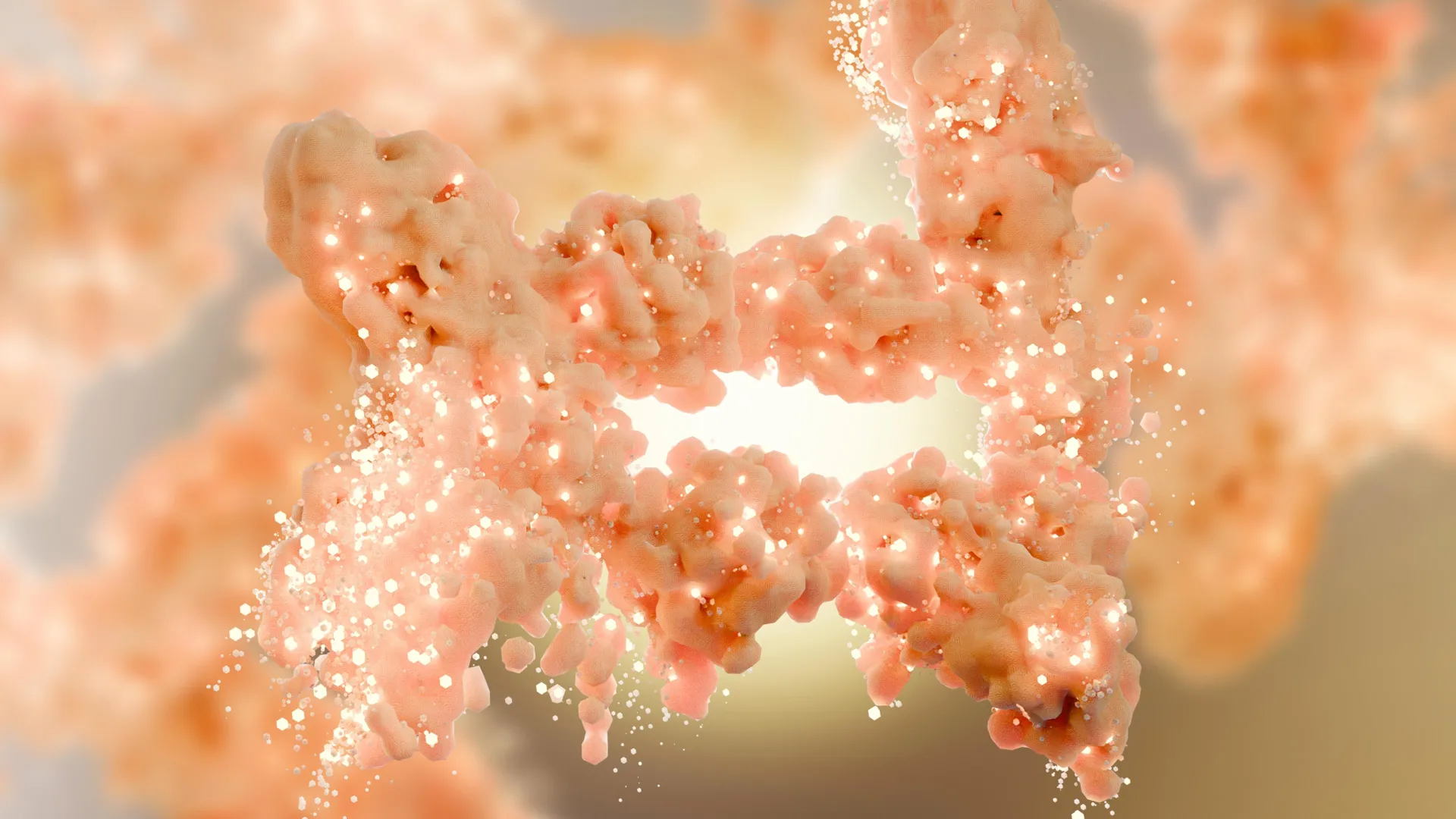
Gorbunova, along with Andrei Seluanov, a professor of biology, and their colleagues, focused on a gene that helps produce HMW-HA. This substance is abundant in naked mole rats and has been tied to their striking resistance to cancer, inflammation, and age-related decline.

Why Naked Mole Rats Fascinate Aging Scientists

Naked mole rats are about the size of mice, yet their lifespans are extraordinary for rodents. They can live up to 41 years, nearly ten times longer than similarly sized rodents.

Their long lives are not the only reason scientists study them. As they age, naked mole rats appear to avoid many conditions that commonly affect other mammals, including neurodegeneration, cardiovascular disease, arthritis, and cancer. For decades, Gorbunova, Seluanov, and other researchers have been investigating how these animals stay so resilient.

One major clue is HMW-HA. Naked mole rats carry roughly ten times more of it than mice and humans. In earlier work, researchers found that when HMW-HA was removed from naked mole rat cells, those cells became more likely to form tumors.

That finding raised a powerful question. If HMW-HA helps naked mole rats resist cancer and age-related damage, could the same mechanism work in a different animal?

Transferring a Naked Mole Rat Longevity Gene

To test the idea, the Rochester team engineered mice to carry the naked mole rat version of the hyaluronan synthase 2 gene. This gene helps make the protein that produces HMW-HA.

All mammals have a version of hyaluronan synthase 2, but the naked mole rat version appears to be especially active. It seems to drive stronger gene expression, leading to greater production of the protective molecule.

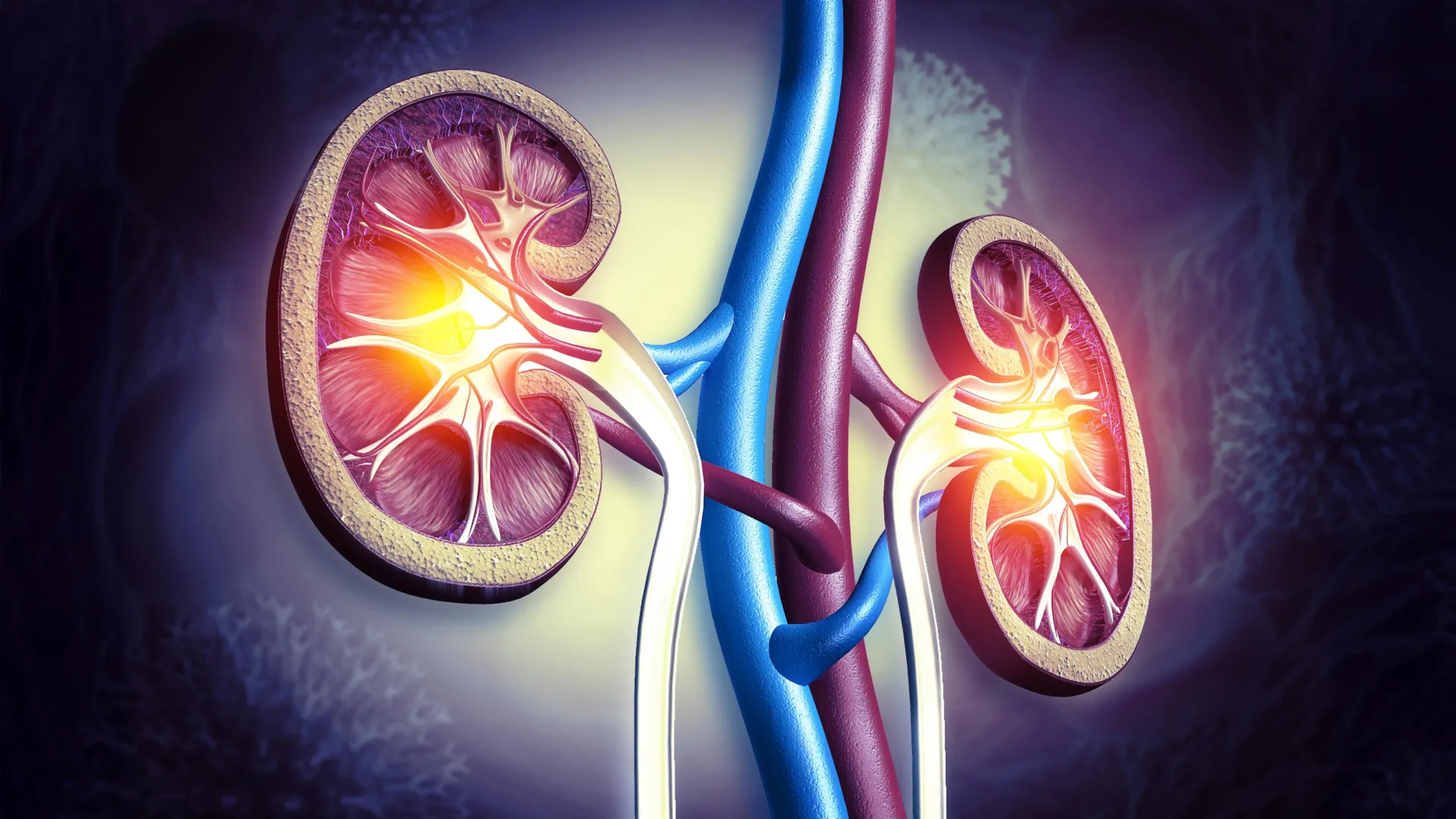
The modified mice developed higher levels of hyaluronan in several tissues. They also showed stronger protection against spontaneous tumors and chemically induced skin cancer.

The effects were not limited to cancer resistance. The mice carrying the naked mole rat gene stayed healthier overall, lived longer than regular mice, had less inflammation in multiple tissues as they aged, and maintained better gut health.

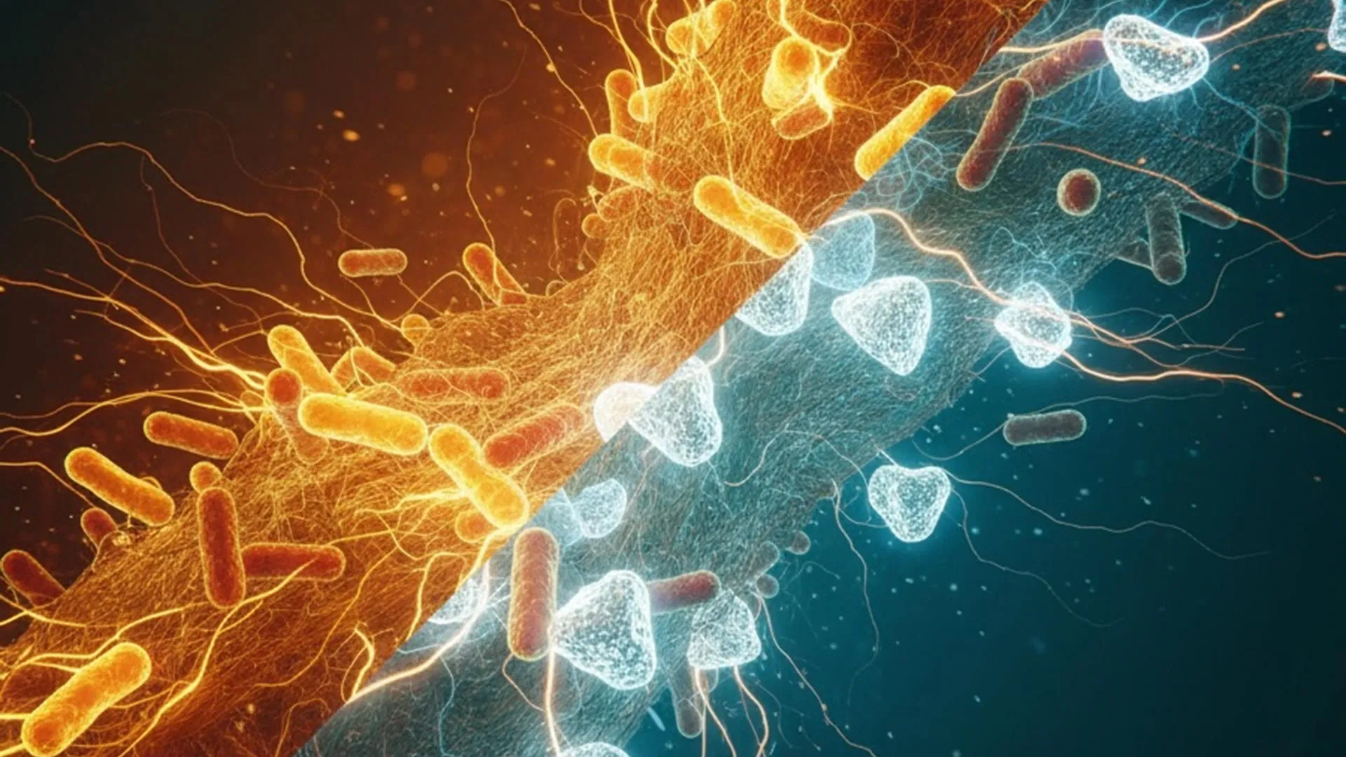
Because chronic inflammation is one of the major biological features of aging, the reduction in inflammation was especially important. The researchers believe HMW-HA may work partly by directly influencing the immune system, although more research is needed to explain exactly how it produces such broad benefits.

A Small Lifespan Gain With Big Implications

The increase in median lifespan was about 4.4 percent, which is modest. But the larger significance is that a longevity mechanism from one mammal was successfully transferred to another.

That makes the finding more than a mouse study about a single gene. It supports the idea that nature’s long-lived species may contain biological tools that can be studied, adapted, and possibly used to improve health in other animals.

“It took us 10 years from the discovery of HMW-HA in the naked mole rat to showing that HMW-HA improves health in mice,” Gorbunova says. “Our next goal is to transfer this benefit to humans.”

The researchers believe there may be two main ways to pursue that goal. One would be to slow the breakdown of HMW-HA in the body. Another would be to increase its production.

“We already have identified molecules that slow down hyaluronan degradation and are testing them in pre-clinical trials,” Seluanov says. “We hope that our findings will provide the first, but not the last, example of how longevity adaptations from a long-lived species can be adapted to benefit human longevity and health.”

Newer Research Adds to the Naked Mole Rat Story

Since the 2023 Nature study, naked mole rats have continued to offer new clues about why they age so differently from other mammals. A 2025 study in Science reported another potential longevity mechanism involving cGAS, a protein better known for its role in immune defense. In humans and mice, cGAS can interfere with some forms of DNA repair, but the naked mole rat version appears to help cells repair DNA damage more effectively. That study found that specific changes in the naked mole rat protein improved genome stability and delayed signs of aging in experimental models.

This newer work does not replace the HMW-HA finding. Instead, it strengthens a broader pattern. Naked mole rats likely owe their unusually long, healthy lives to several overlapping defenses, including cancer resistance, inflammation control, DNA repair, and tissue protection.

For human aging research, that matters. A single molecule is unlikely to become a simple fountain of youth. But each discovery gives scientists another possible route for targeting the biological processes that drive age-related disease.

The 2023 gene transfer study remains a striking proof of concept. A survival strategy that evolved in one of nature’s strangest mammals helped mice resist disease, age more smoothly, and live longer. The next challenge is determining whether those same biological tricks can be safely adapted to improve human healthspan.

Share Button

Brain scans reveal a shocking difference between psychopaths and other people

Neuroscientists have identified a measurable brain difference between people with psychopathic traits and those with few or none. In a study published in the Journal of Psychiatric Research, researchers from Nanyang Technological University, Singapore (NTU Singapore), the University of Pennsylvania, and California State University found that a brain region involved in reward and motivation was larger in individuals with psychopathic traits.

Using magnetic resonance imaging (MRI), the team found that the striatum was about 10 percent larger on average in psychopathic individuals compared with a control group. The striatum sits deep in the forebrain and plays a role in movement planning, decision-making, motivation, reinforcement, and how the brain responds to rewards.

Psychopathy is generally associated with an egocentric and antisocial personality pattern. People with strong psychopathic traits often show reduced empathy, little remorse for harmful actions, and, in some cases, a greater likelihood of criminal behavior. Not everyone with psychopathic traits commits crimes, and not every person who commits a crime is a psychopath, but research has consistently linked psychopathy with a higher risk of violent behavior.

A Larger Reward Center in the Brain

Earlier research had suggested that the striatum may be unusually active in psychopaths, but it was less clear whether the size of this brain region was also involved. The Journal of Psychiatric Research findings added evidence that psychopathy is not shaped only by social and environmental experiences. Biology may also play a role.

To investigate the link, the researchers scanned the brains of 120 people in the United States. They also interviewed the participants using the Psychopathy Checklist — Revised, a widely used psychological assessment designed to measure psychopathic traits.

Assistant Professor Olivia Choy, from NTU’s School of Social Sciences, a neurocriminologist who co-authored the study, said: “Our study’s results help advance our knowledge about what underlies antisocial behavior such as psychopathy. We find that in addition to social environmental influences, it is important to consider that there can be differences in biology, in this case, the size of brain structures, between antisocial and non-antisocial individuals.”

The findings may help researchers better understand how biology contributes to antisocial and criminal behavior. Over time, that knowledge could help refine theories of behavior and inform future approaches to policy, prevention, and treatment.

What the Striatum May Reveal About Risk and Reward

The striatum is part of the basal ganglia, a group of neuron clusters located deep in the brain. The basal ganglia receive information from the cerebral cortex, which helps control thinking, social behavior, and the ability to decide which sensory information deserves attention.

Over the past two decades, scientists have increasingly recognized that the striatum is not only involved in movement and reward. It may also be tied to social behavior and difficulties in social functioning.

By comparing MRI scans with psychopathy assessment results, the researchers found that a larger striatum was linked to a stronger need for stimulation, including thrill-seeking, excitement, and impulsive behavior. In the published study, stimulation-seeking and impulsivity partly explained the relationship between striatal volume and psychopathy, accounting for 49.4 percent of the association.

Professor Adrian Raine from the Departments of Criminology, Psychiatry, and Psychology at University of Pennsylvania, who co-authored the study, said: “Because biological traits, such as the size of one’s striatum, can be inherited to child from parent, these findings give added support to neurodevelopmental perspectives of psychopathy — that the brains of these offenders do not develop normally throughout childhood and adolescence.”

Psychopathic Traits Outside Prison Populations

One important feature of the study was that it included people from the community rather than focusing only on prison populations. That helped the researchers examine psychopathic traits in a broader group of individuals.

Professor Robert Schug from the School of Criminology, Criminal Justice, and Emergency Management at California State University, Long Beach, who co-authored the study, said: “The use of the Psychopathy Checklist — Revised in a community sample remains a novel scientific approach: Helping us understand psychopathic traits in individuals who are not in jails and prisons, but rather in those who walk among us each day.”

The researchers also examined 12 women in the study sample. They reported that, for the first time, psychopathy was linked to an enlarged striatum in adult females as well as males. The female sample was small, so the finding needs further study, but it suggested that the same brain pattern may not be limited to men.

In typical human development, the striatum tends to shrink as a child matures. That pattern raises the possibility that psychopathy may be connected to differences in brain development across childhood and adolescence.

Brain Development and Environment May Both Matter

Asst Prof Choy added: “A better understanding of the striatum’s development is still needed. Many factors are likely involved in why one individual is more likely to have psychopathic traits than another individual. Psychopathy can be linked to a structural abnormality in the brain that may be developmental in nature. At the same time, it is important to acknowledge that the environment can also have effects on the structure of the striatum.”

Prof Raine added: “We have always known that psychopaths go to extreme lengths to seek out rewards, including criminal activities that involve property, sex, and drugs. We are now finding out a neurobiological underpinning of this impulsive and stimulating behavior in the form of enlargement to the striatum, a key brain area involved in rewards.”

The study was published in the Journal of Psychiatric Research under the title “Larger striatal volume is associated with increased adult psychopathy.”

Later Research Points to a Wider Brain Network

Since the 2022 paper, later research has continued to explore how psychopathy relates to brain structure and brain networks. A 2025 study in European Archives of Psychiatry and Clinical Neuroscience examined 39 adult men diagnosed with psychopathy and found that antisocial lifestyle traits were associated with reduced volumes in several brain regions, including parts of the basal ganglia, thalamus, basal forebrain, pons, cerebellum, orbitofrontal cortex, dorsolateral-frontal cortex, and insular cortex. The researchers concluded that these findings point to disruptions in frontal-subcortical circuits involved in behavioral control.

Another 2025 analysis in Neuroscience and Biobehavioral Reviews looked across 38 functional neuroimaging studies of psychopathy. Although individual studies often pointed to different brain locations, the findings appeared to map onto a shared functional brain network involving the default mode network and subcortical regions. The authors argued that psychopathy may be better understood through a network-level view of the brain rather than by focusing on one region alone.

Together, these later findings add nuance to the 2022 striatum study. The enlarged striatum finding remains an important clue, especially because of the striatum’s role in reward, stimulation, and impulsivity. However, psychopathy likely reflects a broader pattern of brain differences involving motivation, emotional processing, impulse control, and social behavior.

Associate Professor Andrea Glenn from the Department of Psychology of The University of Alabama, who was not involved in the 2022 study, said: “By replicating and extending prior work, this study increases our confidence that psychopathy is associated with structural differences in the striatum, a brain region that is important in a variety of processes important for cognitive and social functioning. Future studies will be needed to understand the factors that may contribute to these structural differences.”

Scientists are still working to understand why the striatum may be enlarged in people with psychopathic traits. Future work may help clarify how genetics, development, life experiences, and environment interact to shape the brain systems involved in reward-seeking, impulse control, and antisocial behavior.

Share Button

Scientists discover the brain’s hidden “stop scratching” switch

When an itch strikes, scratching usually brings relief after a few moments. Scientists have now uncovered part of the biological system that tells the brain when enough scratching has occurred. The discovery reveals how the nervous system naturally limits scratching and may help explain why this process breaks down in people with chronic itch disorders. The findings were presented at the 70th Biophysical Society Annual Meeting.

Researchers from the laboratory of Roberta Gualdani at the University of Louvain in Brussels identified an unexpected role for a molecule known as TRPV4 in itch triggered by mechanical stimulation, such as scratching.

“We were initially studying TRPV4 in the context of pain,” Gualdani explained. “But instead of a pain phenotype, what emerged very clearly was a disruption of itch, specifically, how scratching behavior is regulated.”

TRPV4 and the Nervous System

TRPV4 is part of a family of ion channels that function like tiny molecular gateways in sensory nerve cells. These channels allow ions to move through cell membranes in response to physical or chemical changes. They help the nervous system detect sensations including temperature, pressure, and tissue stress.

Scientists have suspected for years that TRPV4 plays a role in sensing mechanical stimulation, but its involvement in itch, particularly chronic itch, has remained unclear and heavily debated.

To investigate more precisely, Gualdani’s team created genetically engineered mice in which TRPV4 was removed only from sensory neurons. Earlier studies had deleted the molecule throughout the entire body, making it difficult to determine exactly where it was acting.

Using genetic analysis, calcium imaging, and behavioral testing, the researchers found that TRPV4 appears in touch sensitive neurons known as Aβ low-threshold mechanoreceptors (Aβ-LTMRs). The channel was also present in certain sensory neurons connected to itch and pain pathways, including neurons expressing TRPV1.

Why Scratching Sometimes Does Not Stop

The team then created a chronic itch condition in mice that resembled atopic dermatitis. The results surprised the researchers. Mice missing TRPV4 in sensory neurons scratched less often overall, but each scratching episode lasted much longer than normal.

“At first glance, that seems paradoxical,” Gualdani said. “But it actually reveals something very important about how itch is regulated.”

According to the study, TRPV4 does not simply create the sensation of itch. Instead, it appears to help activate a negative feedback signal in mechanosensory neurons. This signal informs the spinal cord and brain that scratching has provided enough relief.

Without that feedback system, the sense of satisfaction from scratching becomes weaker, causing scratching to continue for extended periods. Researchers say TRPV4 may therefore function as part of the nervous system’s internal “stop scratching” mechanism.

“When we scratch an itch, at some point we stop because there’s a negative feedback signal that tells us we’re satisfied,” Gualdani explained. “Without TRPV4, the mice don’t feel this feedback, so they continue scratching much longer than normal.”

Implications for Chronic Itch Treatments

The findings also suggest that TRPV4 has a more complicated role in itch than previously believed. In skin cells, the channel may help trigger itch sensations. In neurons, however, it appears to help control and limit scratching behavior.

That distinction could be important for future drug development.

“This means that broadly blocking TRPV4 may not be the solution,” Gualdani noted. “Future therapies may need to be much more targeted — perhaps acting only in the skin, without interfering with the neuronal mechanisms that tell us when to stop scratching.”

Chronic itch affects millions of people living with conditions such as eczema, psoriasis, and kidney disease, but treatment options remain limited. Researchers believe that understanding how the body controls itch, including the signals that tell us when to stop scratching, could eventually lead to more effective therapies.

Share Button

Scientists stunned as volcano cloud destroys methane in the atmosphere

In January 2022, the underwater volcano Hunga Tonga-Hunga Ha’apai in the South Pacific produced one of the most powerful eruptions in modern history. But scientists have now discovered that the eruption also triggered an unexpected atmospheric reaction that partially removed methane, a potent greenhouse gas, from the air. Researchers say the finding could eventually help scientists develop new strategies to slow global warming.

Using satellite observations, scientists detected unusually high levels of formaldehyde inside the enormous volcanic plume created by the eruption. That discovery immediately caught their attention because formaldehyde is produced when methane breaks down in the atmosphere.

“When we analyzed the satellite images, we were surprised to see a cloud with a record-high concentration of formaldehyde. We were able to track the cloud for 10 days, all the way to South America. Because formaldehyde only exists for a few hours, this showed that the cloud must have been destroying methane continuously for more than a week,” explains Dr. Maarten van Herpen from Acacia Impact Innovation BV, first author of the study, which has just been published in Nature Communications.

“It is known that volcanoes emit methane during eruptions, but until now it was not known that volcanic ash is also capable of partially cleaning up this pollution,” he adds.

Volcano Ash, Sea Salt, and Sunlight Triggered Chemical Reaction

The researchers believe the eruption activated a rare chemical process that they had previously identified in an entirely different environment.

In earlier research published in 2023, scientists discovered that dust blowing from the Sahara Desert across the Atlantic Ocean can combine with salt from sea spray to create tiny particles called iron salt aerosols. When sunlight strikes these particles, chlorine atoms are released. Those chlorine atoms react with methane and help break it apart in the atmosphere. The discovery significantly changed scientists’ understanding of atmospheric chemistry in the troposphere.

“What is new — and completely surprising — is that the same mechanism appears to occur in a volcanic plume high up in the stratosphere, where the physical conditions are entirely different,” says Professor Matthew Johnson from the Department of Chemistry at the University of Copenhagen, one of the researchers behind both discoveries.

During the Tonga eruption, massive amounts of salty seawater were blasted into the stratosphere together with volcanic ash. Researchers think sunlight interacting with this mixture created highly reactive chlorine that then helped destroy methane released during the eruption. The unusually high formaldehyde levels detected by satellites served as evidence that methane breakdown was taking place.

Scientists Say Global Methane Estimates May Need Revision

The discovery also suggests that scientists may need to rethink the global methane budget, which estimates how much methane enters and leaves Earth’s atmosphere.

“We now know that atmospheric dust — for example from a volcanic eruption — impacts the methane budget, meaning the budget of how much methane is added to the atmosphere and how much is removed. Because dust has not previously been taken into account, it is important that we correct the data on which these estimates are based,” says Matthew Johnson.

Why Methane Matters for Climate Change

Methane is responsible for about one third of current global warming. Over a 20-year period, methane traps roughly 80 times more heat than CO2. Unlike carbon dioxide, however, methane does not remain in the atmosphere for centuries. It typically breaks down within about 10 years.

Because methane has a shorter atmospheric lifetime, reducing methane pollution could produce climate benefits relatively quickly. Scientists sometimes describe methane reduction as an “emergency brake” for climate change because lowering methane levels could help slow warming within the next decade and potentially reduce the risk of climate tipping points. Researchers stress, however, that cutting CO2 emissions remains critical for long-term climate stability.

Discovery Could Inspire Future Climate Technologies

The team says the findings may help advance efforts to artificially accelerate methane removal from the atmosphere. Scientists around the world are currently exploring several possible approaches, but accurately measuring methane removal has been a major challenge.

“How do you prove that methane has been removed from the atmosphere? How do you know your method works? It’s very difficult. But here we address that problem by showing that methane breakdown can in fact be observed using satellites,” says Dr. Jos de Laat from the Royal Netherlands Meteorological Institute, senior author of the study.

The research relied on the TROPOMI instrument aboard the European Space Agency’s Sentinel-5P satellite, which tracks greenhouse gases and air pollution around the globe every day.

“Retrieving formaldehyde from TROPOMI in a stratospheric volcanic plume is far outside the instrument’s standard operating conditions — we had to carefully correct the satellite’s sensitivity for the unusual altitude of the signal and account for interference from the high sulfur dioxide concentrations. Getting these corrections right was essential to confirm that what we were seeing was real,” said Dr. Isabelle De Smedt, Royal Belgian Institute for Space Aeronomy.

Researchers believe the discovery could eventually inspire practical engineering solutions aimed at reducing methane pollution.

“It’s an obvious idea for industry to try to replicate this natural phenomenon ­ — but only if it can be proven to be safe and effective. Our satellite method could offer a way to help figure out how humans might slow global warming,” concludes Matthew Johnson.

About the Study

  • Researchers estimate that the Tonga eruption released roughly 300 gigagrams (Gg) of methane, an amount comparable to the annual methane emissions produced by more than two million cows. At the same time, the volcanic plume removed about 900 megagrams (Mg) of methane per day, equal to the daily emissions from approximately two million cows.
  • The study was published in Nature Communications.
  • The research team included Maarten van Herpen (Acacia Impact Innovation BV, Netherlands); Isabelle De Smedt (Royal Belgian Institute for Space Aeronomy, Belgium); Daphne Meidan and Alfonso Saiz-Lopez (CSIC, Spain); Matthew Johnson (University of Copenhagen, Denmark); Thomas Röckmann (Utrecht University, Netherlands); and Jos de Laat (Royal Netherlands Meteorological Institute, Netherlands).
  • The work was supported by Spark Climate Solutions.
Share Button

Scientists reversed liver aging with young gut bacteria in stunning study

Scientists may have discovered a powerful new link between the gut microbiome, aging, and liver cancer. New research presented at Digestive Disease Week® (DDW) 2026 suggests that restoring gut bacteria to a more youthful state could help protect the liver, reduce age related damage, and potentially lower cancer risk.

The findings come from a mouse study focused on the microbiome, the vast community of bacteria and other microbes living in the digestive system. Researchers found that giving older mice back their own younger gut microbes produced striking effects throughout the body, especially in the liver.

Young Gut Microbiome Protected Aging Mice

To test the idea, scientists collected fecal samples from eight young mice and preserved them for later use. As the mice aged, the researchers transplanted the stored samples back into the same animals through a process known as fecal microbiota transplantation, or FMT.

Another group of eight aging mice served as controls and received sterilized fecal material instead. Researchers also included a small group of young mice to provide baseline comparisons.

By the end of the study, none of the mice that received their restored youthful microbiome developed liver cancer. In contrast, liver cancer appeared in 2 out of 8 untreated aging mice. The treated mice also showed lower levels of inflammation and reduced liver injury.

“We’re learning from this work that the aging microbiome actively contributes to liver dysfunction and cancer risk rather than simply reflecting the aging process,” said Qingjie Li, PhD, associate professor in the Division of Gastroenterology and Hepatology at The University of Texas Medical Branch, and lead researcher on the study. “The microbiome has a broader influence on the body’s cancer defenses than previously understood.”

Researchers Found Changes in a Cancer Related Gene

After completing the in vivo study, the research team closely examined liver tissue from the mice. They discovered important differences involving MDM2, a gene already associated with liver cancer development.

Young mice showed low levels of the MDM2 protein, while untreated older mice had much higher levels. Older mice that received the restored microbiome had suppressed MDM2 levels that more closely resembled those seen in younger animals.

“Restoring a more youthful microbiome can reverse several core features of aging at both the molecular and functional level, including inflammation, fibrosis, mitochondrial decline, telomere attrition, and DNA damage,” Dr. Li said.

Earlier Heart Research Led to the Discovery

The liver findings emerged unexpectedly from previous research examining the microbiome’s effects on heart health. In that earlier cardiac study, scientists observed that altering gut bacteria appeared to improve heart function.

However, when the researchers later analyzed tissue samples, they noticed even stronger effects in the liver. That observation prompted the team to investigate the connection more deeply.

To reduce the chances of immune complications or infection, the researchers used each mouse’s own preserved microbiome rather than relying on donor samples. They said this approach also creates a clearer proof of concept for possible future human studies.

Dr. Li stressed that the findings are limited to animal research and cannot yet be applied to people. Still, he said the team hopes to begin first in human clinical trials in the near future.

Share Button

New obesity discovery rewrites decades of fat science

For decades, scientists believed they understood one of the body’s key fat-burning proteins. Known as hormone-sensitive lipase, or HSL, the enzyme was thought to work mainly as the body’s emergency fuel switch, helping release stored fat when energy runs low.

But researchers uncovered something unexpected. HSL was not just working on the surface of fat droplets inside fat cells. It was also operating deep inside the nucleus of those cells, where DNA is stored and important genetic activity is controlled. The discovery revealed an entirely different side to a protein scientists had studied since the 1960s.

The findings, published in Cell Metabolism, helped solve a long-standing mystery in obesity research and opened new directions for understanding diabetes, heart disease, and other metabolic disorders.

Fat Cells Do Far More Than Store Calories

Fat cells, also called adipocytes, are often viewed as passive storage containers for excess calories. In reality, they are highly active cells that help regulate the body’s entire energy system.

Inside adipocytes, fat is stored in structures called lipid droplets. When the body needs fuel between meals or during fasting, hormones such as adrenaline trigger the release of that stored energy. HSL plays a central role in this process by breaking down triglycerides into fatty acids that other organs can use for fuel.

Scientists long assumed that removing HSL would prevent fat breakdown and lead to obesity. Surprisingly, that is not what happened.

Studies in both mice and people with mutations in the HSL gene showed the opposite effect. Instead of accumulating extra fat, they developed lipodystrophy, a rare condition in which the body loses healthy fat tissue.

That contradiction puzzled researchers for years.

Obesity and Dangerous Fat Loss Share Similar Problems

Although obesity and lipodystrophy seem completely different, they can produce many of the same health complications.

In obesity, fat tissue becomes enlarged and dysfunctional. In lipodystrophy, the body lacks enough properly functioning fat tissue. In both cases, adipocytes fail to regulate energy normally, which can contribute to insulin resistance, type 2 diabetes, fatty liver disease, inflammation, and cardiovascular problems.

This overlap suggested that healthy fat tissue is not simply about how much fat the body carries. The quality and function of fat cells may be just as important.

Researchers at the Institute of Cardiovascular and Metabolic Diseases (I2MC) at the University of Toulouse wanted to understand why the loss of HSL caused fat tissue to break down instead of build up. What they found changed the scientific picture of fat metabolism.

Scientists Discover HSL Inside the Cell Nucleus

The research team, led by Dominique Langin, discovered that HSL was located in an unexpected place inside adipocytes: the nucleus.

The nucleus acts as the cell’s control center. It contains DNA and regulates which genes are switched on or off. Proteins found in the nucleus often help control cell growth, repair, metabolism, and communication.

“In the nucleus of adipocytes, HSL is able to associate with many other proteins and take part in a program that maintains an optimal amount of adipose tissue and keeps adipocytes ‘healthy’,” explained Jérémy Dufau, co-author of the study.

Researchers found that nuclear HSL appears to help regulate important cellular systems, including mitochondrial activity and the extracellular matrix, which provides structural support for tissues.

Mitochondria are often called the power plants of cells because they generate energy. The extracellular matrix helps maintain the shape and integrity of tissues. Problems in either system have been linked to obesity, inflammation, and metabolic disease.

A Protein With Two Very Different Jobs

The study showed that HSL behaves differently depending on where it is located inside the cell.

On lipid droplets, HSL acts as an enzyme that helps release stored fat during fasting or exercise. In the nucleus, however, it appears to work more like a regulator that helps maintain healthy adipose tissue.

Researchers also discovered that the amount of HSL inside the nucleus changes in response to the body’s metabolic state.

During fasting, adrenaline activates HSL and pushes it out of the nucleus so it can help mobilize fat stores. In obese mice fed a high-fat diet, nuclear HSL levels increased.

The protein’s movement appears to be controlled by signaling pathways involving TGF-β and SMAD3, molecules already known to influence inflammation, tissue remodeling, and metabolic disease.

Scientists also found evidence that nuclear HSL interacts with proteins involved in gene expression and RNA processing, suggesting it may directly influence how fat cells function at a genetic level.

Why the Discovery Matters

The findings helped explain why complete HSL deficiency causes lipodystrophy instead of obesity. Without HSL in the nucleus, fat cells may lose their ability to stay healthy and properly maintain adipose tissue.

“HSL has been known since the 1960s as a fat-mobilizing enzyme. But we now know that it also plays an essential role in the nucleus of adipocytes, where it helps maintain healthy adipose tissue,” Langin said.

The discovery may also help researchers better understand why some obesity treatments succeed while others fail. Many current therapies focus mainly on reducing fat mass. But the study suggests preserving healthy fat tissue function could be equally important.

Scientists are increasingly recognizing that adipose tissue acts as a complex endocrine organ that communicates with the brain, liver, muscles, and immune system through hormones and signaling molecules. Dysfunctional fat tissue can disrupt the body far beyond weight gain alone.

Obesity Remains a Global Health Challenge

The research arrives as obesity rates continue to rise worldwide. According to global estimates, billions of people are now overweight or obese, increasing the risk of diabetes, heart disease, stroke, sleep apnea, and some cancers.

Researchers hope that understanding how proteins like HSL regulate fat cell health could eventually lead to more targeted therapies for metabolic disease.

Instead of simply trying to eliminate fat, future treatments may focus on restoring the normal function of adipocytes and protecting the biological systems that keep fat tissue healthy in the first place.

Share Button

Worldwide race to trace passengers from hantavirus-hit cruise ship

About 12 countries have so far been linked to the hantavirus outbreak on the MV Hondius, as the UN health agency confirms at least five cases.

Share Button

A common constipation drug shows surprising power to protect kidneys

Chronic kidney disease (CKD) affects hundreds of millions of people worldwide and is one of the leading causes of kidney failure. As the disease progresses, many patients eventually need dialysis to survive. While current treatments can help slow damage, there are still no approved medications that directly restore kidney function.

Researchers at Tohoku University Graduate School of Medicine uncovered an unexpected possibility involving a drug that has long been used to treat constipation. In a clinical trial, the medication lubiprostone appeared to slow the decline of kidney function in patients with moderate CKD, raising hopes for an entirely new approach to kidney disease treatment.

“We noticed that constipation is a symptom that often accompanies CKD, and decided to investigate this link further,” explains Abe. “Essentially, constipation disrupts the intestinal microbiota, which worsens kidney function. Working backwards, we hypothesized that we could improve kidney function by treating constipation.”

The Surprising Gut Kidney Connection

Doctors have increasingly focused on what researchers call the “gut kidney axis,” the complex relationship between intestinal bacteria and kidney health. People with CKD often experience constipation and imbalances in gut microbes, which can contribute to inflammation and the buildup of harmful compounds in the body.

Earlier research had hinted that improving gut health might help protect the kidneys, but evidence in humans remained limited. To explore the idea further, researchers launched the multicenter Phase II clinical trial known as the LUBI-CKD TRIAL across nine medical institutions in Japan.

The study enrolled 150 patients with moderate chronic kidney disease. Participants received either lubiprostone or a placebo, allowing scientists to compare how the treatment affected kidney function over time.

The results surprised the researchers. Patients who received either 8 µg or 16 µg doses of lubiprostone showed a slower decline in kidney function compared with those in the placebo group. Kidney performance was measured using estimated glomerular filtration rate (eGFR), one of the most widely used indicators of kidney health.

Researchers reported that the protective effect appeared dose dependent, meaning higher doses were linked to greater benefits. The 16 µg group showed particularly promising preservation of kidney function signals during the 24 week trial period.

How a Constipation Drug May Protect the Kidneys

Scientists then investigated why the drug appeared to help the kidneys.

Their analysis pointed to changes in the gut microbiome. Lubiprostone increased the production of spermidine, a naturally occurring compound tied to healthier mitochondrial activity. Mitochondria are often described as the power plants of cells because they generate the energy cells need to function properly.

The researchers found that improved mitochondrial function may help shield kidney tissue from further damage. They also identified changes in bacterial pathways connected to polyamine production, adding more evidence that gut microbes may directly influence kidney health.

Interestingly, the treatment did not significantly reduce certain uremic toxins that scientists originally expected to change. Instead, the kidney benefits seemed tied more closely to microbiome remodeling and mitochondrial support. That finding could reshape how researchers think about treating CKD in the future.

Why Researchers Are Excited About the Findings

The study has drawn attention because lubiprostone is already an approved medication for chronic constipation, potentially making future clinical use faster than developing a completely new drug from scratch.

Researchers also believe the discovery may have implications beyond kidney disease. Because mitochondrial dysfunction is involved in many chronic illnesses, scientists are exploring whether similar gut targeted approaches could eventually help other disorders as well.

The research team is now planning larger Phase 3 trials to confirm whether the benefits hold up in broader patient populations. Scientists are also searching for biomarkers that could predict which patients are most likely to respond to treatment.

Although more research is still needed, the findings have added momentum to a rapidly growing area of medicine focused on the connection between gut bacteria, cellular energy production, and chronic disease progression. For people living with CKD, even modest slowing of kidney decline could potentially delay dialysis and improve quality of life.

Share Button

Scientists discover a new way to prevent gum disease without killing good bacteria

Bacteria are constantly evolving to survive. One major consequence is that many harmful microbes are becoming resistant to antibiotics and disinfectants, creating serious challenges for medicine and public health. But not all bacteria are dangerous. In fact, many are essential to keeping the human body healthy. Now, scientists are exploring whether it may be possible to influence bacterial behavior instead of simply trying to destroy bacteria outright.

Inside the human mouth, bacteria are in near constant communication. Roughly 700 bacterial species live there, and many exchange chemical messages through a process called quorum sensing. Some of these microbes communicate using signaling molecules known as N-acyl homoserine lactones (AHLs).

Researchers from the College of Biological Sciences and the School of Dentistry set out to investigate how these bacterial signals shape the oral microbiome and whether interrupting those signals could help prevent harmful plaque buildup while preserving healthy bacteria. Their findings, published in npj Biofilms and Microbiomes, could eventually influence treatments far beyond dentistry.

Scientists Target Bacterial Communication

The research team discovered several important patterns in how mouth bacteria interact:

  • Bacteria living in dental plaque produce AHL signals in aerobic environments (such as above the gumline), and those signals can still affect bacteria in anaerobic environments (beneath the gumline).
  • Removing AHL signals using specialized enzymes called lactonases increased populations of bacteria associated with good oral health.
  • The findings suggest that carefully selected enzymes may be able to reshape dental plaque communities and support a healthier oral microbiome.

“Dental plaque develops in a sequence, much like a forest ecosystem,” said Mikael Elias, associate professor in the College of Biological Sciences and senior author of the study. “Pioneer species like Streptococcus and Actinomyces are the initial settlers in simple communities — they’re generally harmless and associated with good oral health. Increasingly diverse late colonizers include the ‘red complex’ bacteria like Porphyromonas gingivalis, which are strongly linked to periodontal disease. By disrupting the chemical signals bacteria use to communicate, one could manipulate the plaque community to remain or return to its health-associated stage.”

Oxygen Levels Change Bacterial Behavior

The researchers also found that oxygen plays a surprisingly important role in determining how these bacterial messages influence plaque growth.

“What’s particularly striking is how oxygen availability changes everything,” said lead author Rakesh Sikdar. “When we blocked AHL signaling in aerobic conditions, we saw more health-associated bacteria. But when we added AHLs under anaerobic conditions, we promoted the growth of disease-associated late colonizers. Quorum sensing may play very different roles above and below the gumline, which has major implications for how we approach treatment of periodontal diseases.”

This discovery suggests that bacterial communication works differently depending on where bacteria live inside the mouth. That insight could help researchers design more targeted approaches to controlling gum disease and maintaining a healthier balance of microbes.

Future Treatments Could Protect Healthy Bacteria

The next phase of the research will examine how bacterial signaling differs across various areas of the mouth and in people with different stages of periodontal disease.

“Understanding how bacterial communities communicate and organize themselves may ultimately give us new tools to prevent periodontal disease — not by waging war on all oral bacteria, but by strategically maintaining a healthy microbial balance,” said Elias.

Researchers believe this strategy could eventually be expanded beyond oral health. Imbalances in the microbiome, known as dysbiosis, have been linked to numerous diseases throughout the body, including certain cancers. Scientists hope these findings could help lay the groundwork for future therapies that guide microbial communities toward healthier states rather than eliminating bacteria altogether.

Funding for the study was provided by the National Institutes of Health.

Share Button

The Global Story

When conversation drifted into fantasy, one man grabbed a hammer and prepared for war.

Share Button