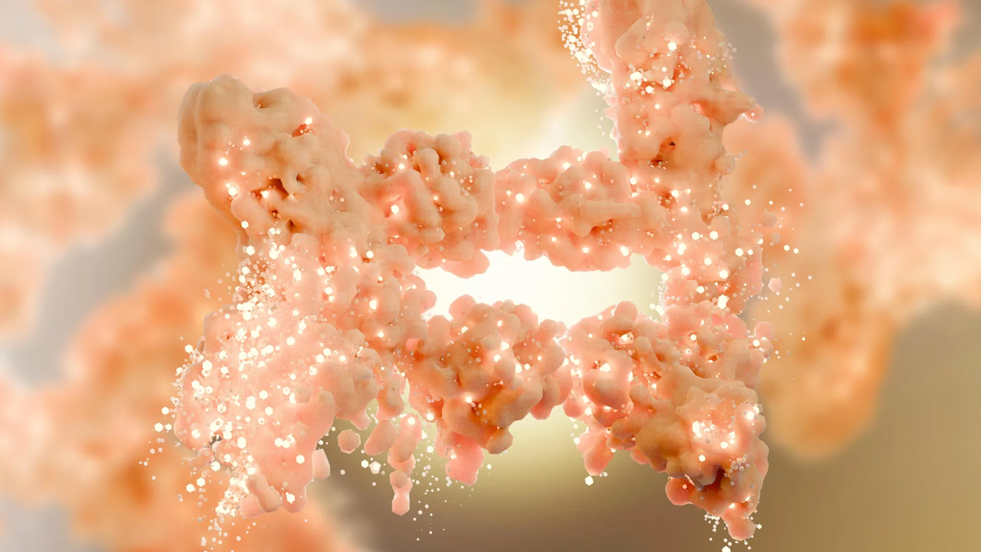
This glowing particle in a laser trap may reveal how lightning begins

Using lasers as precision tools to study how clouds become electrically active may sound futuristic, but researchers at the Institute of Science and Technology Austria (ISTA) have turned it into practical laboratory work. By capturing and charging tiny airborne particles with focused beams of light, scientists can watch how their electrical state changes over time. Their findings, recently reported in Physical Review Letters, could help reveal what triggers lightning.

Aerosols are tiny droplets or solid particles suspended in the air, and they surround us constantly. Some are large enough to see, such as springtime pollen, while others, like viruses that circulate during flu season, are far too small for the human eye. A few can even be sensed by taste, including the fine salt particles carried on ocean winds.

PhD student Andrea Stöllner, a member of the Waitukaitis and Muller groups at ISTA, studies the behavior of ice crystals that form within clouds. To better understand how these crystals gather charge, she works with model aerosols made from very small, transparent silica spheres.

Together with former ISTA postdoc Isaac Lenton, ISTA Assistant Professor Scott Waitukaitis and collaborators, Stöllner has created a technique that uses two intersecting laser beams to trap, stabilize, and electrically charge a single silica particle. This setup opens the door to new investigations into how cloud electrification begins and how lightning is sparked.

Building a Stable Laser Trap

Andrea Stöllner works at a large laboratory table filled with polished metal components. Green laser beams cross the space, bouncing from mirror to mirror. A slow, steady hissing noise comes from the table, similar to air leaking from a tire. “It’s an anti-vibration table,” Stöllner says, pointing out how it protects the lasers from small disturbances in the room or from nearby equipment, which is essential for extremely precise measurements.

The beams travel through a series of aligned parts before converging into two narrow streams that enter a sealed container. Where they meet, they create a concentrated point of light that can hold small particles in place. These “optical tweezers” keep drifting aerosols suspended long enough to study them. When a particle is caught, a bright green flash appears, confirming that the trap has successfully grabbed a glowing, perfectly round aerosol particle.

“The first time I caught a particle, I was over the moon,” Stöllner recalls of her breakthrough moment two years earlier, just before Christmas. “Scott Waitukaitis and my colleagues rushed into the lab and took a short glimpse at the captured aerosol particle. It lasted exactly three minutes, then the particle was gone. Now we can hold it in that position for weeks.”

Achieving this level of control took nearly four years. The experiment began with an earlier version developed by Lenton. “Originally, our setup was built to just hold a single particle, analyze its charge, and figure out how humidity changes its charges,” Stöllner says. “But we never came this far. We found out that the laser we are using is itself charging our aerosol particles.”

How Lasers Knock Electrons Loose

Stöllner and her colleagues discovered that the particles gain charge through a “two-photon process.”

Aerosol particles usually carry almost no net charge, with electrons (negatively charged entities) orbiting within each atom. Laser beams are made of photons (particles of light traveling at the speed of light). When two photons strike the particle at the same moment and are absorbed together, they can remove a single electron. Losing that electron gives the particle one unit of positive charge, and with continued exposure, the particle becomes progressively more positively charged.

For Stöllner, identifying this process has opened new opportunities. “We can now precisely observe the evolution of one aerosol particle as it charges up from neutral to highly charged and adjust the laser power to control the rate.”

As the charge builds, the particle also begins to lose charge again in sudden, short bursts. These spontaneous discharges hint at behaviors that may occur naturally in the atmosphere.

High above, cloud particles may undergo similar cycles of charge buildup and release.

Searching for Lightning’s First Spark

Thunderstorm clouds contain a mix of ice crystals and larger chunks of ice. As these collide, they trade electrical charges. Over time, the cloud becomes so electrically imbalanced that lightning forms. One idea is that the earliest spark of a lightning bolt could arise directly from charged ice crystals. Yet the exact mechanism behind lightning formation remains unresolved. Other theories propose that cosmic rays start the process because the charged particles they produce accelerate within existing electric fields. According to Stöllner, the current scientific view is that, in both scenarios, the electric field inside clouds appears too weak to initiate lightning on its own.

“Our new setup allows us to explore the ice crystal theory by closely examining a particle’s charging dynamics over time,” Stöllner explains. While natural ice crystals in clouds are much larger than the silica particles used in the lab, the team hopes that understanding these small-scale effects will reveal the larger processes that create lightning. “Our model ice crystals are showing discharges and maybe there’s more to that. Imagine if they eventually create super tiny lightning sparks — that would be so cool,” she adds with a smile.

Share Button

This tiny plant survived the vacuum of space and still grows

Mosses are well known for surviving in places that challenge most life, including the Himalayan peaks, the scorching deserts of Death Valley, the Antarctic tundra, and the cooling surfaces of active volcanoes. Their remarkable durability led researchers to test moss sporophytes, the reproductive structures that hold spores, in an even harsher setting: outer space. According to a study published in the journal iScience on November 20, more than 80% of these spores endured 9 months outside the International Space Station (ISS) and returned to Earth still capable of reproduction. This marks the first evidence that an early land plant can survive long-term exposure to space conditions.

“Most living organisms, including humans, cannot survive even briefly in the vacuum of space,” says lead author Tomomichi Fujita of Hokkaido University. “However, the moss spores retained their vitality after nine months of direct exposure. This provides striking evidence that the life that has evolved on Earth possesses, at the cellular level, intrinsic mechanisms to endure the conditions of space.”

Asking Whether Moss Could Survive Beyond Earth

Fujita began exploring the possibility of “space moss” while studying plant evolution. He was impressed by mosses’ ability to colonize the harshest environments on Earth. “I began to wonder: could this small yet remarkably robust plant also survive in space?”

To investigate, Fujita’s team exposed Physcomitrium patens, also known as spreading earthmoss, to a simulated space environment featuring intense UV radiation, extremely high and low temperatures, and vacuum-like conditions.

Testing Moss Structures Under Extreme Stress

The researchers compared three moss forms: protenemata (juvenile moss), brood cells (stress-induced stem cells), and sporophytes (encapsulated spores). They aimed to identify which structure had the greatest likelihood of enduring space.

“We anticipated that the combined stresses of space, including vacuum, cosmic radiation, extreme temperature fluctuations, and microgravity, would cause far greater damage than any single stress alone,” says Fujita.

Their experiments showed that UV radiation posed the biggest threat, and sporophytes clearly outperformed the other structures. Juvenile moss did not survive strong UV exposure or extreme temperatures. Brood cells fared better but still fell short. By contrast, the encased spores showed ~1,000x greater UV tolerance and remained capable of germination even after enduring −196°C for more than a week or 55°C for an entire month.

Why Encased Spores Withstand Harsh Conditions

The team concluded that each spore’s surrounding structure likely absorbs harmful UV light and provides physical and chemical shielding. They suggest that this protective feature may have helped ancient bryophytes, the plant group that includes mosses, move from water to land roughly 500 million years ago and survive repeated mass extinctions.

To determine whether this adaptation held up in real space, the researchers sent sporophytes into orbit.

Launching Moss to the ISS for a Real-World Trial

In March 2022, hundreds of sporophytes traveled to the ISS aboard the Cygnus NG-17 spacecraft. After their arrival, astronauts mounted the samples on the exterior of the station, exposing them to space for 283 days. The specimens later returned to Earth on SpaceX CRS-16 in January 2023 and were brought back to the lab for analysis.

“We expected almost zero survival, but the result was the opposite: most of the spores survived,” says Fujita. “We were genuinely astonished by the extraordinary durability of these tiny plant cells.”

Strong Survival and Healthy Return to Earth

More than 80% of the spores endured the full trip, and all but 11% of those survivors successfully germinated in the laboratory. Measurements of chlorophyll showed normal levels for nearly all pigments, except for a 20% drop in chlorophyll a, a light-sensitive compound. Despite this reduction, the spores remained healthy.

“This study demonstrates the astonishing resilience of life that originated on Earth,” says Fujita.

The team also used their data to build a mathematical model estimating how long the spores might last in similar conditions. Their calculation suggested a potential survival span of up to 5,600 days, or about 15 years, although they stressed that more data is needed for a firm conclusion.

Implications for Growing Life Beyond Earth

The researchers hope these findings support future studies on how extraterrestrial soils might sustain plant life and encourage efforts to use mosses in developing agricultural systems for off-world environments.

“Ultimately, we hope this work opens a new frontier toward constructing ecosystems in extraterrestrial environments such as the Moon and Mars,” says Fujita. “I hope that our moss research will serve as a starting point.”

This work was supported by DX scholarship Hokkaido University, JSPS KAKENHI, and the Astrobiology Center of National Institutes of Natural Sciences.

Share Button

JFK’s granddaughter raises awareness of rare leukaemia with poignant essay

Doctors say Tatiana Schlossberg’s struggle – even with the best care as a member of a prominent family – emphasises the need for more research.

Share Button

Obesity jab drug fails to slow Alzheimer’s

There were initial hopes that the drug in weight loss jab Wegovy could slow progression of dementia.

Share Button

Immune cells use a surprising trick to heal muscle faster

At the cellular scale, the way muscle tissue repairs itself becomes surprisingly complex. The body does not respond the same way to all forms of damage. A sudden muscle tear from a sports injury differs greatly from the slow decline in muscle strength seen in conditions such as muscular dystrophy.

A research team at Cincinnati Children’s has uncovered a shared and unexpected repair process that may help the body recover from several kinds of muscle damage. The findings were published online on Nov. 21, 2025, in Current Biology. The project was led by first author Gyanesh Tripathi, PhD, and corresponding author Michael Jankowski, PhD, who oversees the Research Division in Cincinnati Children’s Department of Anesthesia and serves as Associate Director of Basic Science Research for the Pediatric Pain Research Center.

The newly identified mechanism involves macrophages, a type of immune cell. These cells are usually known for acting like tiny cleanup crews that remove bacteria, dead cells, and other unwanted material.

A Neuron-Like Repair Signal

“The biggest surprise about this was finding that a macrophage has a synaptic-like property that delivers an ion to a muscle fiber to facilitate its repair after an injury,” Jankowski says. “It’s literally like the way a neuron works, and it’s working in an extremely fast synaptic-like fashion to regulate repair.”

Scientists have long known that macrophages respond to muscle injury by releasing cytokines and chemokines that create inflammation, influence pain, and help drive the growth and regeneration of muscle fibers.

Searching for Pain Relief Leads to a Different Breakthrough

The research team originally aimed to uncover ways to ease pain during recovery after surgery. They were looking for clues that could eventually reduce the need for pain medications that carry significant side effects.

Although they did not find a new approach for pain relief, they identified a process that makes muscle repair occur more quickly. This discovery may support the development of future treatments for muscle wasting and acute injuries. The findings also suggest that macrophages might eventually serve as specialized “delivery vehicles” for cell-based therapies targeting a wider range of medical conditions.

“These are infiltrating macrophages, a very specific type. They’re not ones already residing in the tissue. These come in after damage occurs,” Jankowski says.

Real-Time Evidence of Rapid Muscle Activation

In experiments that used mouse models of two different injury types, researchers examined how macrophages interact with the myofibers that form muscle tissue. They were even able to capture key moments of this activity as it occurred.

By using brief bursts of a designer chemical to activate the macrophages, the team observed these immune cells forming synaptic-like contacts with myofibers. The macrophages then released calcium ions directly to the muscle fibers, accelerating early stages of healing. Within 10 to 30 seconds, the researchers measured bursts of electrical activity inside the damaged muscle.

“This occurs in a very rapid fashion. You can activate the macrophage and make the muscle twitch subtly almost immediately,” Jankowski says.

Healing Effects Seen in Both Injury and Disease Models

The same type of macrophage-driven signaling also helped mice with disease-like muscle damage. After recognizing the injury, the immune cells gathered at the site and triggered waves of activity in the muscle fibers. After 10 days, mice that received this treatment had substantially more new muscle fibers than mice in the control group.

“A similar synaptic-like response worked in both scenarios,” Jankowski says.

Next Research Steps

More work is needed to determine whether human macrophages behave the same way when muscle is injured. If they do, researchers will still need to learn how to guide or control the process in ways that could be safely used as therapy.

The team is also interested in an unexpected outcome: although the infiltrating macrophages sped up healing, they did not appear to reduce acute pain. Understanding why this occurs may help explain why about 20% of children who undergo surgery continue to experience lingering pain afterward.

Looking ahead, the researchers want to explore whether macrophages can deliver other helpful signals or materials to muscle cells.

Cincinnati Children’s co-authors include Adam Dourson, PhD, Fabian Montecino-Morales, PhD, Jennifer Wayland, MS, Sahana Khanna, Megan Hofmann, Hima Bindu Durumutla, MS, Thirupugal Govindarajan, PhD, Luis Queme, MD, PhD, and Douglas Millay, PhD. The Bioanalysis and Imaging Facility at Cincinnati Children’s also contributed to the work.

Funding for this research came from grants provided by the National Institutes of Health (R01NS105715, R01NS113965, R61/R33AR078060, R01AR068286, R01AG082697) and the Cincinnati Children’s Hospital Research Foundation.

Share Button

Covid inquiry hears impact on firms and staff

Business owners describe breaking into tears as they were forced to lay off staff, while workers feared for their jobs

Share Button

Boy with rare condition amazes doctors after world-first gene therapy

Oliver has an inherited condition called Hunter syndrome, which causes progressive damage to the body and brain.

Share Button

What are the symptoms of prostate cancer and what should you check for?

One in eight men will be diagnosed with prostate cancer in their lifetime.

Share Button

Global surge in ultra-processed foods sparks urgent health warning

Experts from around the world are raising alarms about the rapid global rise of ultra-processed foods, warning that UPFs are reshaping diets and driving a surge in chronic health problems.

  • A major three paper Series in The Lancet finds that ultra-processed foods (UPFs) are rapidly replacing fresh and minimally processed meals around the world. The evidence links rising UPF intake to poorer diet quality and higher risks of multiple chronic diseases.
  • The authors explain that although more research on UPFs will continue to be valuable, the current science is already strong enough to justify immediate public health action. Waiting for further studies would allow UPFs to gain an even stronger hold in global diets.
  • The Series stresses that improving diets cannot fall solely on individual behavior. Real progress requires coordinated policies that limit UPF production, marketing, and availability, while also addressing high levels of fat, sugar and salt in the food supply and expanding access to healthy food.
  • The authors describe UPFs as products of an industrial food system built around corporate profit rather than nutrition or sustainability. They warn that only a united international response can counter the political influence of UPF companies, which remains the biggest obstacle to effective dietary policy reform.

Rising UPF Consumption Sparks Global Health Concerns

A new three paper Series in The Lancet, written by 43 international experts, warns that the rapid spread of ultra-processed foods (UPFs) across global diets is creating a serious public health challenge. The authors detail how UPF companies use a range of strategies to increase sales and block policies designed to protect consumers. The Series offers a plan for stronger government action, greater community involvement, and broader access to affordable, nutritious foods.

Professor Carlos Monteiro, University of Sao Paulo, Brazil, explains, “The growing consumption of ultra-processed foods is reshaping diets worldwide, displacing fresh and minimally processed foods and meals. This change in what people eat is fueled by powerful global corporations who generate huge profits by prioritizing ultra-processed products, supported by extensive marketing and political lobbying to stop effective public health policies to support healthy eating.”

Calls for Strong, Coordinated Policy Action

Professor Camila Corvalan, University of Chile, Chile, adds, “Addressing this challenge requires governments to step up and introduce bold, coordinated policy action — from including markers of UPFs in front-of-package labels to restricting marketing and implementing taxes on these products to fund greater access to affordable, nutritious foods.”

Dr. Phillip Baker, University of Sydney, Australia, continues, “We need a strong global public health response — like the coordinated efforts to challenge the tobacco industry. Including safeguarding policy spaces from political lobbying and building powerful coalitions to advocate for healthy, fair and sustainable food systems and stand-up to corporate power.”

UPFs, based on the Nova classification, are industrially produced branded foods created from low cost ingredients such as hydrogenated oils, protein isolates or glucose/fructose syrup, along with cosmetic additives (e.g. dyes, artificial sweeteners, emulsifiers). These products are intentionally formulated and promoted to replace fresh foods and traditional meals, while maximizing profits for manufacturers (for a detailed definition see paper 1, panel 1).

Research Shows Clear Links Between UPFs and Chronic Disease

The first paper in The Lancet Series reviews scientific evidence gathered since the Nova classification was developed by Prof Carlos Monteiro and colleagues in 2009. The findings consistently show that UPFs are crowding out traditional dietary patterns, lowering overall diet quality, and contributing to higher risks of many chronic diseases.

National surveys also reveal substantial increases in UPF consumption (paper 1, figure 1). The proportion of dietary energy from UPFs tripled in Spain (11% to 32%) and China (4% to 10%) over the past three decades, and rose from 10% to 23% in Mexico and Brazil during the previous forty years. In the USA and UK, levels have remained above 50% for the past two decades, with slight increases over time.

Growing Body of Evidence Underscores Health Risks

The Series reports that diets high in UPFs are associated with overeating, poor nutrient balance (too much sugar and unhealthy fats, too little fibre and protein), and greater exposure to potentially harmful additives. A systematic review of 104 long-term studies found that 92 showed higher risks for at least one chronic disease, with meta-analyses identifying significant associations with 12 health conditions including obesity, type 2 diabetes, cardiovascular disease, depression, and premature death (paper 1, figure 4, appendix p23-24).

While the authors acknowledge scientific debates about Nova and UPF definitions — including the need for more long-term trials, clearer mechanisms, and recognition of product subgroups with differing nutritional qualities — they emphasize that further research should not delay immediate public health action.

Professor Mathilde Touvier, French National Institute for Health and Medical Research (Inserm), France, states, “While healthy debate about UPFs within the scientific community is welcomed, this should be distinguished from attempts by vested interests to undermine the current evidence. The growing body of research suggests diets high in ultra-processed foods are harming health globally and justifies the need for policy action.”

Policy Solutions to Reduce UPFs and Improve Diet Quality

The second paper in the Series outlines policy options to curb UPF production, marketing, and consumption, holding major companies accountable for promoting unhealthy diets (paper 2, table 1). These recommendations are intended to strengthen existing legislation targeting high fat, salt and sugar (HFSS) foods.

Professor Barry Popkin, University of North Carolina, US, says “We call for including ingredients that are markers of UPFs (eg, colors, flavors, and sweeteners) in front-of-package labels, alongside excessive saturated fat, sugar, and salt, to prevent unhealthy ingredient substitutions, and enable more effective regulation.”

Marketing Restrictions, School Policies, and Fresh Food Access

The authors recommend stronger marketing limits, particularly for promotions aimed at children, digital advertising, and brand-level marketing. They also suggest banning UPFs in public settings such as schools and hospitals, and capping shelf space for UPFs in supermarkets. One example of successful reform is Brazil’s national school feeding program, which has removed most UPFs and will require 90% of school food to be fresh or minimally processed by 2026 (paper 2, panel 4).

Alongside regulation, the authors highlight the need to expand access to fresh foods. Taxing selected UPFs could help support subsidies for healthier options, particularly for low-income households.

Professor Marion Nestle, New York University, US, notes, “Improving diets worldwide requires policies tailored to each country’s unique situation and how entrenched UPFs have become in people’s daily eating habits. While priorities may differ, urgent action is needed everywhere to regulate ultra-processed foods alongside existing efforts to reduce high fat, salt, and sugar content.”

Associate Professor Gyorgy Scrinis, University of Melbourne, Australia, adds, “Importantly, policies must ensure that fresh and minimally processed foods are accessible and affordable — not just for those with time to cook, but for busy families and individuals who rely on convenient options. Only by combining stricter regulation on poor quality food products with realistic support for more nutritious choices can we truly promote better diets for all.”

How Corporate Power Drives the Global UPF Boom

The third paper shows that the sharp rise in UPF consumption is being driven primarily by global food corporations rather than individual behavior. These companies use low cost ingredients, large-scale production methods, and highly persuasive marketing to encourage widespread consumption.

With global annual sales reaching $1.9 trillion, UPFs represent the most profitable segment of the food industry. Manufacturers of these products have delivered more than half of the $2.9 trillion in shareholder payouts made by publicly listed food companies since 1962. The profits help fuel expansion, marketing power, and political influence, reinforcing corporate dominance over modern food systems.

The Series explains that UPF companies rely on sophisticated political strategies to protect their interests — blocking regulations, influencing scientific debates, shaping public opinion, supporting hundreds of interest groups, lobbying, donating to political campaigns, and engaging in litigation to delay policy action (paper 3, table 1 and figure 2).

Professor Simon Barquera, the National Institute of Public Health of Mexico, Mexico, states, “Powerful corporations — not individuals’ choices — are behind the global rise of ultra-processed foods. Through interest groups, these corporations often position themselves as part of the solution, but their actions tell a different story — one focused on protecting profits and resisting effective regulation.”

Urgent Need for a Unified Global Response

The authors call for a global public health movement to protect policy-making from industry interference, end ties between industry and health organizations, and strengthen networks advocating for reduced UPF consumption.

Professor Karen Hoffman, University of the Witwatersrand, South Africa, says, “Just as we confronted the tobacco industry decades ago, we need a bold, coordinated global response now to curb the overproportionate power of UPF corporations and build food systems that prioritize people’s health and well-being.”

They argue that transforming food systems requires a new vision that elevates local food producers, preserves cultural food traditions, promotes gender equity, and ensures that economic benefits flow to communities rather than to distant shareholders.

Dr. Phillip Baker concludes, “We are currently living in a world where our food options are increasingly dominated by UPFs, contributing to rising global levels of obesity, diabetes and mental ill-health. Our Series highlights that a different path is possible — one where governments regulate effectively, communities mobilize, and healthier diets are accessible and affordable for all.”

The Lancet Series on Ultra-Processed Foods and Human Health, was supported by funding from Bloomberg Philanthropies.

Share Button

New obesity discovery rewrites decades of fat metabolism science

Our fat cells, known as adipocytes, do far more than store extra body weight. They serve as an important energy reserve for the body. Inside each adipocyte, fat is packed into lipid droplets that can be tapped when fuel is needed — for example, during the hours between meals. To release this stored energy, the body relies on a protein called HSL, which functions much like a switch. When energy is running low, hormones such as adrenaline activate HSL, prompting it to free fat that can then supply various organs.

Without HSL, it would be reasonable to expect fat to build up, as though the body had lost access to its energy supply. Surprisingly, this is not what happens. Research involving both mice and patients with mutations in the HSL gene shows that the lack of this protein does not lead to excess fat or obesity. Instead, affected individuals experience a loss of fat mass, a condition known as lipodystrophy.

Although obesity and lipodystrophy appear to be complete opposites, both involve fat cells that do not function properly. As a result, each condition can contribute to metabolic disturbances and cardiovascular problems.

HSL Found in an Unexpected Location Inside Fat Cells

To understand this surprising behavior, a team led by Dominique Langin, professor at the University of Toulouse within the I2MC, took a closer look at where HSL is found inside adipocytes. The protein is well known for its role at the surface of lipid droplets, where it helps break down stored fat. However, the study revealed that HSL also resides inside the nucleus of fat cells. “In the nucleus of adipocytes, HSL is able to associate with many other proteins and take part in a program that maintains an optimal amount of adipose tissue and keeps adipocytes ‘healthy’,” explains Jérémy Dufau, co-author of the study, who completed his doctoral thesis on this topic.

The researchers also found that nuclear HSL levels are tightly controlled. Adrenaline, which activates the form of HSL located on lipid droplets, also encourages the protein to leave the nucleus. This process occurs naturally during fasting. In contrast, obese mice show elevated levels of HSL within the nucleus, suggesting a shift in this regulatory system.

A Revised Understanding of HSL’s Role in Metabolism

“HSL has been known since the 1960s as a fat-mobilizing enzyme. But we now know that it also plays an essential role in the nucleus of adipocytes, where it helps maintain healthy adipose tissue,” says Dominique Langin. This additional responsibility helps explain why the absence of HSL results in lipodystrophy, and it offers new insights into metabolic disorders such as obesity and related health complications.

This discovery appears at a critical time. In France, one in two adults is overweight or obese, and globally the number reaches two and a half billion people. Obesity increases the risk of a range of diseases, including diabetes and heart problems, and often reduces overall quality of life. Continued scientific research is crucial to improving prevention efforts and patient care.

Share Button