This new iron supplement heals anemia without hurting your gut

Iron-deficiency anemia is a widespread health problem that often leads to fatigue, headaches, or even cravings for ice. Traditional oral iron supplements can help, but they often leave behind unabsorbed iron that irritates the digestive tract and triggers inflammation. Researchers reporting in ACS Applied Materials & Interfaces have developed a new type of supplement that blends iron with prebiotics and probiotics. In animal studies, this innovative formula successfully restored healthy blood iron levels in anemic mice while preventing inflammation and keeping the gut microbiome balanced.

“By advancing biomaterial-based iron delivery, this research offers a transformative approach to address anemia, directly contributing to improved nutrition and long-term public health,” explains Poonam Sagar, an author of the study.

Anemia develops when the body lacks enough red blood cells to transport oxygen efficiently. It can result from infections, inherited conditions, or, most commonly, a shortage of dietary iron. Doctors typically prescribe oral iron tablets to treat the condition. However, the body absorbs only a small fraction of the iron they contain. The remaining iron can upset the balance of gut bacteria and cause inflammation, which is why probiotics are sometimes prescribed alongside iron to protect digestive health.

In earlier research, scientists had already tried combining iron with probiotics. Sagar, Nitin Kumar Singhal, and their team expanded on this concept by adding prebiotics (nutrients that feed beneficial bacteria) to create a three-part supplement designed to be more effective and gentler on the gut.

The new formulation combines dietary fiber extracted from millet (a grain), the probiotic Lactobacillus rhamnosus, and an iron-containing complex. The researchers first tested the supplement’s compatibility with human cells, then evaluated its effects in mice with iron-deficiency anemia. After two weeks, the treated mice showed:

  • Restored hemoglobin levels (the main iron-rich molecule in red blood cells).
  • Iron excretion levels similar to healthy control mice, showing improved absorption.
  • Increased activity of genes involved in iron transport and metabolism.
  • Very low signs of inflammation in the colon.
  • A recovery of beneficial gut bacteria populations that had been depleted by anemia.

While more research is needed, the team believes this approach could lead to a new generation of iron supplements that restore iron safely, improve gut health, and reduce unwanted side effects.

The study received support from the Ministry of Science and Technology of India and the National Agri-Food Biotechnology Institute.

Share Button

Why medical students are choosing Bulgaria over UK

The BBC has spoken to students choosing Bulgaria due to UK’s strict cap on medical school places.

Share Button

Breakthrough cancer therapy stops tumor growth without harming healthy cells

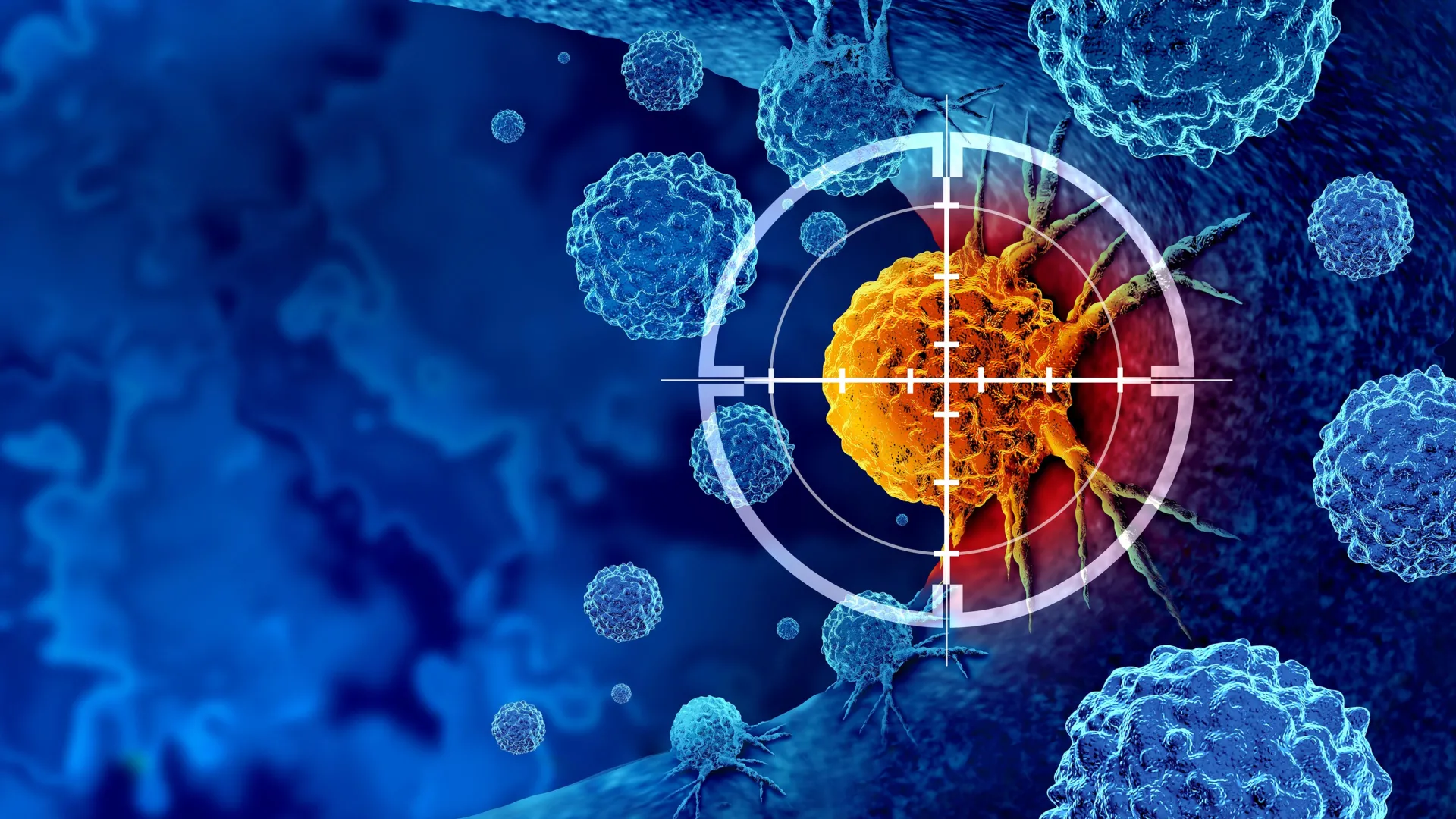
Scientists at the Francis Crick Institute and Vividion Therapeutics have discovered chemical compounds that can precisely prevent the cancer-driving gene RAS from connecting with a key pathway responsible for tumor growth.

The potential treatment is now moving into its first human clinical trial. If proven safe and effective, it could become a way to treat a wide range of cancers while minimizing harm to healthy cells.

The RAS gene plays a central role in controlling how cells grow and divide, but mutations in this gene occur in roughly one in five cancers. When mutated, RAS becomes permanently active, continually sending signals that push cells to keep growing and multiplying.

Inside the cell, RAS sits on the membrane and acts as the starting signal in a chain of growth processes. Completely shutting down RAS or the enzymes it controls has proven difficult, because these same pathways are essential for normal cell function. One of the enzymes linked to RAS, called PI3K, also helps regulate blood sugar through insulin. Blocking PI3K entirely can lead to side effects such as hyperglycemia.

In their study, published on October 9 in Science, the team combined chemical screening with biological testing to identify compounds that stop RAS and PI3K from interacting, while leaving normal cell activity intact.

Researchers at Vividion Therapeutics pinpointed a set of small molecules that permanently attach to the surface of PI3K near the spot where RAS would normally bind. Using an assay created by the Crick researchers, they confirmed that these compounds successfully blocked the RAS-PI3K interaction but still allowed PI3K to perform its other roles, including those related to insulin signaling.

The Crick team and their collaborators at Vividion then tested one of the compounds in mice with RAS-mutated lung tumors. The treatment stopped tumor growth, and the researchers found no signs of elevated blood sugar levels.

Next, they tried combining the new compound with one or two additional drugs that target enzymes within the same pathway. Together, the treatments produced stronger and longer-lasting tumor suppression than any of the drugs used alone.

The scientists also tested the compound in mice with tumors carrying mutations in another cancer-linked gene, HER2, which is often overactive in breast cancer and also connects with PI3K. Tumor growth was again halted, even though the effect did not rely on RAS. This finding suggests that the new compound could potentially help stop the growth of a broader range of cancers.

The drug has now entered the first clinical trial in humans to test for safety and side effects in people with both RAS and HER2 mutations. The trial will also assess if the potential treatment is more effective in combination with other drugs targeting RAS.

Julian Downward, Principal Group Leader of the Oncogene Biology Laboratory at the Crick, said: “Given the RAS gene is mutated across a wide range of cancers, we’ve been exploring how to stop it interacting with cell growth pathways for many years, but side effects have held back the development of treatments.

“Our collaborative effort has overcome this challenge by targeting the PI3K and RAS interaction specifically, leaving PI3K free to bind with its other targets. It’s exciting to see these clinical trials starting, highlighting the power of understanding chemistry and fundamental biology to get to something with potential to help people with cancer.”

“This discovery is a great example of how new discovery approaches can open up completely novel ways to tackle cancer,” said Matt Patricelli, Ph.D., Chief Scientific Officer of Vividion. “By designing molecules that stop RAS and PI3K from connecting, while still allowing healthy cell processes to continue, we’ve found a way to selectively block a key cancer growth signal. It’s incredibly rewarding to see this science now progressing in the clinic, where it has the potential to make a real difference for patients.”

Share Button

Even “diet” soda may be quietly damaging your liver, scientists warn

A large-scale investigation has found that people who regularly consume both sugar-sweetened beverages (SSBs) and low- or no-sugar-sweetened beverages (LNSSBs) face a significantly greater likelihood of developing metabolic dysfunction-associated steatotic liver disease (MASLD).1

Presented at UEG Week 2025, the research followed 123,788 adults from the UK Biobank who had no signs of liver disease at the start of the study. Participants’ drink habits were recorded through repeated 24-hour dietary questionnaires, allowing researchers to explore how both SSB and LNSSB consumption related to MASLD, liver fat buildup, and deaths linked to liver disease.

Individuals who drank more than 250 grams of either type of beverage per day had notably higher risks: a 60% increased likelihood of developing MASLD for those consuming LNSSBs (HR: 1.599) and a 50% increase for those drinking SSBs (HR: 1.469). During a median follow-up of 10.3 years, 1,178 participants developed MASLD and 108 died from liver-related causes. Although SSBs were not significantly connected to liver-related mortality, LNSSB consumption was. Both kinds of drinks were also associated with higher levels of liver fat.

MASLD, previously known as non-alcoholic fatty liver disease (NAFLD), occurs when excess fat builds up in the liver. Over time, this can trigger inflammation (hepatitis) and lead to symptoms such as abdominal pain, fatigue, and loss of appetite.2 Now the most common chronic liver condition worldwide, MASLD affects more than 30% of people and is rapidly becoming a leading cause of liver-related deaths.3

Lead researcher Lihe Liu explained, “SSBs have long been under scrutiny, while their ‘diet’ alternatives are often seen as the healthier choice. Both, however, are widely consumed and their effects on liver health have not been well understood.”

“Our study shows that LNSSBs were actually linked to a higher risk of MASLD, even at modest intake levels such as a single can per day. These findings challenge the common perception that these drinks are harmless and highlight the need to reconsider their role in diet and liver health, especially as MASLD emerges as a global health concern.”

Liu also discussed the possible biological reasons behind the findings: “The higher sugar content in SSBs can cause rapid spikes in blood glucose and insulin, promote weight gain and increase uric acid levels, all of which contribute to liver fat accumulation. LNSSBs, on the other hand, may affect liver health by altering the gut microbiome, disrupting the feeling of fullness, driving sweet cravings and even stimulating insulin secretion.”

The authors emphasized that these findings support limiting both SSBs and LNSSBs as part of a comprehensive prevention strategy, targeting not only liver disease but also cardio-renal-metabolic health. Replacing either beverage with water significantly reduced MASLD risk — by 12.8% for SSBs and 15.2% for LNSSBs — while substitution between the two types of beverages offered no risk reduction.

Liu added, “The safest approach is to limit both sugar-sweetened and artificially sweetened drinks. Water remains the best choice as it removes the metabolic burden and prevents fat accumulation in the liver, whilst hydrating the body.”

The researchers now aim to explore causal mechanisms more deeply through long-term, randomized and genetic trials with a focus on how sugar and its substitutes interact with the gut microbiome and influence liver disease.

References:

  1. Liu, L et al. Sugar- and low/non-sugar-sweetened beverages and risks of metabolic dysfunction-associated steatotic liver disease and liver-related mortality: A prospective analysis of the UK Biobank. Presented at UEG Week 2025; 7 October 2025; Berlin, Germany.
  2. Girish, V. and John, S. Metabolic Dysfunction-Associated Steatotic Liver Disease (MASLD). (2025). PMID: 31082077
  3. Younossi, Z. M. et al. The global epidemiology of nonalcoholic fatty liver disease (NAFLD) and nonalcoholic steatohepatitis (NASH): a systematic review. (2023). Journals. DOI: 10.1097/HEP.0000000000000004
Share Button

Einstein’s overlooked idea could explain how the Universe really began

How did the universe come into existence, and what early processes shaped everything that followed? A new study published in Physical Review Research takes aim at this fundamental question. Scientists from Spain and Italy have introduced a model that reimagines what happened moments after the universe was born. Their approach could upend long-standing ideas about the forces and events that governed the universe’s earliest evolution.

To explore these beginnings, the researchers ran advanced computer simulations that question the traditional “inflation” theory. According to that theory, the universe expanded at an extraordinary rate within a tiny fraction of a second after it came into existence. The inflation model relies on several interconnected variables, all of which must align to make the theory work.

The newly proposed model offers a simpler explanation. It suggests that gravitational waves — predicted by general relativity — may be the true driving force behind the universe’s formation, giving rise to galaxies, stars, planets, and ultimately life on Earth. The researchers link this idea to a mathematical construct known as De Sitter space, named for Dutch mathematician Willem De Sitter, who collaborated with Albert Einstein in the 1920s on understanding the structure of the cosmos.

“For decades, we have tried to understand the early moments of the Universe using models based on elements we have never observed,” said Dr. Raúl Jiménez, who studies experimental sciences & mathematics at ICREA in Spain and is a co-author on the study. “What makes this proposal exciting is its simplicity and verifiability. We are not adding speculative elements but rather demonstrating that gravity and quantum mechanics may be sufficient to explain how the structure of the cosmos came into being.”

The concept of gravitational waves dates back to 1893 and 1905, when Oliver Heaviside and Henri Poincaré first proposed related ideas. Albert Einstein expanded on this in 1916, describing gravitational waves as ripples in the fabric of space-time in his general theory of relativity. These waves can originate from powerful cosmic events such as supernovae, merging black holes, and colliding neutron stars. Because they are incredibly faint, detecting them requires highly sensitive instruments. It was not until September 2015 that scientists at the Laser Interferometer Gravitational-Wave Observatory (LIGO), with facilities in Washington and Louisiana, achieved the first confirmed detection.

The birth of the universe continues to be one of science’s greatest puzzles. The Big Bang theory remains the prevailing explanation, yet many questions persist — especially about what might have occurred before that explosive beginning.

Carl Sagan once reflected on humanity’s deep connection to the cosmos, saying, “The cosmos is within us. We are made of star-stuff. We are a way for the universe to know itself.”

We may never know exactly how the universe began and the processes responsible for you reading this article right now. But like the simplicity this study presents, perhaps this study is simply a way for us to know the universe itself a little bit better.

What new discoveries about the origins of the universe will researchers make in the coming years and decades? Only time will tell, and this is why we science!

As always, keep doing science & keep looking up!

Adapted from an article originally published on Universe Today.

Share Button

Scientists just debunked the calcium and dementia myth

New findings from Edith Cowan University (ECU), Curtin University, and the University of Western Australia show no evidence that taking calcium alone increases the risk of developing dementia over time. The results help ease earlier fears that calcium supplements might have harmful effects on the brain health of older women.

The investigation drew on data from an earlier project involving 1,460 older women who were randomly assigned to receive either calcium supplements or a placebo for five years. Researchers found that the supplements did not raise the likelihood of dementia in the long term.

“Calcium supplements are often recommended to prevent or manage osteoporosis,” said ECU PhD student Ms. Negar Ghasemifard.

About 20 percent of women over 70 live with osteoporosis, and calcium is widely advised to help prevent bone fractures.

“Previous research has raised concerns around the impacts that calcium supplements could have on cognitive health, particularly dementia. Results from our study provides reassurance to patients and clinicians regarding the safety of calcium supplements in the context of dementia risk for older women,” Ms. Ghasemifard said.

According to ECU Senior Research Fellow Dr. Marc Sim, even after adjusting for supplement use, diet, lifestyle factors, and genetic risk, the outcomes did not change.

“Previous research suggesting potential links between calcium supplement use and the risk for dementia was purely observational in nature. Our research, in comparison, consisted of a post-hoc analysis from a 5-year double-blind, placebo controlled randomized clinical trial on calcium supplements to prevent fracture. Whilst our study is still epidemiology, its design does reduce the likelihood of unmeasured confounding”

“Some 730 older women were given calcium supplements over five years, and a further 730 were given placebo. This study design offers more accurate data on dosage and duration, and we had a long follow-up period of 14.5 years, which strengthens our results,” Dr. Sim said.

Although the findings suggest calcium does not increase the risk of dementia in older women, particularly those over 80, further studies are still needed, said Professor Simon Laws, Director of ECU’s Centre for Precision Health.

“Whether this extrapolates to other demographics, such as men or even women commencing supplementation earlier in life, remains unknown. To confirm the current findings, particularly regarding brain health, and to address these population gaps, future clinical trials of calcium supplements, with or without vitamin D, would need to be undertaken. These should include specific and robust assessments of brain health as the primary outcome measures.”

Professor Blossom Stephan, a Dementia Australia Honorary Medical Advisor said the research highlighted a very important finding that provides reassurance to clinicians and patients about the long-term safety of calcium supplementation.

“Given calcium’s critical role in multiple physiological functions, including bone health, these results provide reassurance that long-term calcium supplementation did not increase dementia risk in older women,” she said.

Share Button

Scientists finally read the hidden DNA code that shapes disease

For centuries, scientists have noticed that certain illnesses seem to pass from one generation to the next, a connection first noted by Hippocrates, who observed that some diseases “ran in families.” Over time, researchers have steadily advanced their ability to uncover the biological roots of these inherited patterns within the human genome.

A team of EMBL researchers and collaborators has now created a tool that takes single-cell analysis to a new level. It can capture both genomic variations and RNA within the same cell, offering greater accuracy and scalability than earlier technologies. This approach allows scientists to identify variations in non-coding regions of DNA, the areas most often linked to disease, giving them a new way to explore how genetic differences contribute to human health. With its precision and ability to process large numbers of cells, the tool marks a major step toward linking specific genetic variants with disease outcomes.

“This has been a long-standing problem, as current single-cell methods to study DNA and RNA in the same cell have had limited throughput, lacked sensitivity, and are complicated,” said Dominik Lindenhofer, the lead author on a new paper about SDR-Seq published in Nature Methods and a postdoctoral fellow in EMBL’s Steinmetz Group. “On a single-cell level, you could read out variants in thousands of cells, but only if they had been expressed — so only from coded regions. Our tool works, irrespective of where variants are located, yielding single-cell numbers that enable analysis of complex samples.”

The important difference between coding and non-coding regions

DNA contains both coding and non-coding regions. The coding parts function like instruction manuals, since their genes are expressed into RNA, which directs cells in building proteins essential to life.

Non-coding regions, on the other hand, contain regulatory elements that guide how cells grow and function. Over 95% of disease-linked DNA variants occur in these non-coding regions, yet existing single-cell methods have not had the sensitivity or scale to study them effectively. Until now, researchers were unable to observe DNA and RNA from the same cell on a large scale, limiting insight into how DNA variants affect gene activity and contribute to disease.

“In this non-coding space, we know there are variants related to things like congenital heart disease, autism, and schizophrenia that are vastly unexplored, but these are certainly not the only diseases like this,” Lindenhofer said. “We needed a tool to do that exploration to understand which variants are functional in their endogenous genomic context and understand how they contribute to disease progression.”

Deciphering barcodes that track single cells

To perform single-cell DNA-RNA sequencing (SDR-seq), researchers used tiny oil-water droplets, each containing a single cell, allowing them to analyze DNA and RNA simultaneously. This method enabled them to examine thousands of cells in a single experiment and directly link genetic changes to patterns of gene activity. Developing this technology required overcoming major challenges and brought together teams from EMBL’s Genome Biology and Structural and Computational Biology units, the Stanford University School of Medicine, and Heidelberg University Hospital.

Collaborators from EMBL’s Judith Zaugg and Kyung-Min Noh groups developed a way to preserve delicate RNA by “fixing” the cells, while computational biologists in Oliver Stegle’s group designed a specialized program to decode the complex DNA barcoding system needed for data analysis. Although this decoding software was built for this specific project, the team believes it could prove valuable for many other studies.

Researchers from Wolfgang Huber’s and Sasha Dietrich’s groups at EMBL and Universitätsklinikum Heidelberg were already examining B-cell lymphoma samples for other studies. These patient samples, rich in genetic variation, provided an ideal test case for the new technology. Using these samples, Lindenhofer observed how variations in DNA were linked to disease processes and found that cancer cells with more variants showed stronger activation signals that support tumor growth.

“We are using these small reaction chambers to read out DNA and RNA in the same single cell,” Lindenhofer said. “This lets us accurately tell whether a variant is on one or both copies of a gene and measure its effects on gene expression in the same single cells. With the B-cell lymphoma cells, we were able to show that depending on the variant makeup of cells, they had different propensities to belong to distinct cellular states. We could also see that increasing variants in a cell actually were associated with a more malignant B-cell lymphoma state.”

The many opportunities from a single-cell sequencing tool

The SDR-seq tool now offers genomic biologists scale, precision, and speed to help better understand genetic variants. While it could eventually play a role in treating a broad range of complex diseases, it may first help in developing better screening tools for diagnosis.

“We have a tool that can link variants to disease,” said Lars Steinmetz, a senior author on the paper, an EMBL group leader, and a genetics professor at Stanford University School of Medicine. “This capability opens up a wide range of biology that we can now discover. If we can discern how variants actually regulate disease and understand that disease process better, it means we have a better opportunity to intervene and treat it.”

Share Button

Exciting results from blood test for 50 cancers

The Galleri test looks for fragments of DNA that have broken off a tumour and are circulating in the blood.

Share Button

This common liver supplement could boost cancer treatment success

Immunotherapy is a cancer treatment that harnesses the body’s own immune defenses to attack tumors. It has shown remarkable success against cancers of the lung, kidney, and bladder but has not worked as well for liver cancer. That gap is troubling because liver cancer cases have nearly tripled over the past four decades.

To explore why liver cancer responds poorly to immunotherapy, scientists at the Salk Institute examined how the immune system interacts with the liver. Using both mouse models and human tumor samples, they discovered that certain bile acids — molecules produced by the liver to aid digestion — can interfere with cancer-fighting immune cells known as T cells.

The team pinpointed several bile acids linked to weakened T cell function and faster tumor growth. By blocking the production of these acids, they were able to slow or stop tumor progression. One bile acid, called ursodeoxycholic acid (UDCA), had the opposite effect, enhancing T cell activity in the liver. When researchers increased UDCA levels through dietary supplements, liver tumors in mice shrank. Because UDCA supplements are already approved for other liver diseases, scientists believe they could potentially make immunotherapy more effective for liver cancer patients.

The study, published in Science, sheds light on why immune cells behave differently depending on the tumor’s location and identifies new molecular targets to strengthen liver cancer therapies.

“How do organ-specific properties and processes influence the immune response?” asks Professor Susan Kaech, senior author of the study and director of Salk’s NOMIS Center for Immunobiology and Microbial Pathogenesis. “Livers have a particularly unique environment, but we didn’t really understand how it was affecting the immune and cancer cells. By investigating these liver-specific features, we have identified several potential ways to regulate bile acids, improve T cell performance, and enhance patient outcomes.”

The liver generates more than 100 types of bile acids, which travel through the intestines to help digest fats. To combat liver cancer, T cells must function effectively within this chemically rich environment. Past studies have linked high bile acid levels to poor health and cancer progression, but researchers had not previously distinguished the effects of individual bile acids.

“Considering how T cell performance varies across different organs, tissues, and tumors puts us at a great vantage point for looking at ways to optimize cancer treatment,” says Siva Karthik Varanasi, former postdoctoral researcher in Kaech’s lab and current assistant professor at the University of Massachusetts Chan Medical School. “By taking this unique approach, we’re able to see that bile acids in the liver are hugely influencing T cells’ ability to do their job and therefore may be a useful therapeutic target.”

To better understand these effects, the Salk team first analyzed human liver cancer biopsies to identify which bile acids were present. They found elevated levels of conjugated bile acids and tested whether these compounds contributed to tumor growth. When they removed a protein called BAAT, which produces conjugated bile acids, the tumor load in mice dropped significantly. This suggests that adjusting BAAT activity in humans could improve their response to immunotherapy.

The researchers then examined 20 distinct bile acids to determine how each affected T cells. Most primary bile acids showed little influence, except for one called TCDCA, which triggered oxidative stress — a harmful molecular imbalance. Secondary bile acids had much stronger effects. One, called LCA, damaged T cell function by causing endoplasmic reticulum stress, while another, UDCA, boosted T cell performance and drew more immune cells to the liver. Increasing UDCA levels through supplementation effectively reduced tumor growth in mice, pointing to a promising strategy for enhancing immunotherapy in liver cancer.

Together, these results suggest that lowering BAAT and increasing UDCA could help control liver tumor growth and strengthen the immune system’s response to treatment.

“We’re already a huge step ahead when it comes to translating our findings to the clinic, because UDCA supplementation is already used to treat liver disease and could easily be tested in liver cancer next,” says Kaech, who also holds the NOMIS Chair at Salk. “We are really excited to also explore the role of the gut microbiome in all of this, since bile acids are a huge part of that picture — how can we manipulate ‘good’ and ‘bad’ bacteria in the microbiome to further regulate bile acid levels? How does the microbiome change during liver cancer? Could probiotics be a therapeutic approach?”

In addition to exploring dietary and microbiome manipulations that could help with liver cancer, the team is curious to see if other conditions could be treated by targeting BAAT. Already, they believe chronic liver disease and obesity may benefit from the same reduction of conjugated bile acids.

Other authors include Dan Chen, Melissa Johnson, Kathryn Lande, Michael LaPorta, Filipe Hoffmann, Thomas Mann, Eduardo Casillas, Kailash Mangalhara, Varsha Mathew, Ming Sun, Yagmur Farsakoglu, Timothy Chen, Bianca Parisi, Shaunak Deota, H. Kay Chung, Satchidananda Panda, April Williams, and Gerald Shadel of Salk; Jin Lee, Yingluo Liu, Cayla Miller, and Gen-Sheng Feng of UC San Diego; Souradipta Ganguly and Debanjan Dhar of UC San Diego and Sanford Burnham Prebys Medical Discovery Institute; Marcos Teneche, Aaron Havas, and Peter Adams of Sanford Burnham Prebys Medical Discovery Institute; Isaac Jensen and Donna Farber of Columbia University; Andrea Schietinger of Memorial Sloan Kettering Cancer Center, Weill Cornell Graduate School of Medical Sciences, and Parker Institute for Cancer Immunotherapy; and Mark Sundrud of Dartmouth College.

The work was supported by the National Institutes of Health (NCI CCSG: P30 014195, S10-OD023689, P30 AG068635, P30 CA014195, P01 AG073084, R01 CA240909-04, R21 AI151562, F31CA278581, CCSG Grant P30CA23100, R01DK137061, R01DK133930, DK120515, R01AI143821, R01AI164772, U01AI163063), Waitt Foundation, Helmsley Charitable Trust, Chapman Foundation, Cancer Research Institute, National Cancer Center, NOMIS Foundation, Salkexcellerators Fellowship, Damon Runyon Fellowship, Audrey Geisel endowed Chair of Biomedical Science, Altman Clinical Translational Research Institute (KL2TR001444), San Diego Digestive Diseases Research Center, and Dartmouth Cancer Center.

Share Button

Student paramedic helps deliver lecturer’s baby

Elise Faragher is on placement in Worcester and lecturer Aaron Collins, and his wife, came in.

Share Button