This common sugar builds stronger cancer-killing T cells

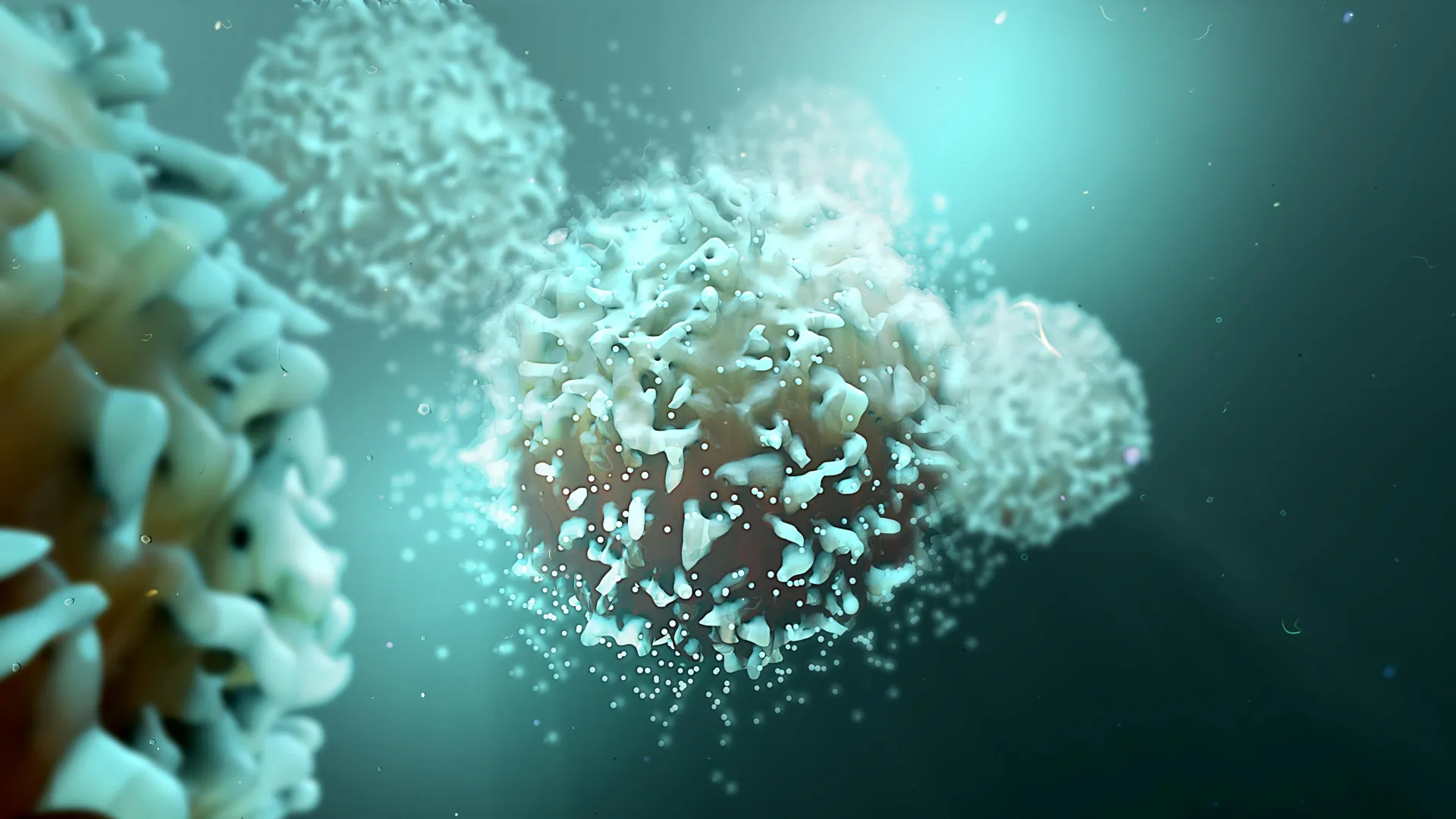
For cancer, and infection-fighting T cells, glucose offers far more than a simple sugar rush.

A new discovery by Van Andel Institute scientists reveals that glucose, an essential cellular fuel that powers immune cells, also aids in T cells’ internal communication and boosts their cancer-fighting properties. The findings may help optimize T cells’ ability to combat cancer and other diseases.

A study describing the work published on September 2 in Cell Metabolism.

“Immune cells are highly influenced by their environment” said Joseph Longo, Ph.D., the study’s first author and a postdoctoral fellow in the lab of Russell Jones, Ph.D. “We knew that T cells need access to glucose to function, but we didn’t know exactly why. It was previously thought that T cells mainly break down glucose for energy, but our new work shows that T cells use glucose as a building block for other molecules that are necessary to support T cells’ anti-cancer properties.”

The findings reveal that T cells allocate significant portions of glucose to build large molecules called glycosphingolipids (GSLs). These sugar-fat compounds are essential for T cell growth and making proteins that T cells use to combat cancer.

GSLs help form fat-rich structures on T cell surfaces called lipid rafts, which bring together cell signaling proteins that instruct the T cell to kill cancer cells. Without GSLs, these signals are weaker, making T cells less effective at destroying tumors.

“Both T cells and cancer cells leverage different nutrients to support varying aspects of their function,” Jones said. “The more we know about these different fuel sources, the better we can support T cells’ innate cancer-fighting abilities while also developing ways to possibly make cancer cells more vulnerable to immune attack.”

Other authors include Lisa M. DeCamp, Brandon M. Oswald, Ph.D., Robert Teis, Alfredo Reyes-Oliveras, Ph.D., Michael S. Dahabieh, Ph.D., Abigail E. Ellis, Michael P. Vincent, Ph.D., Hannah Damico, M.B., Kristin L. Gallik, Ph.D., Nicole M. Foy, Shelby E. Compton, Ph.D., Colt D. Capan, M.S., Kelsey S. Williams, Ph.D., Corinne R. Esquibel, Ph.D., Zachary B. Madaj, M.S., Hyoungjoo Lee, Ph.D., Connie Krawczyk, Ph.D., Brian B. Haab, Ph.D., and Ryan D. Sheldon, Ph.D., of VAI; and Dominic G. Roy, Ph.D., of Université de Montréal.

Research reported in this publication was supported by the National Institute of Allergy and Infectious Diseases of the National Institutes of Health under award no. R01AI165722 (Jones). The content is solely the responsibility of the authors and does not necessarily represent the official views of the National Institutes of Health.

Share Button

Dinosaur teeth reveal secrets of Jurassic life 150 million years ago

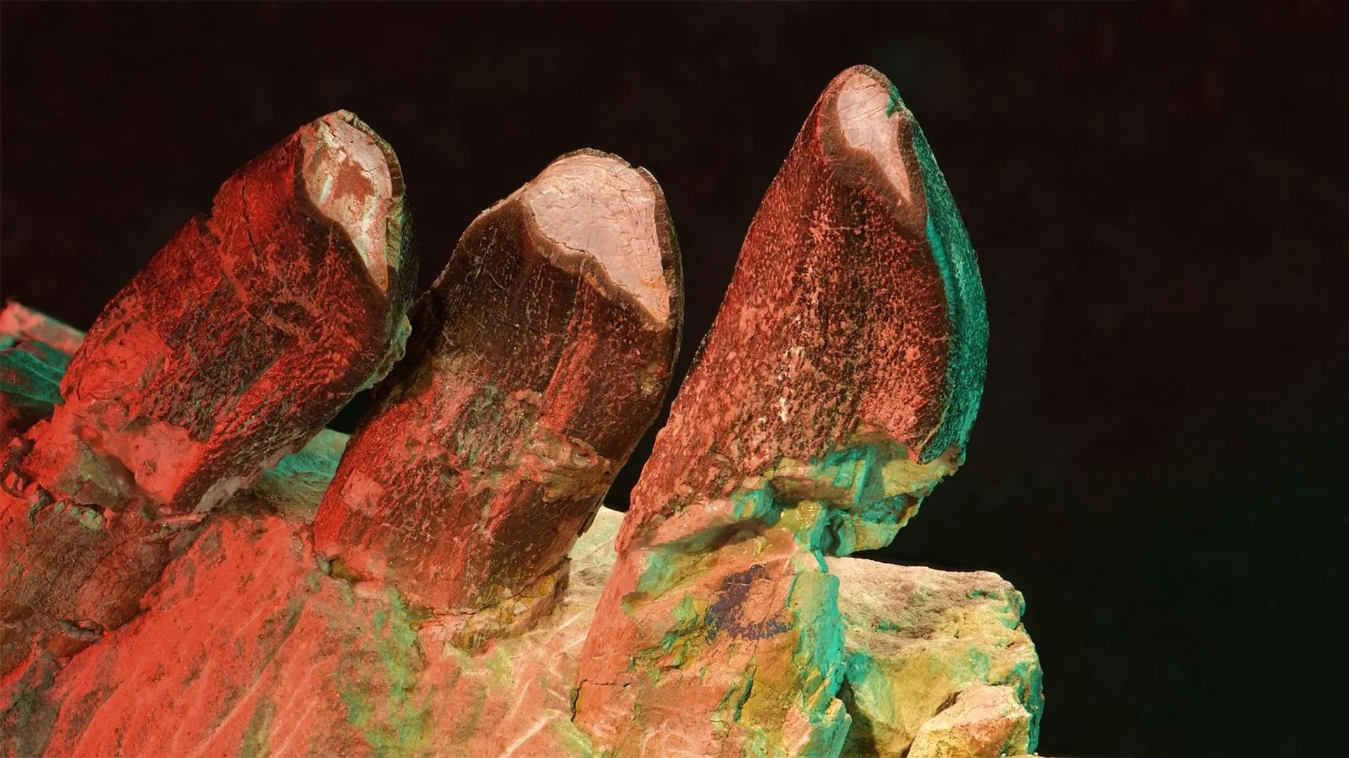
What did long-necked dinosaurs eat – and where did they roam to satisfy their hunger? A team of researchers has reconstructed the feeding behavior of sauropods using cutting-edge dental wear analysis. Their findings, published in Nature Ecology and Evolution, show that microscopic enamel wear marks provide surprising insights into migration, environmental conditions, and niche distribution within ecosystems from 150 million years ago.

How did massive dinosaurs live during the Jurassic period? What did they eat, how did they share their habitats – and did they perhaps migrate seasonally? These questions have been explored by an international team of researchers led by Dr Daniela E. Winkler, a postdoctoral researcher at Kiel University, Dr Emanuel Tschopp, visiting scientist at the LIB and research associate at Freie Universität Berlin, and André Saleiro, PhD student at NOVA University Lisbon. Their new study employs an unusual method: using wear marks on fossilised teeth as a window into the past.

“I still find it fascinating that microscopic scratches on fossil teeth can tell us so much about diet and even behavior,” says Winkler, an expert in the applied methodology. The technique, known as Dental Microwear Texture Analysis (DMTA), was originally developed by a research group led by LIB scientist Professor Thomas Kaiser for studying mammals. The current study, published in Nature Ecology and Evolution, marks the first systematic application of the method to sauropods. The analyses were carried out in the laboratories of the LIB.

Tooth Enamel as an Environmental Archive

The team analyzed 322 high-resolution 3D scans of tooth surfaces from three geological formations that are famous for their dinosaur fossils: the Lourinhã Formation in Portugal, the Morrison Formation in the USA, and the Tendaguru Formation in Tanzania. All the teeth came from a total of 39 individuals. Samples were taken directly from original teeth or from high-resolution silicone molds. “We’re talking about structures at the micrometer scale,” Winkler explains. “These tiny wear marks results from the interaction between tooth and food – they reveal what the animals had eaten in the last days or weeks of their lives.”

Surprising Differences between Species and Regions

The statistical analyses revealed clear differences between various sauropod groups and their localities oder geographic regions. Particularly striking was the high variability in wear patterns among the flagellicaudatans – a group of long-tailed sauropods that includes the well-known Diplodocus. This heterogeneity suggests that these animals had access to a variety of food sources and displayed generalist feeding behavior.

A particular surprise was that Camarasaurus specimens from both Portugal and the USA had highly uniform wear patterns. Such consistency in microwear is unlikely to be explained solely by uniform plant availability – rather, it indicates that these dinosaurs deliberately sought out the same preferred food sources throughout the year. “The climate at the time in both Portugal and the USA was highly seasonal, so certain plants likely weren’t available year-round,” explains Emanuel Tschopp. “The consistency in Camarasaurus tooth wear suggests they may have migrated seasonally to access the same resources.”

Things were different with the titanosauriforms from Tanzania, whose teeth showed significantly more intense and complex wear. The researchers interpret this as a result of specific environmental conditions: the Tendaguru Formation featured tropical to semi-arid climates and nearby was a large desert belt, from where quartz sand was probably often blown onto plants these sauropods ate. This sand-contaminated diet likely caused the highly abrasive wear patterns seen on the teeth.

Climate, Not Plant Variety, as the Key Factor

There were also clear differences between the regions themselves: teeth from Tanzania were consistently more heavily worn than those from Portugal or the USA. The crucial influencing factor? Climate.

“One of the most interesting aspects of this work is that we were able to relate differences in dental wear patterns to palaeogeography and the habitat preferences of different sauropod faunas,” concludes André Saleiro. These findings also guide his future research: “The study showed me how to approach my ongoing work on niche partitioning in herbivorous dinosaurs – by focusing on specific palaeo-environments to better understand the ecological relationships within species groups, and how these differences evolved across ecosystems.”

For Emanuel Tschopp, this is also one of the most exciting elements of the research: “With these microscopic traces, we can suddenly make behavioral statements about these enormous extinct animals. Migration, specialization, niche use – it all becomes tangible.” Another notable aspect: wear patterns differed depending on the area of the tooth – on the side (buccal) or on the chewing surface (occlusal). These differences were accounted for in the analysis to avoid distortion.

Relevance for Biodiversity Research

This study provides not only new insights into the lives of individual dinosaur species but also contributes to a broader understanding of paleoecological relationships. Niche partitioning, climate-driven adaptations, and potential competition avoidance can thus be identified even in fossilized ecosystems.

“We demonstrate that ecological principles like niche formation and migration behavior were important not just today, but already 150 million years ago,” says Winkler. Tschopp adds: “The sauropods of the Morrison Formation show enormous species diversity – and that diversity was only possible because the species behaved differently and occupied different dietary niches.”

Looking Ahead: More Teeth, More Knowledge

The research is far from over. Future studies aim to explore whether juvenile and adult sauropods differed in their diets, or how dwarf species such as Europasaurus from Lower Saxony adapted to their specific island environment. Saleiro is already working on an expanded dataset for the Portuguese fauna, including other herbivorous dinosaurs.

“What excites me is that we can keep refining this method – and every new sample adds another piece to the puzzle,” says Winkler. “Our tools are getting better – and so is our understanding of what life back then was really like.” Tschopp agrees: “We’re still at the beginning with this method – but combining paleontology, modern technology and interdisciplinary collaboration opens up fascinating insights into ancient worlds.”

Share Button

Disabled NHS medic told she’s ‘not a real doctor’

Disabled and neurodivergent doctors say the lack of reasonable adjustments is pushing some out.

Share Button

Strange new bacteria found in Amazon sand flies. Could it spread to humans?

A new species of bacteria of the genus Bartonella has been found in the Amazon National Park in the state of Pará, Brazil, in phlebotomine insects, also known as sand flies. This type of insect is generally associated with transmitting leishmaniasis, but according to the researchers, the DNA of the newly discovered microorganism is similar to that of two other Andean species of bacteria, B. bacilliformis and B. ancashensis. These bacteria cause Carrión’s disease (also known as Peruvian wart and Oroya fever) and are both transmitted by phlebotomine sand flies.

There is currently no evidence in Brazil that this new species of bacteria can cause disease. However, since species of the genus Bartonella are responsible for several diseases in other countries, further studies are needed.

The research was conducted by Marcos Rogério André in partnership with Eunice Aparecida Bianchi Galati. Both researchers are affiliated with Brazilian institutions: the Faculty of Agricultural and Veterinary Sciences of São Paulo State University (FCAV-UNESP) in Jaboticabal campus and the School of Public Health of the University of São Paulo (FSP-USP). The study was supported by FAPESP through two projects (22/08543-2 and 22/16085-4).

It was published in the scientific journal Acta Tropica and included the participation of researchers Paulo Vitor Cadina Arantes, Israel de Souza Pinto, Daniel Antônio Braga Lee, Anna Cláudia Baumel Mongruel, and Rosângela Zacarias Machado.

What is the disease?

Bartonellosis is a term that refers to a group of diseases caused by bacteria of the genus Bartonella. These bacteria are transmitted by various vectors. In addition to sand flies, they can be transmitted by fleas and lice.

Symptoms usually include infections that take a long time to clear up in both humans and animals. These bacteria can remain in the body for a long time without being detected and end up harming patients with preexisting immune problems.

“Bartonellosis is a neglected disease. The disease best known to health professionals is cat scratch disease, caused by Bartonella henselae. It’s important to understand the real prevalence of these diseases, especially in isolated regions with low human development indices, where populations don’t have easy access to health services,” explains André.

The objective of the study was to investigate the presence of Bartonella spp. DNA in 297 specimens of female sand flies (Diptera: Psychodidae) collected in the Amazon National Park in the state of Pará. “This park has caves and receives many visitors, so it’s important to study it,” says the researcher.

The phlebotomine sand flies were collected between February 2022 and February 2023. Every month, the researchers collected samples along two trails near the banks of the Uruá and Tracoá rivers, which are located within the conservation unit.

“The discovery of Bartonella species in phlebotomine sand flies here in Brazil may indicate that B. bacilliformis and B. ancashensis, which cause Carrión’s disease or Peruvian wart, can adapt to non-Andean species and be transmitted in areas outside the Andes. This isn’t too much of an extrapolation, as two species that have been identified as vectors of B. bacilliformis, Pintomyia robusta and Pintomyia maranonensis in Peru, are very similar to species found in Brazil, namely Pintomyia serrana and Pintomyia nevesi,” explains Galati.

In recent years, the group has been studying the diversity of bacteria found in this genus and the diseases they cause in both humans and animals. According to the scientists, the sequences found in the Amazon differ from those found in Peru; however, the results corroborate data collected in a previous study.

According to André, this second article by the research group confirms evidence found in previous studies, such as the discovery of new species of Bartonella in Acre, another Brazilian state in the Amazon region. Therefore, they decided to expand the investigation and analyze samples from Pará and other locations.

“We’re detecting a strain here in Brazil that’s never been described and is very similar to two species of the Bartonella genus that cause disease in Andean countries. Despite this similarity, we don’t yet have information on whether it can cause disease with distinct symptoms. That’s why we need to study them further,” the professor points out.

To continue mapping the insects and the bacteria with which they may be infected, the researchers are collecting samples in various biomes.

“The next steps are to continue investigations involving more populations of phlebotomine sand flies and other diptera from different biomes in search of these strains, as well as to look for other strains,” says Galati.

According to the researcher, the next step would be to investigate what animals these insects feed on to find “reservoirs.”

“I have a project funded by FAPESP in which I was able to store many specimens of phlebotomine sand flies from the Atlantic Forest of São Paulo, and the idea is to explore this material in partnership with Professor André,” reveals Galati.

Although the results are preliminary, the project has helped the researchers uncover the possibility of finding disease agents that had not yet been detected.

According to André, since this is a new finding, it would be beneficial for doctors and researchers to collaborate on investigating this group of bacteria in individuals with fever of unknown origin.

“Could people with fever who are often sent home and who have repeated episodes of fever be infected with this pathogen? Could patients with Leishmania also be co-infected with this new species of Bartonella?” asks the professor.

Share Button

MIT scientists uncover shocking origin of the moon’s magnetic scars

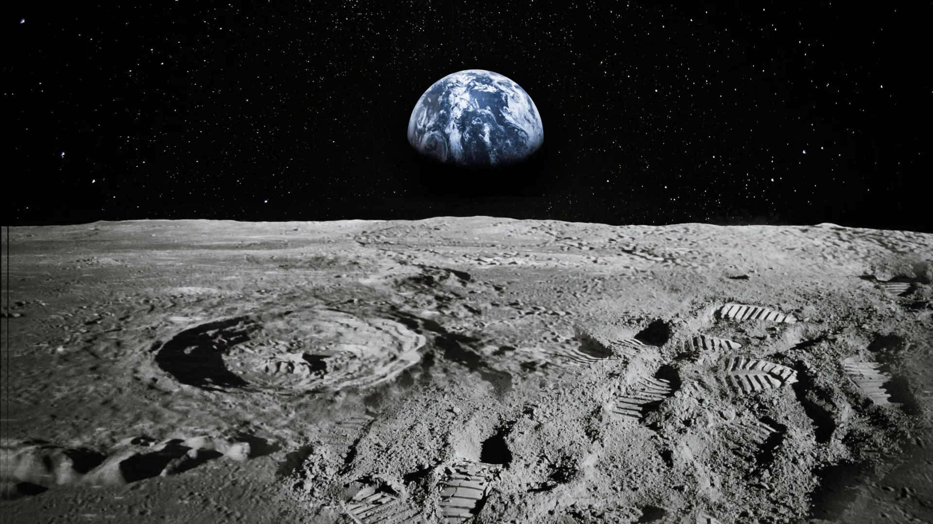
Where did the moon’s magnetism go? Scientists have puzzled over this question for decades, ever since orbiting spacecraft picked up signs of a high magnetic field in lunar surface rocks. The moon itself has no inherent magnetism today.

Now, MIT scientists may have solved the mystery. They propose that a combination of an ancient, weak magnetic field and a large, plasma-generating impact may have temporarily created a strong magnetic field, concentrated on the far side of the moon.

In a study appearing in the journal Science Advances, the researchers show through detailed simulations that an impact, such as from a large asteroid, could have generated a cloud of ionized particles that briefly enveloped the moon. This plasma would have streamed around the moon and concentrated at the opposite location from the initial impact. There, the plasma would have interacted with and momentarily amplified the moon’s weak magnetic field. Any rocks in the region could have recorded signs of the heightened magnetism before the field quickly died away.

This combination of events could explain the presence of highly magnetic rocks detected in a region near the south pole, on the moon’s far side. As it happens, one of the largest impact basins — the Imbrium basin — is located in the exact opposite spot on the near side of the moon. The researchers suspect that whatever made that impact likely released the cloud of plasma that kicked off the scenario in their simulations.

“There are large parts of lunar magnetism that are still unexplained,” says lead author Isaac Narrett, a graduate student in the MIT Department of Earth, Atmospheric and Planetary Sciences (EAPS). “But the majority of the strong magnetic fields that are measured by orbiting spacecraft can be explained by this process — especially on the far side of the moon.”

Narrett’s co-authors include Rona Oran and Benjamin Weiss at MIT, along with Katarina Miljkovic at Curtin University, Yuxi Chen and Gábor Tóth at the University of Michigan at Ann Arbor, and Elias Mansbach PhD ’24 at Cambridge University. Nuno Loureiro, professor of nuclear science and engineering at MIT, also contributed insights and advice.

Beyond the sun

Scientists have known for decades that the moon holds remnants of a strong magnetic field. Samples from the surface of the moon, returned by astronauts on NASA’s Apollo missions of the 1960s and 70s, as well as global measurements of the moon taken remotely by orbiting spacecraft, show signs of remnant magnetism in surface rocks, especially on the far side of the moon.

The typical explanation for surface magnetism is a global magnetic field, generated by an internal “dynamo,” or a core of molten, churning material. The Earth today generates a magnetic field through a dynamo process, and it’s thought that the moon once may have done the same, though its much smaller core would have produced a much weaker magnetic field that may not explain the highly magnetized rocks observed, particularly on the moon’s far side.

An alternative hypothesis that scientists have tested from time to time involves a giant impact that generated plasma, which in turn amplified any weak magnetic field. In 2020, Oran and Weiss tested this hypothesis with simulations of a giant impact on the moon, in combination with the solar-generated magnetic field, which is weak as it stretches out to the Earth and moon.

In simulations, they tested whether an impact to the moon could amplify such a solar field, enough to explain the highly magnetic measurements of surface rocks. It turned out that it wasn’t, and their results seemed to rule out plasma-induced impacts as playing a role in the moon’s missing magnetism.

A spike and a jitter

But in their new study, the researchers took a different tack. Instead of accounting for the sun’s magnetic field, they assumed that the moon once hosted a dynamo that produced a magnetic field of its own, albeit a weak one. Given the size of its core, they estimated that such a field would have been about 1 microtesla, or 50 times weaker than the Earth’s field today.

From this starting point, the researchers simulated a large impact to the moon’s surface, similar to what would have created the Imbrium basin, on the moon’s near side. Using impact simulations from Katarina Miljkovic, the team then simulated the cloud of plasma that such an impact would have generated as the force of the impact vaporized the surface material. They adapted a second code, developed by collaborators at the University of Michigan, to simulate how the resulting plasma would flow and interact with the moon’s weak magnetic field.

These simulations showed that as a plasma cloud arose from the impact, some of it would have expanded into space, while the rest would stream around the moon and concentrate on the opposite side. There, the plasma would have compressed and briefly amplified the moon’s weak magnetic field. This entire process, from the moment the magnetic field was amplified to the time that it decays back to baseline, would have been incredibly fast — somewhere around 40 minutes, Narrett says.

Would this brief window have been enough for surrounding rocks to record the momentary magnetic spike? The researchers say, yes, with some help from another, impact-related effect.

They found that an Imbrium-scale impact would have sent a pressure wave through the moon, similar to a seismic shock. These waves would have converged to the other side, where the shock would have “jittered” the surrounding rocks, briefly unsettling the rocks’ electrons — the subatomic particles that naturally orient their spins to any external magnetic field. The researchers suspect the rocks were shocked just as the impact’s plasma amplified the moon’s magnetic field. As the rocks’ electrons settled back, they assumed a new orientation, in line with the momentary high magnetic field.

“It’s as if you throw a 52-card deck in the air, in a magnetic field, and each card has a compass needle,” Weiss says. “When the cards settle back to the ground, they do so in a new orientation. That’s essentially the magnetization process.”

The researchers say this combination of a dynamo plus a large impact, coupled with the impact’s shockwave, is enough to explain the moon’s highly magnetized surface rocks — particularly on the far side. One way to know for sure is to directly sample the rocks for signs of shock, and high magnetism. This could be a possibility, as the rocks lie on the far side, near the lunar south pole, where missions such as NASA’s Artemis program plan to explore.

“For several decades, there’s been sort of a conundrum over the moon’s magnetism — is it from impacts or is it from a dynamo?” Oran says. “And here we’re saying, it’s a little bit of both. And it’s a testable hypothesis, which is nice.”

The team’s simulations were carried out using the MIT SuperCloud. This research was supported, in part, by NASA.

Share Button

A common supplement could reverse the hidden harm of sucralose

Sucralose is a popular sugar substitute for people who are cutting calories or managing blood sugar levels, but new research by the University of Pittsburgh and UPMC Hillman Cancer Center suggests that the artificial sweetener may not be the best choice for patients undergoing cancer immunotherapy.

Publishing recently in Cancer Discovery, a publication of the American Association for Cancer Research, the study found that patients with melanoma and non-small cell lung cancer who consumed high levels of sucralose had worse response to immunotherapy and poorer survival than those with diets low in the artificial sweetener.

Strikingly, supplements that boosted levels of the amino acid arginine mitigated the negative effects of sucralose on immunotherapy in mice, an approach that could now be tested in clinical trials.

“It’s easy to say, ‘Stop drinking diet soda,’ but when patients are being treated for cancer, they are already dealing with enough, so asking them to drastically alter their diet may not be realistic,” said lead author Abby Overacre, Ph.D., assistant professor in the Department of Immunology at Pitt and UPMC Hillman. “We need to meet patients where they are. That’s why it’s so exciting that arginine supplementation could be a simple approach to counteract the negative effects of sucralose on immunotherapy.”

Senior author Diwakar Davar, M.D., associate professor of medicine at Pitt and a medical oncologist and hematologist at UPMC Hillman, collaborating with Overacre and their team, used mouse models to show that the negative impacts of sucralose are driven by disruption to gut bacteria.

Sucralose shifted the composition of the mouse gut microbiome, increasing bacterial species that degrade arginine, which reduced levels of this amino acid in the blood, tumor fluid and stool.

Immune checkpoint inhibitor immunotherapies such as anti-PD1 work by ramping up T cell activity so that they can more effectively kill cancer cells. Arginine is essential for T cell function, especially in cancer.

“When arginine levels were depleted due to sucralose-driven shifts in the microbiome, T cells couldn’t function properly,” said Overacre. “As a result, immunotherapy wasn’t as effective in mice that were fed sucralose.”

In mouse models of adenocarcinoma and melanoma, adding sucralose to the diet inhibited anti-PD1 therapy, leading to larger tumors and poorer survival. But when the researchers gave sucralose-fed mice arginine or citrulline, which is metabolized into arginine in the body, the effectiveness of immunotherapy was restored.

To assess the relevance of these findings for humans, the researchers looked at 132 patients with advanced melanoma or non-small cell lung cancer who received anti-PD1 therapy alone or in combination with chemotherapy. Patients filled out detailed diet history questionnaires that included questions about how often they consumed artificial sweeteners in coffee, tea and diet soda.

“We found that sucralose impeded the effectiveness of immunotherapies across a range of cancer types, stages and treatment modalities,” said Davar. “These observations raise the possibility of designing prebiotics, such as targeted nutrient supplementation for patients who consume high levels of sucralose.”

The researchers hope to launch a clinical trial investigating whether citrulline supplements — which boost arginine levels more than arginine itself — affect the gut microbiome and anti-tumor immune response in patients.

They are also interested in looking at how other sugar substitutes, such as aspartame, saccharin, xylitol and stevia, impact the immune system and response to immunotherapy.

Other authors on the study were Kristin Morder, M.S., Madison Nguyen, Drew Wilfahrt, Ph.D., Zakaria Dahmani, Ansen Burr, M.D., Ph.D., Bingxian Xie, Ph.D., Michael Morikone, Ph.D., Hector Nieves-Rosado, M.D., Ph.D., William Gunn, M.S., Drew Hurd, Hong Wang, Ph.D., Steven Mullett, Kaitlin Bossong, Stacy Gelhaus, Ph.D., Dhivyaa Rajasundaram, Ph.D., Lawrence Kane, Ph.D., and Greg Delgoffe, Ph.D., and Jishnu Das, Ph.D., all of Pitt or UPMC.

This research was supported by the National Institutes of Health (DP2AI177967, S10OD023402, S10OD032141, R01CA206517, R01AI138504, T32GM008208, U01 CA271407, R01 CA257265, U01 CA268806 and P50 CA254865), the Damon Runyon Cancer Research Foundation and Gateway for Cancer Research (G-22-800).

Share Button

Experts warn: Smartphones before 13 could harm mental health for life

Owning a smartphone before age 13 is associated with poorer mind health and wellbeing in early adulthood, according to a global study of more than 100,000 young people.

Published recently in the peer-reviewed Journal of Human Development and Capabilities, the study found that 18- to 24-year-olds who had received their first smartphone at age 12 or younger were more likely to report suicidal thoughts, aggression, detachment from reality, poorer emotional regulation, and low self-worth.

The data also shows evidence that these effects of smartphone ownership at an early age are in large part associated with early social media access and higher risks of cyberbullying, disrupted sleep, and poor family relationships by adulthood.

A team of experts from Sapien Labs, which hosts the world’s largest database on mental wellbeing, the Global Mind Project — where the data for this research was pooled from — are calling for urgent action to protect the mind health of future generations.

“Our data indicate that early smartphone ownership — and the social media access it often brings — is linked with a profound shift in mind health and wellbeing in early adulthood,” says lead author neuroscientist Dr Tara Thiagarajan, who is the founder and Chief Scientist of Sapien Labs.

“These correlations are mediated through several factors, including social media access, cyberbullying, disrupted sleep, and poor family relationships leading to symptoms in adulthood that are not the traditional mental health symptoms of depression and anxiety and can be missed by studies using standard screeners. These symptoms of increased aggression, detachment from reality and suicidal thoughts can have significant societal consequences as their rates grow in younger generations.

“Based on these findings, and with the age of first smartphones now well under age 13 across the world, we urge policymakers to adopt a precautionary approach, similar to regulations on alcohol and tobacco, by restricting smartphone access for under 13s, mandating digital literacy education and enforcing corporate accountability.”

Since the early 2000s, smartphones have reshaped how young people connect, learn and form identities. But alongside these opportunities come growing concerns over how AI-driven social media algorithms may amplify harmful content and encourage social comparison — while also impacting on other activities such as face-to-face interaction and sleep.

Although many social media platforms set a minimum user age of 13, enforcement is inconsistent. Meanwhile, the average age of first smartphone ownership continues to fall, with many children spending hours a day on their devices.

Currently, it is a mixed picture internationally around the banning on phones in schools, at least. In recent years, several countries have banned or restricted cell phone use in institutions, including France, the Netherlands, Italy, and New Zealand. Results of these moves are limited, however a study commissioned by the Dutch government has found improved focus among students. This month, policymakers in New York have announced it was to become the largest US state yet to ban smartphones in schools, joining locations such as Alabama, Arkansas, Nebraska, North Dakota, Oklahoma and West Virginia which have all passed legislation requiring schools to have policies that at least limit access to smartphones.

Overall, previous studies into screen time, social media and smartphone access and various mental health outcomes have shown negative effects, but also mixed, often conflicting results — making it hard for policymakers, schools, and families to navigate this issue. Possibly this may have to do with the use of screeners that miss the critical associated symptoms.

For this new analysis, the team at Sapien drew data from their Global Mind Project, and then used the Mind Health Quotient (MHQ) — a self-assessment tool that measures social, emotional, cognitive, and physical wellbeing — to generate an overall ‘mind health’ score.

Their results showed:

· The specific symptoms most strongly linked with earlier smartphone ownership include suicidal thoughts, aggression, detachment from reality, and hallucinations.

· Young adults who received their first smartphone before age 13 had lower MHQ scores, with scores progressively declining the younger the age of first ownership. For example, those who owned a smartphone at age 13 scored an average of 30, dropping to just 1 for those who had one at age five.

· Correspondingly, the percentage considered distressed or struggling (with scores indicating they had five or more severe symptoms) rose by 9.5% for females and 7% for males. This pattern was consistent across all regions, cultures and languages, pointing to a critical window of heightened vulnerability.

· That younger ownership is also associated with diminished self-image, self-worth and confidence, and emotional resilience among females, and lower stability and calmness, self-worth and empathy among males.

Further analysis indicated that early access to social media explains about 40% of the association between earlier childhood smartphone ownership and later mind health, with poor family relationships (13%), cyberbullying (10%) and disrupted sleep (12%) also playing significant downstream roles.

The researchers acknowledge the COVID-19 pandemic may have magnified these patterns, but the consistency of these trends across all global regions suggests a broader developmental impact of early smartphone access.

While current evidence does not yet prove direct causation between early smartphone ownership and later mind health and wellbeing, a limitation of the paper, the authors argue that the scale of the potential harm is too great to ignore and justifies a precautionary response.

They recommend four key areas for policymakers to address:

· A requirement of mandatory education on digital literacy and mental health.

· To strengthen the active identification of social media age violations and ensure meaningful consequences for technology companies.

· Restricting access to social media platforms.

· Implementing graduated access restrictions for smartphones.

“Altogether, these policy recommendations aim to safeguard mind health during critical developmental windows,” states Dr Thiagarajan, whose research specialism focuses on the impact of environment on the brain and mind, with an interest in understanding and enabling the productive evolution of the human mind and human systems.

“Their implementation requires substantial political and societal will, effective enforcement, and a multi-stakeholder approach, but successful precedents do exist. For example, in the United States, underage alcohol access and consumption is regulated through a combination of parental, commercial, and corporate accountability.”

Concluding she states: “Our evidence suggests childhood smartphone ownership, an early gateway into AI-powered digital environments, is profoundly diminishing mind health and wellbeing in adulthood with deep consequences for individual agency and societal flourishing.

“I was initially surprised by how strong the results are. However when you give it due consideration, it does begin to make sense that the younger developing mind is more compromised by the online environment given their vulnerability and lack of worldly experience.

“That said, I think it is also important to point out that smartphones and social media are not the only assault to mental health and crisis facing younger adults. It explains some of the overall decline but not all of it. “Now, while more research is needed to unravel the causal mechanisms, waiting for irrefutable proof in the face of these population-level findings unfortunately risks missing the window for timely, preventative action.”

This paper is part of a special cohesive set, entitled ‘The Policy Forum’, in the upcoming publication of Journal of Human Development and Capabilities.

Share Button

The fat you can’t see may be damaging your heart, even if you exercise

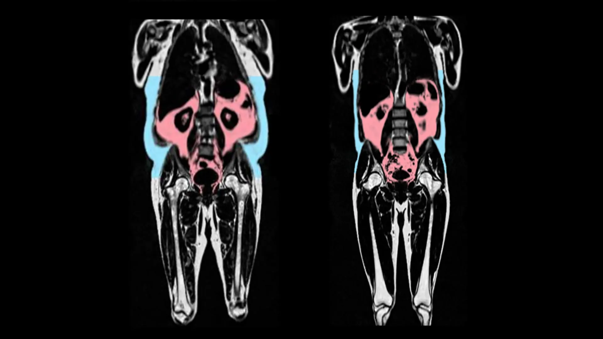
Excessive amounts of visceral fat — the hidden fat surrounding organs — is linked with faster aging of the heart, a new study has found.

Aging is the biggest risk factor for heart disease, but why some people age faster than others isn’t fully understood. The scientists leading the research say that visceral body fat could play an important role in accelerating aging of the heart and blood vessels. This type of fat is known to be harmful to health and this study now links it to faster heart aging.

The study, led by scientists from the Medical Research Council (MRC) Laboratory of Medical Sciences, in London, UK, also found differences between men and women, and discovered that fat around the hips and thighs could potentially slow heart aging in women.

In the study, published in the European Heart Journal, the scientists analyzed data from 21,241 participants in UK Biobank, which includes whole body imaging to map the amount of fat and where it is located in the body.

The UK Biobank data also includes detailed imaging of the heart and blood vessels. Artificial intelligence was used to analyse these images to capture signs of organ aging — such as tissues becoming stiff and inflamed. An individual was given a “heart age” which can be compared to their actual age at the time of the scan.

The researchers found that faster heart aging was linked to having more visceral adipose tissue. Visceral adipose tissue is fat found deep inside the abdomen around organs such as the stomach, intestines, and liver. This type of fat cannot be seen from the outside, and some people can have large amounts of visceral fat despite having a healthy weight.

The researchers found signs on blood tests that visceral fat is linked to increased inflammation in the body – which is a potential cause of premature aging.

They also found differences between the sexes. Male-type fat distribution (fat around the belly, often called ‘apple’ shaped) was particularly predictive of early aging in men.

In contrast, a genetic predisposition to female-type fat (fat on the hips and thighs, often called “pear” shaped) was protective against heart aging in women.

The researchers also found a link between higher oestrogen levels in premenopausal women and a slowing of heart aging, which they suggest could indicate a role for hormones in protecting against heart aging.

Professor Declan O’Regan, who led the research at the MRC Laboratory of Medical Sciences and Imperial College London, and is the British Heart Foundation Professor of Cardiovascular AI, said: “We have known about the apple and pear distinction in body fat, but it hasn’t been clear howit leads to poor health outcomes. Our research shows that “bad” fat, hidden deep around the organs, accelerates aging of the heart. But some types of fat could protect against aging- specifically fat around the hips and thighs in women.”

“We also showed that BMI wasn’t a good way of predicting heart age which underscores the importance of knowing where fat is stored in the body and not just total body weight.”

“The goal of our research is to find ways to increase healthy lifespan. While being active is important, we found that hidden fat could still be harmful even in fit people. In the future we plan to investigate how drug therapies, such as GLP-1 inhibitors (e.g. Ozempic) could improve not just diabetes and obesity, but target the aging effects of hidden visceral fat.”

This study was funded by the Medical Research Council, British Heart Foundation, and the National Institute for Health and Care Research Imperial College Biomedical Research Centre.

Professor Bryan Williams OBE, chief scientific and medical officer at the British Heart Foundation said: “We already know excess visceral fat around the heart and liver can lead to increased blood pressure and high cholesterol, so it is concerning that it could also help to speed up aging of the heart and blood vessels.

“As the pattern of fat distribution typically seen in women’s bodies is linked to oestrogen, that hormone may be key to future therapies developed to tackle heart aging.

“Eating a healthier diet and becoming more active can help to reduce visceral fat levels.”
 

Share Button

Baby pterosaurs died in ancient storms—and their fossils reveal the truth

The cause of death for two baby pterosaurs has been revealed by University of Leicester paleontologists in a post-mortem 150 million years in the making.

Detailed in a new study in the journal Current Biology, their findings show how these flying reptiles were tragically struck down by powerful storms that also created the ideal conditions to preserve them and hundreds more fossils like them.

The Mesozoic, or age of reptiles, is often imagined as a time of giants. Towering dinosaurs, monstrous marine reptiles, and vast-winged pterosaurs dominate museum halls and the public consciousness. But this familiar picture is skewed. Just as today’s ecosystems are mostly populated by small animals, so too were ancient ones. The difference? Fossilization tends to favor the largest and the most robust organisms. Small, fragile creatures rarely make it into the paleontological record.

On rare occasions, however, nature conspires to preserve the delicate and the diminutive inhabitants of these lost worlds. One of the most famous examples is the 150-million-year-old Solnhofen Limestones of southern Germany. These lagoonal deposits are renowned for their exquisitely preserved fossils, including many specimens of pterosaurs, the flying reptiles of the Mesozoic.

Yet here lies a mystery: while Solnhofen has yielded hundreds of pterosaur fossils, nearly all are very small, very young individuals, perfectly preserved. By contrast, larger, adult pterosaurs are rarely found, and when they are, they’re represented only by fragments (often isolated skulls or limbs). This pattern runs counter to expectations: larger, more robust animals should stand a better chance of fossilization than delicate juveniles.

Lead author of the study Rab Smyth, from the University of Leicester’s Centre for Palaeobiology and Biosphere Evolution, was funded by the Natural Environment Research Council through the CENTA Doctoral Training Partnership

Rab said: “Pterosaurs had incredibly lightweight skeletons. Hollow, thin-walled bones are ideal for flight but terrible for fossilization. The odds of preserving one are already slim and finding a fossil that tells you how the animal died is even rarer.”

The discovery of two baby pterosaurs with broken wings has helped to solve this mystery. These tiny fossils, though easily overlooked, are powerful evidence of ancient tropical storms and how they shaped the fossil record.

Ironically nicknamed Lucky and Lucky II by the researchers, the two individuals belong to Pterodactylus, the first pterosaur ever scientifically named. With wingspans of less than 20 cm (8 inches) these hatchlings are among the smallest of all known pterosaurs. Their skeletons are complete, articulated and virtually unchanged from when they died. Except for one detail. Both show the same unusual injury: a clean, slanted fracture to the humerus. Lucky’s left wing and Lucky II’s right wing were both broken in a way that suggests a powerful twisting force, likely the result of powerful gusts of wind rather than a collision with a hard surface.

Catastrophically injured, the pterosaurs plunged into the surface of the lagoon, drowning in the storm driven waves and quickly sinking to the seabed where they were rapidly buried by very fine limy muds stirred up by the death storms. This rapid burial allowed for the remarkable preservation seen in their fossils.

Like Lucky I and II, which were only a few days or weeks old when they died, there are many other small, very young pterosaurs in the Solnhofen Limestones, preserved in the same way as the Luckies, but without obvious evidence of skeletal trauma. Unable to resist the strength of storms these young pterosaurs were also flung into the lagoon. This discovery explains why smaller fossils are so well preserved – they were a direct result of storms – a common cause of death for pterosaurs that lived in the region.

Larger, stronger individuals, it seems, were able to weather the storms and rarely followed the Luckies stormy road to death. They did eventually die though but likely floated for days or weeks on the now calm surfaces of the Solnhofen lagoon, occasionally dropping parts of their carcasses into the abyss as they slowly decomposed.

“For centuries, scientists believed that the Solnhofen lagoon ecosystems were dominated by small pterosaurs,” said Smyth. “But we now know this view is deeply biased. Many of these pterosaurs weren’t native to the lagoon at all. Most are inexperienced juveniles that were likely living on nearby islands that were unfortunately caught up in powerful storms.”

Co-author Dr David Unwin from the University of Leicester added: “When Rab spotted Lucky we were very excited but realized that it was a one-off. Was it representative in any way? A year later, when Rab noticed Lucky II we knew that it was no longer a freak find but evidence of how these animals were dying. Later still, when we had a chance to light-up Lucky II with our UV torches, it literally leapt out of the rock at us — and our hearts stopped. Neither of us will ever forget that moment.”

This research was supported by the Central England NERC Training Alliance (CENTA), under grant number NE/S007350/1.

Share Button

Why the world is watching RFK’s fight with US health agency

The US health secretary’s plans could have a huge impact on health policy not just in the US, but across the globe.

Share Button