NASA’s x-ray telescope finds bizarre features in a cosmic hand

In 2009, NASA’s Chandra X-ray Observatory released a captivating image: a pulsar and its surrounding nebula that is shaped like a hand.

i>

  • X-rays from Chandra have been combined with radio data obtained by the Australian Telescope Compact Array to reveal new structures.
  • At the center of this image is a pulsar, a rapidly spinning neutron star, which is responsible for creating the nebula.
  • This system was created when a massive star collapsed and exploded after it burned through its sustainable nuclear fuel.
  • In 2009, NASA’s Chandra X-ray Observatory released a captivating image: a pulsar and its surrounding nebula that is shaped like a hand.

    Since then, astronomers have used Chandra and other telescopes to continue to observe this object. Now, new radio data from the Australia Telescope Compact Array (ATCA), has been combined with Chandra’s X-ray data to provide a fresh view of this exploded star and its environment, to help understand its peculiar properties and shape.

    At the center of this new image lies the pulsar B1509-58, a rapidly spinning neutron star that is only about 12 miles in diameter. This tiny object is responsible for producing an intricate nebula (called MSH 15-52) that spans over 150 light-years, or about 900 trillion miles. The nebula, which is produced by energetic particles, resembles a human hand with a palm and extended fingers pointing to the upper right in X-rays.

    The collapse of a massive star created the pulsar when much of the star crashed inward once it burned through its sustainable nuclear fuel. An ensuing explosion sent the star’s outer layers outward into space as a supernova.

    The pulsar spins around almost seven times every second and has a strong magnetic field, about 15 trillion times stronger than the Earth’s. The rapid rotation and strong magnetic field make B1509-58 one of the most powerful electromagnetic generators in the Galaxy, enabling it to drive an energetic wind of electrons and other particles away from the pulsar, creating the nebula.

    In this new composite image, the ATCA radio data (represented in red) has been combined with X-rays from Chandra (shown in blue, orange and yellow), along with an optical image of hydrogen gas (gold). The areas of overlap between the X-ray and radio data in MSH 15-52 show as purple. The optical image shows stars in the field of view along with parts of the supernova’s debris, the supernova remnant RCW 89.

    Radio data from ATCA now reveals complex filaments that are aligned with the directions of the nebula’s magnetic field, shown by the short, straight, white lines in a supplementary image. These filaments could result from the collision of the pulsar’s particle wind with the supernova’s debris.

    By comparing the radio and X-ray data, researchers identified key differences between the sources of the two types of light. In particular, some prominent X-ray features, including the jet towards the bottom of the image and the inner parts of the three “fingers” towards the top, are not detected in radio waves. This suggests that highly energetic particles are leaking out from a shock wave — similar to a supersonic plane’s sonic boom — near the pulsar and moving along magnetic field lines to create the fingers.

    The radio data also shows that RCW 89’s structure is different from typical young supernova remnants. Much of the radio emission is patchy and closely matches clumps of X-ray and optical emission. It also extends well beyond the X-ray emission. All of these characteristics support the idea that RCW 89 is colliding with a dense cloud of nearby hydrogen gas.

    However, the researchers do not fully understand all that the data is showing them. One area that is perplexing is the sharp boundary of X-ray emission in the upper right of the image that seems to be the blast wave from the supernova — see the labeled feature. Supernova blast waves are usually bright in radio waves for young supernova remnants like RCW 89, so it is surprising to researchers that there is no radio signal at the X-ray boundary.

    MSH 15-52 and RCW 89 show many unique features not found in other young sources. There are, however, still many open questions regarding the formation and evolution of these structures. Further work is needed to provide better understanding of the complex interplay between the pulsar wind and the supernova debris.

    A paper describing this work, led by Shumeng Zhang of the University of Hong Kong, with co-authors Stephen C.Y. Ng of the University of Hong Kong and Niccolo’ Bucciantini of the Italian National Institute for Astrophysics, has been published in The Astrophysical Journal.

    NASA’s Marshall Space Flight Center in Huntsville, Alabama, manages the Chandra program. The Smithsonian Astrophysical Observatory’s Chandra X-ray Center controls science operations from Cambridge, Massachusetts, and flight operations from Burlington, Massachusetts.


    Share Button

    Baby dies of whooping cough after mother not vaccinated while pregnant

    The first death from the illness this year comes as vaccination rates among children have declined.

    Share Button

    Scientists reveal breakthrough blood pressure treatment that works when others fail

    A new treatment has been shown to significantly lower blood pressure in people whose levels stay dangerously high, despite taking several existing medicines, according to the results of a Phase III clinical trial led by a UCL Professor.

    Globally around 1.3 billion people have high blood pressure (hypertension), and in around half of cases the condition is uncontrolled or treatment resistant. These individuals face a much greater risk of heart attack, stroke, kidney disease, and early death. In the UK the number of people with hypertension is around 14 million.

    The international BaxHTN trial, led by Professor Bryan Williams (UCL Institute of Cardiovascular Science) and sponsored by AstraZeneca, assessed the new drug baxdrostat – which is taken as a tablet – with participation from nearly 800 patients across 214 clinics worldwide.

    The study was supported by the NIHR Biomedical Research Centre at UCLH.

    Results were presented on August 30th at the European Society of Cardiology (ESC) Congress 2025 in Madrid and are being simultaneously published in the New England Journal of Medicine.

    The trial results showed that, after 12 weeks, patients taking baxdrostat (1 mg or 2 mg once daily in pill form) saw their blood pressure fall by around 9-10 mmHg more than placebo – a reduction large enough to cut cardiovascular risk. About 4 in 10 patients reached healthy blood pressure levels, compared with fewer than 2 in 10 on placebo.

    Principal Investigator, Professor Williams, who is presenting the results at ESC, said: “Achieving a nearly 10 mmHg reduction in systolic blood pressure with baxdrostat in the BaxHTN Phase III trial is exciting, as this level of reduction is linked to substantially lower risk of heart attack, stroke, heart failure and kidney disease.”

    How baxdrostat works

    Blood pressure is strongly influenced by a hormone called aldosterone, which helps the kidneys regulate salt and water balance.

    Some people produce too much aldosterone, causing the body to hold onto salt and water. This aldosterone dysregulation pushes blood pressure up and makes it very difficult to control.

    Addressing aldosterone dysregulation has been a key effort in research over many decades, but it has been so far difficult to achieve.

    Baxdrostat works by blocking aldosterone production, directly addressing this driver of high blood pressure (hypertension).

    Professor Williams, Chair of Medicine at UCL, said: “These findings are an important advance in treatment and in our understanding of the cause of difficult to control blood pressure.

    “Around half of people treated for hypertension do not have it controlled, however this is a conservative estimate and the number is likely higher, especially as the target blood pressure we try to reach is now much lower than it was previously.*

    “In patients with uncontrolled or resistant hypertension, the addition of baxdrostat 1mg or 2mg once daily to background antihypertensive therapy led to clinically meaningful reductions in systolic blood pressure, which persisted up to 32 weeks with no unanticipated safety findings.

    “This suggests that aldosterone is playing an important role in causing difficult to control blood pressure in millions of patients and offers hope for more effective treatment in the future.”

    Historically higher income Western countries were reported to have far higher levels of hypertension; however, largely due to changing diets (adding less salt to food), the numbers of people living with the condition is now far higher in Eastern and lower income countries. More than half of those affected live in Asia, including 226 million people in China and 199 million in India**.

    Professor Williams added: “The results suggest that this drug could potentially help up to half a billion people globally – and as many as 10 million people in the UK alone, especially at the new target level for optimal blood pressure control.”

    *The ESC 2024 hypertension guidelines recommended a target blood pressure of less than 130/80 mmHg. Prior to 2024 the target had been 140/90 mmHg.

    ** Figures from Blood Pressure UK

    Share Button

    Common heart drug taken by millions found useless, possibly risky

    Beta blockers—drugs commonly prescribed for a range of cardiac conditions, including heart attacks—provide no clinical benefit for patients who have had an uncomplicated myocardial infarction with preserved heart function. Beta blockers have been the standard treatment for these patients for 40 years.

    This is a breakthrough discovery from the “REBOOT Trial” with senior investigator Valentin Fuster, MD, PhD, President of Mount Sinai Fuster Heart Hospital and General Director of Spain’s Centro Nacional de Investigaciones Cardiovasculares (CNIC). The study results, which could overturn a standard treatment paradigm, were presented on Saturday, August 30, during a “Hot Line” session at the European Society of Cardiology Congress in Madrid, and simultaneously published in The New England Journal of Medicine.

    Additionally, a REBOOT substudy, published on August 30 in the European Heart Journal, shows that women treated with beta blockers had a higher risk of death, heart attack, or hospitalization for heart failure compared to women not receiving the drug. Men did not have this increased risk.

    “This trial will reshape all international clinical guidelines. It joins other previous landmark trials led by CNIC and Mount Sinai—such as SECURE with the polypill and DapaTAVI, with SLT2 inhibition associated to TAVI—that have already transformed some global approaches to cardiovascular disease,” says Dr. Fuster.

    The SECURE trial showed a polypill, a single pill that that combines three medications – which contains aspirin, ramipril, and atorvastatin – reduces cardiovascular events by 33 percent in patients treated with this after a heart attack. The DapaTAVI trial showed both dapagliflozin and the related medication empagliflozin – drugs used to treat diabetes- improves the prognosis of patients with aortic stenosis treated by transcatheter aortic valve implantation.

    “REBOOT will change clinical practice worldwide,” says Principal Investigator Borja Ibáñez, MD, CNIC’s Scientific Director, who presented the results. “Currently, more than 80 percent of patients with uncomplicated myocardial infarction are discharged on beta blockers. The REBOOT findings represent one of the most significant advances in heart attack treatment in decades.”

    Although generally considered safe, beta blockers can cause side effects such as fatigue, bradycardia (low heart rate), and sexual dysfunction. For more than 40 years, beta blockers have been prescribed as a standard treatment after a heart attack, but their benefit in the context of modern treatments was unproven. The REBOOT trial, is the largest clinical trial on this subject. The international study was coordinated by CNIC in collaboration with the Mario Negri Institute for Pharmacological Research in Milan.

    Researchers enrolled 8,505 patients across 109 hospitals in Spain and Italy. Participants were randomly assigned to receive or not receive beta blockers after hospital discharge. All patients otherwise received the current standard of care and were followed for a median of nearly four years. The results showed no significant differences between the two groups in rates of death, recurrent heart attack, or hospitalization for heart failure.

    A REBOOT subgroup analysis found that women treated with beta blockers experienced more adverse events. Results show women treated with beta-blockers had a 2.7 percent higher absolute risk of mortality compared to those not treated with beta-blockers during the 3.7 years of follow-up of the study. The elevated risk when treated with beta-blockers was restricted to women with a complete normal cardiac function after a heart attack (left ventricular ejection fraction of 50 percent or higher). Those with a mild deterioration in cardiac function did not have an excess risk of adverse outcomes when treated with beta-blockers.

    “After a heart attack, patients are typically prescribed multiple medications, which can make adherence difficult,” explains Dr. Ibáñez. “Beta blockers were added to standard treatment early on because they significantly reduced mortality at the time. Their benefits were linked to reduced cardiac oxygen demand and arrhythmia prevention. But therapies have evolved. Today, occluded coronary arteries are reopened rapidly and systematically, drastically lowering the risk of serious complications such as arrhythmias. In this new context—where the extent of heart damage is smaller—the need for beta blockers is unclear. While we often test new drugs, it’s much less common to rigorously question the continued need for older treatments.”

    That was the motivation behind REBOOT.

    “The trial was designed to optimize heart attack care based on solid scientific evidence and without commercial interests. These results will help streamline treatment, reduce side effects, and improve quality of life for thousands of patients every year,” Dr. Ibanez adds.

    REBOOT was conducted without pharmaceutical industry funding.

    Share Button

    Vaccination plea after baby’s ‘terrifying’ measles

    Eight areas in the North East and Cumbria have seen a slight or moderate increase in MMR jab rates.

    Share Button

    The trade in US body parts that’s completely legal – but ripe for exploitation

    Human remains can mean profits. But there is an emotional debate about the ethics of the industry.

    Share Button

    AI stethoscope could detect major heart conditions in seconds

    The new technology could be a “game-changer” resulting in patients being treated sooner, experts say.

    Share Button

    New AI model predicts which genetic mutations truly drive disease

    When genetic testing reveals a rare DNA mutation, doctors and patients are frequently left in the dark about what it actually means. Now, researchers at the Icahn School of Medicine at Mount Sinai have developed a powerful new way to determine whether a patient with a mutation is likely to actually develop disease, a concept known in genetics as penetrance.

    The team set out to solve this problem using artificial intelligence (AI) and routine lab tests like cholesterol, blood counts, and kidney function. Details of the findings were reported in the August 28 online issue of Science. Their new method combines machine learning with electronic health records to offer a more accurate, data-driven view of genetic risk.

    Traditional genetic studies often rely on a simple yes/no diagnosis to classify patients. But many diseases, like high blood pressure, diabetes, or cancer, don’t fit neatly into binary categories. The Mount Sinai researchers trained AI models to quantify disease on a spectrum, offering more nuanced insight into how disease risk plays out in real life.

    “We wanted to move beyond black-and-white answers that often leave patients and providers uncertain about what a genetic test result actually means,” says Ron Do, PhD, senior study author and the Charles Bronfman Professor in Personalized Medicine at the Icahn School of Medicine at Mount Sinai. “By using artificial intelligence and real-world lab data, such as cholesterol levels or blood counts that are already part of most medical records, we can now better estimate how likely disease will develop in an individual with a specific genetic variant. It’s a much more nuanced, scalable, and accessible way to support precision medicine, especially when dealing with rare or ambiguous findings.”

    Using more than 1 million electronic health records, the researchers built AI models for 10 common diseases. They then applied these models to people known to have rare genetic variants, generating a score between 0 and 1 that reflects the likelihood of developing the disease.

    A higher score, closer to 1, suggests a variant may be more likely to contribute to disease, while a lower score indicates minimal or no risk. The team calculated “ML penetrance” scores for more than 1,600 genetic variants.

    Some of the results were surprising, say the investigators. Variants previously labeled as “uncertain” showed clear disease signals, while others thought to cause disease had little effect in real-world data.

    “While our AI model is not meant to replace clinical judgment, it can potentially serve as an important guide, especially when test results are unclear. Doctors could in the future use the ML penetrance score to decide whether patients should receive earlier screenings or take preventive steps, or to avoid unnecessary worry or intervention if the variant is low-risk,” says lead study author Iain S. Forrest, MD, PhD, in the lab of Dr. Do at the Icahn School of Medicine at Mount Sinai. “If a patient has a rare variant associated with Lynch syndrome, for instance, and it scores high, that could trigger earlier cancer screening, but if the risk appears low, jumping to conclusions or overtreatment might be avoided.”

    The team is now working to expand the model to include more diseases, a wider range of genetic changes, and more diverse populations. They also plan to track how well these predictions hold up over time, whether people with high-risk variants actually go on to develop disease, and whether early action can make a difference.

    Ultimately, our study points to a potential future where AI and routine clinical data work hand in hand to provide more personalized, actionable insights for patients and families navigating genetic test results,” says Dr. Do. “Our hope is that this becomes a scalable way to support better decisions, clearer communication, and more confidence in what genetic information really means.”

    The paper is titled “Machine learning-based penetrance of genetic variants.”

    The study’s authors, as listed in the journal, are Iain S. Forrest, Ha My T. Vy, Ghislain Rocheleau, Daniel M. Jordan, Ben O. Petrazzini, Girish N. Nadkarni, Judy H. Cho, Mythily Ganapathi, Kuan-Lin Huang, Wendy K. Chung, and Ron Do.

    This work was supported in part by the following grants: National Institute of General Medical Sciences of the National Institutes of Health (NIH) (T32-GM007280); the National Institute of General Medical Sciences of the NIH (R35-GM124836); the National Institute of Diabetes and Digestive and Kidney Diseases (U24-DK062429); the National Human Genome Research Institute of the NIH (R01-HG010365); the National Institute of General Medical Sciences of the NIH (R35-GM138113); and the National Institute of Diabetes and Digestive and Kidney Diseases (U24-DK062429).

    * Mount Sinai Health System member hospitals: The Mount Sinai Hospital; Mount Sinai Brooklyn; Mount Sinai Morningside; Mount Sinai Queens; Mount Sinai South Nassau; Mount Sinai West; and New York Eye and Ear Infirmary of Mount Sinai

    Share Button

    Cells “vomit” waste in a hidden healing shortcut that could also fuel cancer

    When injured, cells have well-regulated responses to promote healing. These include a long-studied self-destruction process that cleans up dead and damaged cells as well as a more recently identified phenomenon that helps older cells revert to what appears to be a younger state to help grow back healthy tissue.

    Now, a new study in mice led by researchers at Washington University School of Medicine in St. Louis and the Baylor College of Medicine reveals a previously unknown cellular purging process that may help injured cells revert to a stem cell-like state more rapidly. The investigators dubbed this newly discovered response cathartocytosis, taking from Greek root words that mean cellular cleansing.

    Published online in the journal Cell Reports, the study used a mouse model of stomach injury to provide new insights into how cells heal, or fail to heal, in response to damage, such as from an infection or inflammatory disease.

    “After an injury, the cell’s job is to repair that injury. But the cell’s mature cellular machinery for doing its normal job gets in the way,” said first author Jeffrey W. Brown, MD, PhD, an assistant professor of medicine in the Division of Gastroenterology at WashU Medicine. “So, this cellular cleanse is a quick way of getting rid of that machinery so it can rapidly become a small, primitive cell capable of proliferating and repairing the injury. We identified this process in the GI tract, but we suspect it is relevant in other tissues as well.”

    Brown likened the process to a “vomiting” or jettisoning of waste that essentially adds a shortcut, helping the cell declutter and focus on regrowing healthy tissues faster than it would be able to if it could only perform a gradual, controlled degradation of waste.

    As with many shortcuts, this one has potential downsides: According to the investigators, cathartocytosis is fast but messy, which may help shed light on how injury responses can go wrong, especially in the setting of chronic injury. For example, ongoing cathartocytosis in response to an infection is a sign of chronic inflammation and recurring cell damage that is a breeding ground for cancer. In fact, the festering mess of ejected cellular waste that results from all that cathartocytosis may also be a way to identify or track cancer, according to the researchers.

    A novel cellular process

    The researchers identified cathartocytosis within an important regenerative injury response called paligenosis, which was first described in 2018 by the current study’s senior author, Jason C. Mills, MD, PhD. Now at the Baylor College of Medicine, Mills began this work while he was a faculty member in the Division of Gastroenterology at WashU Medicine and Brown was a postdoctoral researcher in his lab.

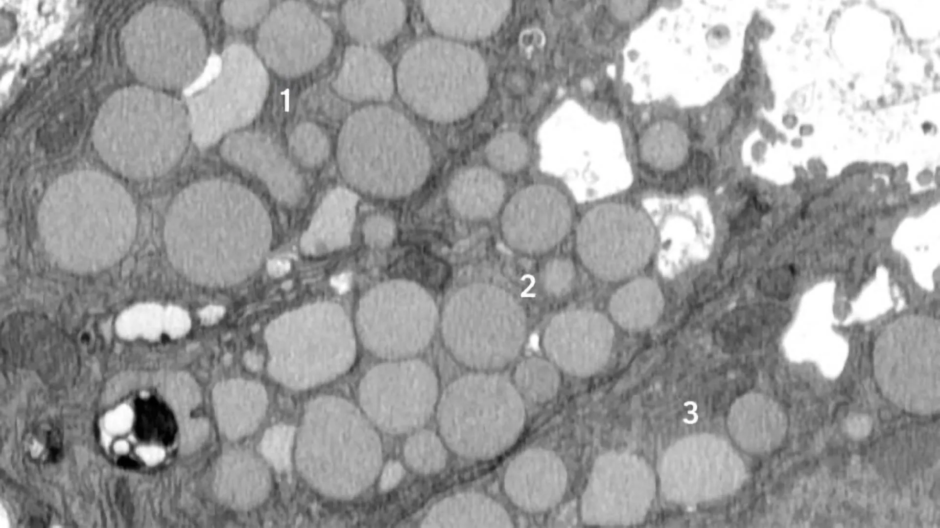
    In paligenosis, injured cells shift away from their normal roles and undergo a reprogramming process to an immature state, behaving like rapidly dividing stem cells, as happens during development. Originally, the researchers assumed the decluttering of cellular machinery in preparation for this reprogramming happens entirely inside cellular compartments called lysosomes, where waste is digested in a slow and contained process.

    From the start, though, the researchers noticed debris outside the cells. They initially dismissed this as unimportant, but the more external waste they saw in their early studies, the more Brown began to suspect that something deliberate was going on. He utilized a model of mouse stomach injury that triggered the reprogramming of mature cells to a stem cell state all at once, making it obvious that the “vomiting” response — now happening in all the stomach cells simultaneously — was a feature of paligenosis, not a bug. In other words, the vomiting process was not just an accidental spill here and there but a newly identified, standard way cells behaved in response to injury.

    Although they discovered cathartocytosis happening during paligenosis, the researchers said cells could potentially use cathartocytosis to jettison waste in other, more worrisome situations, like giving mature cells that ability to start to act like cancer cells.

    The downside to downsizing

    While the newly discovered cathartocytosis process may help injured cells proceed through paligenosis and regenerate healthy tissue more rapidly, the tradeoff comes in the form of additional waste products that could fuel inflammatory states, making chronic injuries harder to resolve and correlating with increased risk of cancer development.

    “In these gastric cells, paligenosis — reversion to a stem cell state for healing — is a risky process, especially now that we’ve identified the potentially inflammatory downsizing of cathartocytosis within it,” Mills said. “These cells in the stomach are long-lived, and aging cells acquire mutations. If many older mutated cells revert to stem cell states in an effort to repair an injury — and injuries also often fuel inflammation, such as during an infection — there’s an increased risk of acquiring, perpetuating and expanding harmful mutations that lead to cancer as those stem cells multiply.”

    More research is needed, but the authors suspect that cathartocytosis could play a role in perpetuating injury and inflammation in Helicobacter pylori infections in the gut. H. pylori is a type of bacteria known to infect and damage the stomach, causing ulcers and increasing the risk of stomach cancer.

    The findings also could point to new treatment strategies for stomach cancer and perhaps other GI cancers. Brown and WashU Medicine collaborator Koushik K. Das, MD, an associate professor of medicine, have developed an antibody that binds to parts of the cellular waste ejected during cathartocytosis, providing a way to detect when this process may be happening, especially in large quantities. In this way, cathartocytosis might be used as a marker of precancerous states that could allow for early detection and treatment.

    “If we have a better understanding of this process, we could develop ways to help encourage the healing response and perhaps, in the context of chronic injury, block the damaged cells undergoing chronic cathartocytosis from contributing to cancer formation,” Brown said.

    This work was supported by the National Institutes of Health (NIH), grant numbers K08DK132496, R21AI156236, P30DK052574, P30DK056338, R01DK105129, R01CA239645, F31DK136205, K99GM159354 and F31CA236506; the Department of Defense, grant number W81XWH-20-1-0630; the American Gastroenterological Association, grant numbers AGA2021-5101 and AGA2024-13-01; and a Philip and Sima Needleman Student Fellowship in Regenerative Medicine. The content is solely the responsibility of the authors and does not necessarily represent the official views of the NIH.

    Share Button

    Why ultra-processed diets make you gain fat even without extra calories

    Over the past 50 years, rates of obesity and type-2 diabetes have soared, while sperm quality has plummeted. Driving these changes could be the increasing popularity of ultra-processed foods, which have been linked to a range of poor health outcomes. However, scientists still aren’t sure whether it’s the industrial nature of the ingredients themselves, the processing of the foods, or whether it’s because they lead people to eat more than they should.

    An international team of scientists has now discovered that people gain more weight on an ultra-processed diet compared to a minimally processed diet, even when they eat the same number of calories. The study in humans also revealed a diet high in ultra-processed foods introduces higher levels of pollutants that are known to affect sperm quality. The findings were published in the journal Cell Metabolism.

    “Our results prove that ultra-processed foods harm our reproductive and metabolic health, even if they’re not eaten in excess. This indicates that it is the processed nature of these foods that makes them harmful,” says Jessica Preston, lead author of the study, who carried out the research during her PhD at the University of Copenhagen’s NNF Center for Basic Metabolic Research (CBMR).

    Same calories, different outcomes

    To get the best possible data, the scientists compared the health impact of unprocessed and ultra-processed diets on the same person. They recruited 43 men aged 20 to 35, who spent three weeks on each of the two diets, with three months ‘washout’ in between. Half started on the ultra-processed and half started on the unprocessed diet. Half of the men also received a high-calorie diet with an extra 500 daily calories, while half received the normal amount of calories for their size, age and physical activity levels. They were not told which diet they were on. Both the unprocessed and ultra-processed diets had the same amount of calories, protein, carbs and fats.

    Men gained around 1 kg more of fat mass while on the ultra-processed diet compared to the unprocessed diet, regardless of whether they were on the normal or excess calorie diet. Several other markers of cardiovascular health were also affected.

    Ultra-processed foods polluted with endocrine disruptors

    The scientists also discovered a worrying increase in the level of the hormone-disrupting phthalate cxMINP, a substance used in plastics, in men on the ultra-processed diet. Men on this diet also saw decreases in their levels of testosterone and follicle-stimulating hormone, which are crucial for sperm production.

    “We were shocked by how many body functions were disrupted by ultra-processed foods, even in healthy young men. The long-term implications are alarming and highlight the need to revise nutritional guidelines to better protect against chronic disease.” says the study’s senior author Professor Romain Barrès from the University of Copenhagen’s NNF Center for Basic Metabolic Research, and the Université Côte d’Azur.

    Share Button