The surprising brain chemistry behind instant friendships

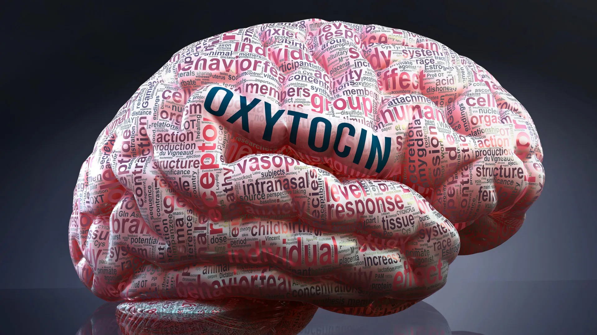
A new UC Berkeley study shows that the so-called love hormone, oxytocin, is also critical for the formation of friendships.

Oxytocin is released in the brain during sex, childbirth, breastfeeding and social interactions and contributes to feelings of attachment, closeness and trust. Never mind that it’s also associated with aggression; the hormone is commonly referred to as the “cuddle” or “happy” hormone, and people are encouraged to boost their oxytocin levels for better well-being by touching friends and loved ones, listening to music and exercising.

But recent studies involving the prairie vole have called this love association into question. They’ve shown that oxytocin, which in the brain acts as a neuromodulator, is not essential for long-term mate bonding, or “social monogamy,” or for parenting behavior, though without it, voles take longer to form such bonds.

Scientists focus on prairie voles because, like humans, they form stable and selective relationships. While most studies focus on mate bonds, the Beery lab at UC Berkeley is particularly interested in selective peer relationships, analogous to human friendships. Such studies could shed light on human psychiatric conditions, such as autism and schizophrenia, that interfere with a person’s ability to form or maintain social bonds.

“Prairie voles are special because they allow us to get at the neurobiology of friendship and how it’s similar to and different from other types of relationships,” said Annaliese Beery, a UC Berkeley associate professor of integrative biology and neuroscience and senior author of the study.

Beery and integrative biology graduate student Alexis Black, one of two first authors of the study, found that prairie voles that lack oxytocin receptors take longer than normal voles to form peer relationships. Prairie voles that are close friends typically huddle side by side, groom and even sit on one another.

“Oxytocin seems to be particularly important in the early formation phase of relationships and especially in the selectivity of those relationships: ‘I prefer you to this stranger,’ for example,” Beery said. “The animals that didn’t have intact oxytocin signaling took longer to form relationships. And then when we challenged those relationships by making new groups, they lost track of their original partners right away.”

The voles, genetically modified in the UC San Francisco laboratory of collaborator and co-author Dr. Devanand Manoli, also lacked the social rewards that normally come from selective attachments — they didn’t work very hard to snuggle up with their friends and were less avoidant of and less aggressive towards strangers.

“In other words, oxytocin is playing a crucial role not so much in how social they are, but more in who they are social with, their selectivity,” she said.

Lacking oxytocin receptors also changed the regulation of oxytocin availability and release in the brain, which the group documented using a novel oxytocin nanosensor in collaboration with postdoctoral fellow Natsumi Komatsu and Markita Landry, a UC Berkeley professor of chemical and biomolecular engineering.

“That helped us understand the feedback consequences of lacking this receptor, and how oxytocin signaling was altered in the brain,” said Beery.

The study was published Aug. 8 in the journal Current Biology.

What social voles tell us about social humans

Beery has long been interested in social relationships in rodents, focusing primarily on the animals’ seldom-studied peer or friendship relationships. While voles are her main focus, she believes studying similar behaviors across multiple species is key to determining what’s species-specific versus generalizable across species.

To complement her laboratory research, she has conducted field studies comparing social behavior and oxytocin receptor distribution in the brain within and across species in a group of South American rodents and North American Belding’s ground squirrels, which vary in whether or not they live in groups. She also recently began field tests of multiple vole species — there are about 50 worldwide — to compare their social behavior.

She suspects that in rodents such as voles, and perhaps in other mammals, the formation of peer relationships may have preceded the evolution of monogamous mating relationships.

“While most rodents prefer to interact with unfamiliar individuals, it turns out that the majority of vole species we’ve tested in our early trials form peer-partner preferences, which is what we call these selective friendships. So there seems to be this widespread tendency to bond,” Beery said. “But only a couple of those species are also monogamous. Someday, I hope to be able to tell you, ‘Do selective peer relationships precede the development of monogamy? Is that why monogamy has evolved so many times in this genus?’ I think this familiarity preference is deeply rooted.”

Beery was a co-author of a 2023 study led by Manoli that threw into question the association of oxytocin with sex and parenting. That study showed that prairie voles unable to respond to oxytocin exhibit the same monogamous mating, attachment and parenting behaviors as regular voles. Those voles had been genetically engineered to have no cellular receptors for oxytocin, and were the same voles used in the current study.

But while oxytocin isn’t essential for eventual bond formation, additional studies by the same group published in 2024 showed that these receptor-deficient (or “null mutant”) prairie voles took about twice as long as normal voles to establish a relationship with a potential mate.

Interested in how the lack of an oxytocin receptor affects voles’ friendship bonds, as opposed to mating bonds, Beery and Black conducted three sets of experiments. In one, they tested how long it took for voles to establish a preference for a partner. Whereas normal voles take about 24 hours of close proximity to form a relationship that makes them choose that partner over a stranger, oxytocin receptor-deficient voles showed no preference in that amount of time, and took up to a week to establish a peer preference.

“Wild-type animals form this incredibly robust preference within one day of co-housing, but the null mutants have no sign of a relationship after 24 hours. After a week, they mostly get there, and the lifetime partners look no different from each other,” Beery said. “Our conclusion from that experiment is that oxytocin isn’t required to have a relationship, but it’s really important in those early phases of a relationship to facilitate it happening quickly and efficiently.”

They then put long-term pair-bonded voles in a party-like, mixed-group situation: an enclosure with other voles and many rooms connected by tubes. In such a situation, normal voles would hang out with known friends until they eventually started to socialize with strangers.

“They can all separate, they can all come together, or they can hang out in any combinations that they want,” she said. “The wild-type animals keep track of who they know. It’s like if I went to a party with a friend, I would stand near that friend for the first part of the party and then I might start to mingle. The voles that lack oxytocin receptors just mixed. It was as if they didn’t even have a partner in there with them.”

In the third experiment, they tested the strength of both peer and mate bonding by having the voles press levers to get access to either a friend/mate or a stranger.

“Female wild-type voles typically press more to get their partner than to get a stranger, in both peer and mate relationships. The oxytocin receptor deficient mutants also press more to get to their mating partner, but not for peer relationships,” Beery said. “That makes sense at some level because we think mate relationships are more rewarding than peer relationships, or at least they depend more on reward-signaling pathways.”

Lack of oxytocin signaling thus not only delays the formation of relationships, but also creates deficits in long-term peer relationships.

On the flip side, voles lacking oxytocin receptors were also less aggressive toward strangers and less avoidant of them.

“You can see contributions of oxytocin signaling to both sides of selectivity,” Beery said. “On the prosocial side, it’s involved in wanting to be with a known friend or peer, while on the antisocial side, it’s aiding in rejecting an unfamiliar animal. We’ve seen effects of oxytocin on both affiliation and aggression in our other studies in prairie voles, and it parallels human findings on a role of oxytocin in in-group/out-group dynamics.”

Oxytocin nanosensors

The researchers used a new oxytocin sensor developed in Landry’s UC Berkeley lab to determine whether lack of an oxytocin receptor caused increases or decreases in oxytocin release. If oxytocin release increased in these voles, it could potentially interact with a receptor for a similar neuropeptide that is also involved in formation of social relationships, compensating for the absence of oxytocin receptors.

Landry, an associate professor in the departments of chemical and biomolecular engineering, neuroscience, and molecular and cell biology and a co-corresponding author of the paper, created these sensors from carbon nanotubes joined with specific single-stranded DNA sequences selected because they latch onto the oxytocin molecule and fluoresce. Komatsu and Landry found no excess of oxytocin in the voles’ brains. In fact, oxytocin was being released in lower amounts from fewer sites in the nucleus accumbens, a key brain region for social reward across species.

Co-authors with Black, Komatsu, Beery, Landry and Manoli are Jiaxuan Zhao, Scarlet Taskey and Nicole Serrano of UC Berkeley, and Ruchira Sharma of UCSF. Beery’s work was supported by the National Science Foundation (CAREER award 2239635) and the National Institutes of Health (R01MH132908). Komatsu is now an assistant professor at the University of Illinois.


Share Button

AI finds hidden safe zones inside a fusion reactor

A public-private partnership between Commonwealth Fusion Systems (CFS), the U.S. Department of Energy’s (DOE) Princeton Plasma Physics Laboratory (PPPL) and Oak Ridge National Laboratory has led to a new artificial intelligence (AI) approach that is faster at finding what’s known as “magnetic shadows” in a fusion vessel: safe havens protected from the intense heat of the plasma.

Known as HEAT-ML, the new AI could lay the foundation for software that significantly speeds up the design of future fusion systems. Such software could also enable good decision-making during fusion operations by adjusting the plasma so that potential problems are thwarted before they start.

“This research shows that you can take an existing code and create an AI surrogate that will speed up your ability to get useful answers, and it opens up interesting avenues in terms of control and scenario planning,” said Michael Churchill, co-author of a paper in Fusion Engineering and Design about HEAT-ML and head of digital engineering at PPPL.

Fusion, the reaction that fuels the sun and stars, could provide potentially limitless amounts of electricity on Earth. To harness it, researchers need to overcome key scientific and engineering challenges. One such challenge is handling the intense heat coming from the plasma, which reaches temperatures hotter than the sun’s core when confined using magnetic fields in a fusion vessel known as a tokamak. Speeding up the calculations that predict where this heat will hit and what parts of the tokamak will be safe in the shadows of other parts is key to bringing fusion power to the grid.

“The plasma-facing components of the tokamak might come in contact with the plasma, which is very hot and can melt or damage these elements,” said Doménica Corona Rivera, an associate research physicist at PPPL and first author on the paper on HEAT-ML. “The worst thing that can happen is that you would have to stop operations.”

PPPL amplifies its impact through public-private partnership

HEAT-ML was specifically made to simulate a small part of SPARC: a tokamak currently under construction by CFS. The Massachusetts company hopes to demonstrate net energy gain by 2027, meaning SPARC would generate more energy than it consumes.

Simulating how heat impacts SPARC’s interior is central to this goal and a big computing challenge. To break down the challenge into something manageable, the team focused on a section of SPARC where the most intense plasma heat exhaust intersects with the material wall. This particular part of the tokamak, representing 15 tiles near the bottom of the machine, is the part of the machine’s exhaust system that will be subjected to the most heat.

To create such a simulation, researchers generate what they call shadow masks. Shadow masks are 3D maps of magnetic shadows, which are specific areas on the surfaces of a fusion system’s internal components that are shielded from direct heat. The location of these shadows depends on the shape of the parts inside the tokamak and how they interact with the magnetic field lines that confine the plasma.

Creating simulations to optimize the way fusion systems operate

Originally, an open-source computer program called HEAT, or the Heat flux Engineering Analysis Toolkit, calculated these shadow masks. HEAT was created by CFS Manager Tom Looby during his doctoral work with Matt Reinke, now leader of the SPARC Diagnostic Team, and was first applied on the exhaust system for PPPL’s National Spherical Torus Experiment-Upgrade machine.

HEAT-ML traces magnetic field lines from the surface of a component to see if the line intersects other internal parts of the tokamak. If it does, that region is marked as “shadowed.” However, tracing these lines and finding where they intersect the detailed 3D machine geometry was a significant bottleneck in the process. It could take around 30 minutes for a single simulation and even longer for some complex geometries.

HEAT-ML overcomes this bottleneck, accelerating the calculations to a few milliseconds. It uses a deep neural network: a type of AI that has hidden layers of mathematical operations and parameters that it applies to the data to learn how to do a specific task by looking for patterns. HEAT-ML’s deep neural network was trained using a database of approximately 1,000 SPARC simulations from HEAT to learn how to calculate shadow masks.

HEAT-ML is currently tied to the specific design of SPARC’s exhaust system; it only works for that small part of that particular tokamak and is an optional setting in the HEAT code. However, the research team hopes to expand its capabilities to generalize the calculation of shadow masks for exhaust systems of any shape and size, as well as the rest of the plasma-facing components inside a tokamak.

DOE supported this work under contracts DE-AC02-09CH11466 and DE-AC05-00OR22725, and it also received support from CFS.

Share Button

UK cases of mosquito-borne virus more than double

More than 70 cases of the chikungunya virus were reported in the UK in the first six months of this year.

Share Button

ADHD drugs have wider life benefits, study suggests

A study has linked ADHD drugs to reduced substance misuse, suicide, transport accidents and criminality.

Share Button

The 30-minute workout that could slash cancer cell growth by 30%

A single bout of either resistance or high intensity interval training could help in the cancer battle, new research from Edith Cowan University (ECU) has found.

ECU PhD student Mr Francesco Bettariga found that a single bout of exercise increased the levels of myokines, a protein produced by muscles which have anti-cancer effects, and which could reduce the proliferation of cancer growth by 20 to 30 per cent.

“Exercise has emerged as a therapeutic intervention in the management of cancer, and a large body of evidence exists that show the safety and effectiveness of exercise as medicine, either during or post cancer treatment,” Mr Bettariga said.

His research with survivors of breast cancer measured myokine levels before, immediately after and 30 minutes post a single bout of either resistance of high intensity interval training and found that both sets of exercise had a resultant increase in myokine levels.

While higher levels of myokines were expected in a healthy population, post a vigorous workout, Mr Bettariga investigated whether breast cancer survivors would see the same results, given the impact that cancer treatments and cancer itself often has on the body.

“The results from the study show that both types of exercise really work to produce these anti-cancer myokines in breast cancer survivors. The results from this study are excellent motivators to add exercise as standard care in the treatment of cancer,” Mr Bettariga said.

He added that the long-term implications of elevated myokine levels should be further investigated, particularly in relation to cancer recurrence.

Further research by Mr Bettariga investigated how changes in body composition, following consistent exercise, could impact inflammation, which plays a key role in breast cancer recurrence and mortality by promoting tumour progression.

Persistent inflammation not only promotes tumour progression by influencing cell proliferation, survival, invasiveness, and metastasis, but also inhibits immune function. Given that the cancer itself and the side-effects of treatments can elevate levels of inflammatory biomarkers, survivors of breast cancer are at increased risk of cancer progression, recurrence and mortality.

“Strategies are needed to reduce inflammation which may provide a less supportive environment for cancer progression, leading to a lower risk of recurrence and mortality in survivors of breast cancer,” Mr Bettariga said.

The new research found that by reducing fat mass and increasing lean mass, through consistent and persistent exercise, cancer survivors had a better chance at reducing inflammation.

“If we are able to improve body composition, we have a better chance of decreasing inflammation because we are improving lean mass and reducing fat mass, which is responsible for releasing anti and pro-inflammatory markers,” Mr Bettariga said.

Unfortunately, quick fixes to reduce fat mass would not have the same beneficial effects, Mt Bettariga stressed.

“You never want to reduce your weight without exercising, because you need to build or preserve muscle mass and produce these chemicals that you can’t do through just diet alone.”

Share Button

Survival rates for most deadly cancers making little progress, experts warn

Half of people with cancer survive but lack of progress for some forms of disease is worrying experts.

Share Button

Driving theory test to include CPR first aid questions

Experts hope to boost cardiac arrest survival rates, with 2.4 million people sitting the test each year.

Share Button

The hidden mental health danger in today’s high-THC cannabis

Science News

from research organizations


Date:
August 12, 2025
Source:
Canadian Medical Association Journal
Summary:
THC levels in cannabis have soared in recent years, raising the risk of psychosis—especially in young, frequent users. Studies reveal a strong connection between cannabis-induced psychosis and schizophrenia, making early cessation and treatment essential.
Share:

FULL STORY


“Cannabis from the 2000s is not the same as in 2025,” said coauthor Dr. Nicholas Fabiano, MD, resident and researcher with the Department of Psychiatry, University of Ottawa, Ottawa, Ontario. “THC content has increased by 5 times. This is likely a significant driver in the increasing link between cannabis use and schizophrenia.”

the last 20 years in Canada from about 4% to 20% in most legal dried cannabis.

  • High-potency and regular cannabis use is linked to increased risk of psychosis — The risk of psychosis is increased in people using high-potency THC (more than 10% THC), people using it frequently, and those who are younger and male. A history of mental disorders (depression, anxiety, etc.) also appears to increase the risk.
  • Cannabis-induced psychosis and cannabis use disorder increase the risk of schizophrenia — A recent study of 9.8 million people in Ontario found a 14.3-fold higher risk of developing a schizophrenia-spectrum disorder in people visiting the emergency department for cannabis use and a 241.6-fold higher risk from visits for cannabis-induced psychosis.
  • Treatment requires stopping cannabis and taking medication — Continued use of cannabis after a first episode of cannabis-induced psychosis is linked to greater risk of returning symptoms. Antipsychotic medication can help people with severe and prolonged symptoms.
  • Behavioral options may help with cannabis cessation — Motivational interviewing or cognitive behavioral therapy by a physician or psychologist can help build skills to resist cravings and follow treatment recommendations.
  • “Cannabis from the 2000s is not the same as in 2025,” said coauthor Dr. Nicholas Fabiano, MD, resident and researcher with the Department of Psychiatry, University of Ottawa, Ottawa, Ontario. “THC content has increased by 5 times. This is likely a significant driver in the increasing link between cannabis use and schizophrenia.”


    Story Source:

    Materials provided by Canadian Medical Association Journal. Note: Content may be edited for style and length.


    Journal Reference:

    1. Sophie Li, Marco Solmi, Daniel T. Myran and Nicholas Fabiano. Cannabis and psychosis. CMAJ, 11 August 2025 DOI: 10.1503/cmaj.250659

    Cite This Page:

    Canadian Medical Association Journal. “The hidden mental health danger in today’s high-THC cannabis.” ScienceDaily. ScienceDaily, 12 August 2025. <www.sciencedaily.com/releases/2025/08/250811104237.htm>.

    Canadian Medical Association Journal. (2025, August 12). The hidden mental health danger in today’s high-THC cannabis. ScienceDaily. Retrieved August 12, 2025 from www.sciencedaily.com/releases/2025/08/250811104237.htm

    Canadian Medical Association Journal. “The hidden mental health danger in today’s high-THC cannabis.” ScienceDaily. www.sciencedaily.com/releases/2025/08/250811104237.htm (accessed August 12, 2025).

    Explore More

    from ScienceDaily


    RELATED STORIES


    Share Button

    Scientists detect virus traces in blood that may unlock long COVID’s mystery

    Researchers from the Translational Genomics Research Institute (TGen), part of City of Hope, and the Lundquist Institute for Biomedical Innovation at Harbor-UCLA Medical Center have identified a potential biomarker for long COVID.

    If the findings of their study are confirmed by other research centers, the biomarker could be the first specific and quantifiable indicator for confirming long COVID. Currently, clinicians confer a diagnosis of long COVID based upon a collection of symptoms that patients develop after SARS-CoV-2 infection.

    “If a patient arrives in clinic and they relate the persistence of typical signs and symptoms of long COVID, 12 weeks or more after COVID -19 infection, I give them a presumptive diagnosis, but I don’t have any blood tests or biomarkers to confirm this diagnosis,” said William Stringer, M.D., a Lundquist Institute investigator and senior author on the study.

    The study results, reported in the journal Infection, detail the detection of SARS-CoV-2 protein fragments within extracellular vesicles (EVs) — tiny, naturally occurring packages that help cells share proteins, metabolites, and other materials. The researchers collected and analyzed blood samples from 14 patients over 12 weeks of aerobic exercise training (56 samples in all) in a clinical trial led by Stringer in long COVID.

    The researchers found 65 distinct protein fragments from SARS-CoV-2 inside the EVs. These fragments come from the virus’s Pp1ab protein, an RNA Replicase enzyme which is key to how the virus copies itself and makes other viral particles. This protein is found uniquely in SARS-CoV-2, and not in uninfected human cells, noted Asghar Abbasi, Ph.D., a Lundquist Institute investigator and first author of the study.

    Significantly, the researchers found that these viral peptides were demonstrated in each subject, but not each blood draw, in the EVs of Long COVID patients and were not detected in a separate control group of pre-pandemic EV samples.

    These findings add to growing evidence that suggests that SARS-CoV-2 may persist in certain body tissues long after the initial infection. Some groups hypothesize these lingering viral reservoirs could play a role in Long COVID. How the virus reaches tissues without its usual entry points — such as the brain — remains an open question, and may be related to EV particles.

    “We thought that maybe if the virus is circulating or moving in the body, we should try to see if EVs are carrying those viral fragments,” Abbasi explained.

    This idea became part of an ongoing clinical trial led by Drs. Abbasi and Stringer, which was already studying EVs to see if they are linked to changes in immune function related to exercise and post-exertional malaise, a common symptom in these patients.

    “While promising, the molecular signal of the viral peptides within the study samples was observed to be subtle and not consistently detected at every blood collection time point,” said Patrick Pirrotte, Ph.D., associate professor at TGen, director of the Integrated Mass Spectrometry Shared Resource at TGen and City of Hope, and co-senior author of the study. “There’s still a lot to unpack that we don’t know at this point.”

    For instance, he added, the researchers don’t know if the exercise itself drives the expression of viral programs intracellularly, and then those viral programs result in proteins that are going to be shed, or if there is a permanent reservoir in those cells, and it’s just a matter of detecting it at a certain time point. Although the identified peptides originated from one of the virus’ largest proteins, the researchers did not detect other comparably large proteins indicative of active viral replication. It’s possible that the peptides contained in the EVs are just molecular “trash” leftover after the formation of new viral proteins.

    “We haven’t run [our tests] on people without long COVID symptoms who are currently, or who were, infected with COVID,” said Stringer. “This raises the question: is this just continuing to take out the trash from the COVID infected cell or is this really ongoing replication someplace? I think that’s the mechanistic issue that needs to be resolved in future studies.”

    The Pulmonary Education and Research Foundation (PERF) and the UCLA David Geffen School of Medicine (DGSoM)-Ventura County Community Foundation (VCCF) funded this research.

    Share Button

    Cats develop dementia in a similar way to humans

    Scientists in Edinburgh believe the discovery could help their research into new treatments for Alzheimer’s.

    Share Button