The hidden mental health danger in today’s high-THC cannabis

Science News

from research organizations


Date:
August 12, 2025
Source:
Canadian Medical Association Journal
Summary:
THC levels in cannabis have soared in recent years, raising the risk of psychosis—especially in young, frequent users. Studies reveal a strong connection between cannabis-induced psychosis and schizophrenia, making early cessation and treatment essential.
Share:

FULL STORY


“Cannabis from the 2000s is not the same as in 2025,” said coauthor Dr. Nicholas Fabiano, MD, resident and researcher with the Department of Psychiatry, University of Ottawa, Ottawa, Ontario. “THC content has increased by 5 times. This is likely a significant driver in the increasing link between cannabis use and schizophrenia.”

the last 20 years in Canada from about 4% to 20% in most legal dried cannabis.

  • High-potency and regular cannabis use is linked to increased risk of psychosis — The risk of psychosis is increased in people using high-potency THC (more than 10% THC), people using it frequently, and those who are younger and male. A history of mental disorders (depression, anxiety, etc.) also appears to increase the risk.
  • Cannabis-induced psychosis and cannabis use disorder increase the risk of schizophrenia — A recent study of 9.8 million people in Ontario found a 14.3-fold higher risk of developing a schizophrenia-spectrum disorder in people visiting the emergency department for cannabis use and a 241.6-fold higher risk from visits for cannabis-induced psychosis.
  • Treatment requires stopping cannabis and taking medication — Continued use of cannabis after a first episode of cannabis-induced psychosis is linked to greater risk of returning symptoms. Antipsychotic medication can help people with severe and prolonged symptoms.
  • Behavioral options may help with cannabis cessation — Motivational interviewing or cognitive behavioral therapy by a physician or psychologist can help build skills to resist cravings and follow treatment recommendations.
  • “Cannabis from the 2000s is not the same as in 2025,” said coauthor Dr. Nicholas Fabiano, MD, resident and researcher with the Department of Psychiatry, University of Ottawa, Ottawa, Ontario. “THC content has increased by 5 times. This is likely a significant driver in the increasing link between cannabis use and schizophrenia.”


    Story Source:

    Materials provided by Canadian Medical Association Journal. Note: Content may be edited for style and length.


    Journal Reference:

    1. Sophie Li, Marco Solmi, Daniel T. Myran and Nicholas Fabiano. Cannabis and psychosis. CMAJ, 11 August 2025 DOI: 10.1503/cmaj.250659

    Cite This Page:

    Canadian Medical Association Journal. “The hidden mental health danger in today’s high-THC cannabis.” ScienceDaily. ScienceDaily, 12 August 2025. <www.sciencedaily.com/releases/2025/08/250811104237.htm>.

    Canadian Medical Association Journal. (2025, August 12). The hidden mental health danger in today’s high-THC cannabis. ScienceDaily. Retrieved August 12, 2025 from www.sciencedaily.com/releases/2025/08/250811104237.htm

    Canadian Medical Association Journal. “The hidden mental health danger in today’s high-THC cannabis.” ScienceDaily. www.sciencedaily.com/releases/2025/08/250811104237.htm (accessed August 12, 2025).

    Explore More

    from ScienceDaily


    RELATED STORIES


    Share Button

    Scientists detect virus traces in blood that may unlock long COVID’s mystery

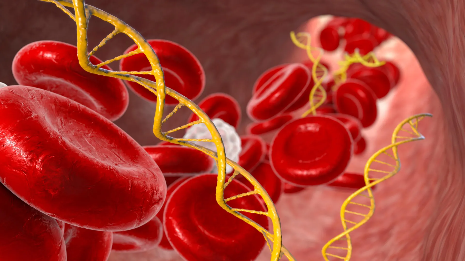
    Researchers from the Translational Genomics Research Institute (TGen), part of City of Hope, and the Lundquist Institute for Biomedical Innovation at Harbor-UCLA Medical Center have identified a potential biomarker for long COVID.

    If the findings of their study are confirmed by other research centers, the biomarker could be the first specific and quantifiable indicator for confirming long COVID. Currently, clinicians confer a diagnosis of long COVID based upon a collection of symptoms that patients develop after SARS-CoV-2 infection.

    “If a patient arrives in clinic and they relate the persistence of typical signs and symptoms of long COVID, 12 weeks or more after COVID -19 infection, I give them a presumptive diagnosis, but I don’t have any blood tests or biomarkers to confirm this diagnosis,” said William Stringer, M.D., a Lundquist Institute investigator and senior author on the study.

    The study results, reported in the journal Infection, detail the detection of SARS-CoV-2 protein fragments within extracellular vesicles (EVs) — tiny, naturally occurring packages that help cells share proteins, metabolites, and other materials. The researchers collected and analyzed blood samples from 14 patients over 12 weeks of aerobic exercise training (56 samples in all) in a clinical trial led by Stringer in long COVID.

    The researchers found 65 distinct protein fragments from SARS-CoV-2 inside the EVs. These fragments come from the virus’s Pp1ab protein, an RNA Replicase enzyme which is key to how the virus copies itself and makes other viral particles. This protein is found uniquely in SARS-CoV-2, and not in uninfected human cells, noted Asghar Abbasi, Ph.D., a Lundquist Institute investigator and first author of the study.

    Significantly, the researchers found that these viral peptides were demonstrated in each subject, but not each blood draw, in the EVs of Long COVID patients and were not detected in a separate control group of pre-pandemic EV samples.

    These findings add to growing evidence that suggests that SARS-CoV-2 may persist in certain body tissues long after the initial infection. Some groups hypothesize these lingering viral reservoirs could play a role in Long COVID. How the virus reaches tissues without its usual entry points — such as the brain — remains an open question, and may be related to EV particles.

    “We thought that maybe if the virus is circulating or moving in the body, we should try to see if EVs are carrying those viral fragments,” Abbasi explained.

    This idea became part of an ongoing clinical trial led by Drs. Abbasi and Stringer, which was already studying EVs to see if they are linked to changes in immune function related to exercise and post-exertional malaise, a common symptom in these patients.

    “While promising, the molecular signal of the viral peptides within the study samples was observed to be subtle and not consistently detected at every blood collection time point,” said Patrick Pirrotte, Ph.D., associate professor at TGen, director of the Integrated Mass Spectrometry Shared Resource at TGen and City of Hope, and co-senior author of the study. “There’s still a lot to unpack that we don’t know at this point.”

    For instance, he added, the researchers don’t know if the exercise itself drives the expression of viral programs intracellularly, and then those viral programs result in proteins that are going to be shed, or if there is a permanent reservoir in those cells, and it’s just a matter of detecting it at a certain time point. Although the identified peptides originated from one of the virus’ largest proteins, the researchers did not detect other comparably large proteins indicative of active viral replication. It’s possible that the peptides contained in the EVs are just molecular “trash” leftover after the formation of new viral proteins.

    “We haven’t run [our tests] on people without long COVID symptoms who are currently, or who were, infected with COVID,” said Stringer. “This raises the question: is this just continuing to take out the trash from the COVID infected cell or is this really ongoing replication someplace? I think that’s the mechanistic issue that needs to be resolved in future studies.”

    The Pulmonary Education and Research Foundation (PERF) and the UCLA David Geffen School of Medicine (DGSoM)-Ventura County Community Foundation (VCCF) funded this research.

    Share Button

    Cats develop dementia in a similar way to humans

    Scientists in Edinburgh believe the discovery could help their research into new treatments for Alzheimer’s.

    Share Button

    When is it too hot to walk your dog?

    How to make sure your pets are cool and comfortable during periods of hot weather.

    Share Button

    Scientists discover the pancake secret that makes vegan eggs irresistible

    As the demand for plant-based meat and dairy options grows, vegan burgers, nuggets, and beverages have been in the spotlight for sustainable protein alternatives. But unlike their meatless burger counterparts, plant-based eggs haven’t received the same attention from researchers — until now.

    Enter Da Eun Kim, a doctoral student at the University of Illinois Urbana-Champaign, and Brenna Ellison, a professor at Purdue University. Together, they set out to answer a deceptively simple question: What makes someone willing to purchase a vegan egg?

    Their new study is one of the first to examine this question. Using an experimental method known as a vignette design, researchers tested how factors such as price, product type, and setting influence people’s behavior.

    “We wanted to measure the consumer’s perception about plant-based eggs,” said Kim, who is a graduate student in the Department of Agricultural and Consumer Economics, part of the College of Agricultural, Consumer and Environmental Sciences at Illinois. “A vignette experiment allowed us to create hypothetical scenarios and isolate what consumers really think.”

    Instead of directly asking people if they would purchase a plant-based egg, the vignette method asked participants to imagine they were about to eat breakfast. This meal would take place either at home or in a restaurant, and consumers had a choice between scrambled eggs or pancakes made with plant-based eggs.

    The results were both surprising and informative.

    “We expected the setting to be more important, as we thought the novelty of the plant-based egg product would lead people to want to eat it in a restaurant where chefs know how to prepare the product in a way that tastes good,” Ellison said. “Surprisingly, the location of where you were eating, whether it be at a restaurant versus at home, didn’t have as much of an effect as we imagined.”

    Additionally, consumers were more likely to purchase plant-based eggs when they were mixed into something familiar, like pancakes, instead of served on their own.

    The team also asked participants to rate how plant-based eggs would compare to traditional ones. Unsurprisingly, expected taste and appearance still favor the classic egg. But the plant-based version came out on top for environmental impact and animal welfare. This reflects a broader trend in food science, where ethical motivations are beginning to influence consumer choices.

    Does this mean every consumer will want to purchase plant-based eggs? “Probably not,” Ellison said. “However, we did find that consumers who had previous experience trying plant-based eggs were more likely to purchase them compared to people without that experience. This suggests that consumers had a positive experience with the product previously.”

    While familiarity with plant-based eggs is helpful, familiarity with the end product, such as pancakes, may also be key. “Introducing them as an ingredient, especially in a product that consumers are comfortable with, is a way to get people over any ‘mental hurdles’ associated with trying plant-based eggs,” Ellison said.

    These results offer a clear message for the industry: give people an easy, tasty, and recognizable way to try something new.

    “There are still sensory barriers,” Kim said. “I’ve tried the liquid version that comes in a bottle, like egg whites. The taste was different, but I was surprised the texture was very similar to traditional eggs.”

    That firsthand experience echoes the study’s findings; while taste and texture still matter, many consumers just need a nudge to take the first bite. And the best way to do that may be through a subtle swap in a familiar dish.

    While plant-based eggs might not be ready to replace the classic scramble at every breakfast table, they are finding their niche. Science shows that people may not be ready to eat them plain — but mix them in, make it easy, and meet consumers where they are.

    Share Button

    Men being over-treated for prostate cancer, says charity

    More low-risk cancers could be monitored rather than treated, avoiding significant side effects, says a charity.

    Share Button

    Knee implant used by NHS known for years to be faulty

    Patients say they were left immobile or addicted to painkillers after receiving the NexGen implant.

    Share Button

    What scientists discovered about french fries and diabetes

    Eating three servings of French fries a week is associated with a 20% increased risk of developing type 2 diabetes, but eating similar amounts of potatoes cooked in other ways — boiled, baked or mashed — does not substantially increase the risk, finds a study published by The BMJ on August 6.

    What’s more, replacing any form of potatoes with whole grains was associated with a lower type 2 diabetes risk, but swapping them for white rice was linked to an increased risk, the results show.

    Potatoes contain several nutrients including fiber, vitamin C, and magnesium, but they also have a high starch content and therefore a high glycemic index, so have been linked to a higher risk of developing type 2 diabetes.

    But neither the preparation method for potatoes nor specific foods that potatoes would replace have been considered, both of which are key to evaluating the overall health impact of potatoes.

    To address this, researchers investigated the association between intake of potatoes prepared by different methods (boiled, baked, or mashed versus French fries) and risk of type 2 diabetes. They also looked at the impact on health of replacing potatoes with other major carbohydrates, such as whole grains and rice.

    Their findings are based on more than 205,000 health professionals from three large US studies carried out between 1984 and 2021. Participants were free of diabetes, heart disease or cancer and completed detailed food questionnaires every four years.

    During almost 40 years of follow up, 22,299 people were diagnosed with type 2 diabetes.

    After adjusting for lifestyle and dietary factors related to diabetes risk, the researchers found that for every three weekly servings of total potato, the rate of type 2 diabetes increased by 5% and for every three weekly servings of French fries, the rate increased by 20%. However, similar intake of baked, boiled, or mashed potatoes was not associated with a significantly increased risk.

    Replacing three weekly servings of total potato with whole grains lowered the type 2 diabetes rate by 8%. Substituting baked, boiled, or mashed potatoes with whole grains lowered the rate by 4%, and replacing French fries lowered the rate by 19%.

    In contrast, replacing total potatoes or baked, boiled, or mashed potatoes with white rice was associated with an increased risk of type 2 diabetes.

    This is an observational study so no firm conclusions can be drawn about cause and effect and the researchers can’t rule out the possibility that other unmeasured factors may have influenced their results. Most participants were also health professionals of European ancestry, so findings may not apply to other populations.

    Nevertheless, they conclude: “Our findings underscore that the association between potato intake and type 2 diabetes risk depends on the specific foods used as replacement. The findings also align with current dietary recommendations that promote the inclusion of whole grains as part of a healthy diet for the prevention of type 2 diabetes.”

    So, are potatoes back on the plate? Well, it depends, say researchers in a linked editorial, who note that it is important to consider preparation method and replacement food when guiding the public or informing policy.

    They point out that with their relatively low environmental impact and their health impact, baked, boiled, or mashed potatoes can be part of a healthy and sustainable diet, though whole grains should remain a priority, but say future studies from more diverse populations that account for both preparation methods and substitution analysis are needed.

    Share Button

    Hubble captures a wild star-birthing storm 160,000 light-years away

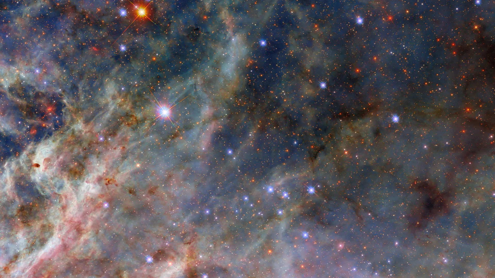
    A scene from a star-forming factory shines in this NASA/ESA Hubble Space Telescope Picture of the Week. This Hubble picture captures incredible details in the dusty clouds in a star-forming region called the Tarantula Nebula. What’s possibly the most amazing aspect of this detailed image is that this nebula isn’t even in our galaxy. Instead, it’s in the Large Magellanic Cloud, a dwarf galaxy that is located about 160,000 light-years away in the constellations Dorado and Mensa.

    The Large Magellanic Cloud is the largest of the dozens of small satellite galaxies that orbit the Milky Way. The Tarantula Nebula is the largest and brightest star-forming region not just in the Large Magellanic Cloud, but in the entire group of nearby galaxies to which the Milky Way belongs.

    The Tarantula Nebula is home to the most massive stars known, some of which are roughly 200 times as massive as our Sun. The scene pictured here is located away from the centre of the nebula, where there is a super star cluster called R136, but very close to a rare type of star called a Wolf-Rayet star. Wolf-Rayet stars are massive stars that have lost their outer shell of hydrogen and are extremely hot and luminous, powering dense and furious stellar winds.

    This nebula is a frequent target for Hubble, whose multiwavelength capabilities are critical for capturing sculptural details in the nebula’s dusty clouds. The data used to create this image come from an observing program called Scylla, named for a multi-headed sea monster from the Greek myth of Ulysses. The Scylla program was designed to complement another Hubble observing program called ULLYSSES (Ultraviolet Legacy Library of Young Stars as Essential Standards). ULLYSSES targets massive young stars in the Small and Large Magellanic Clouds, while Scylla investigates the structures of gas and dust that surround these stars.

    Share Button

    How NASA’s Lunar Trailblazer was lost before reaching the Moon

    The small satellite was to map lunar water, but operators lost contact with the spacecraft the day after launch and were unable to recover the mission.

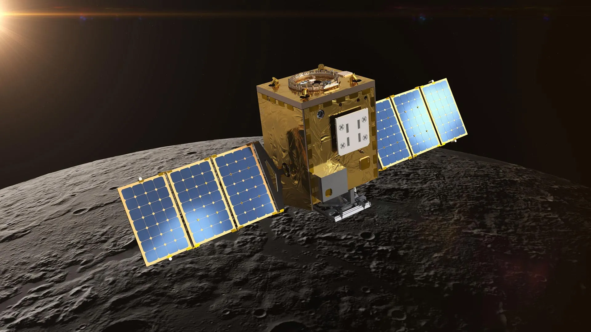
    NASA’s Lunar Trailblazer ended its mission to the Moon on July 31. Despite extensive efforts, mission operators were unable to establish two-way communications after losing contact with the spacecraft the day following its Feb. 26 launch.

    The mission aimed to produce high-resolution maps of water on the Moon’s surface and determine what form the water is in, how much is there, and how it changes over time. The maps would have supported future robotic and human exploration of the Moon as well as commercial interests while also contributing to the understanding of water cycles on airless bodies throughout the solar system.

    Lunar Trailblazer shared a ride on the second Intuitive Machines robotic lunar lander mission, IM-2, which lifted off at 7:16 p.m. EST on Feb. 26 aboard a SpaceX Falcon 9 rocket from the agency’s Kennedy Space Center in Florida. The small satellite separated as planned from the rocket about 48 minutes after launch to begin its flight to the Moon. Mission operators at Caltech’s IPAC in Pasadena established communications with the small spacecraft at 8:13 p.m. EST. Contact was lost the next day.

    Without two-way communications, the team was unable to fully diagnose the spacecraft or perform the thruster operations needed to keep Lunar Trailblazer on its flight path.

    “At NASA, we undertake high-risk, high-reward missions like Lunar Trailblazer to find revolutionary ways of doing new science,” said Nicky Fox, associate administrator, Science Mission Directorate at NASA Headquarters in Washington. “While it was not the outcome we had hoped for, mission experiences like Lunar Trailblazer help us to learn and reduce the risk for future, low-cost small satellites to do innovative science as we prepare for a sustained human presence on the Moon. Thank you to the Lunar Trailblazer team for their dedication in working on and learning from this mission through to the end.”

    The limited data the mission team had received from Lunar Trailblazer indicated that the spacecraft’s solar arrays were not properly oriented toward the Sun, which caused its batteries to become depleted.

    For several months, collaborating organizations around the world — many of which volunteered their assistance — listened for the spacecraft’s radio signal and tracked its position. Ground radar and optical observations indicated that Lunar Trailblazer was in a slow spin as it headed farther into deep space.

    “As Lunar Trailblazer drifted far beyond the Moon, our models showed that the solar panels might receive more sunlight, perhaps charging the spacecraft’s batteries to a point it could turn on its radio,” said Andrew Klesh, Lunar Trailblazer’s project systems engineer at NASA’s Jet Propulsion Laboratory in Southern California. “The global community’s support helped us better understand the spacecraft’s spin, pointing, and trajectory. In space exploration, collaboration is critical — this gave us the best chance to try to regain contact.”

    However, as time passed, Lunar Trailblazer became too distant to recover as its telecommunications signals would have been too weak for the mission to receive telemetry and to command.

    Technological Legacy

    The small satellite’s High-resolution Volatiles and Minerals Moon Mapper (HVM3) imaging spectrometer was built by JPL to detect and map the locations of water and minerals. The mission’s Lunar Thermal Mapper (LTM) instrument was built by the University of Oxford in the United Kingdom and funded by the UK Space Agency to gather temperature data and determine the composition of silicate rocks and soils to improve understanding of why water content varies over time.

    “We’re immensely disappointed that our spacecraft didn’t get to the Moon, but the two science instruments we developed, like the teams we brought together, are world class,” said Bethany Ehlmann, the mission’s principal investigator at Caltech. “This collective knowledge and the technology developed will cross-pollinate to other projects as the planetary science community continues work to better understand the Moon’s water.”

    Some of that technology will live on in the JPL-built Ultra Compact Imaging Spectrometer for the Moon (UCIS-Moon) instrument that NASA recently selected for a future orbital flight opportunity. The instrument, which has has an identical spectrometer design as HVM3, will provide the Moon’s highest spatial resolution data of surface lunar water and minerals.

    More About Lunar Trailblazer

    Lunar Trailblazer was selected by NASA’s SIMPLEx (Small Innovative Missions for Planetary Exploration) competition, which provides opportunities for low-cost science spacecraft to ride-share with selected primary missions. To maintain the lower overall cost, SIMPLEx missions have a higher risk posture and less-stringent requirements for oversight and management. This higher risk acceptance bolsters NASA’s portfolio of targeted science missions designed to test pioneering mission approaches.

    Caltech, which manages JPL for NASA, led Lunar Trailblazer’s science investigation, and Caltech’s IPAC led mission operations, which included planning, scheduling, and sequencing of all spacecraft activities. Along with managing Lunar Trailblazer, NASA JPL provided system engineering, mission assurance, the HVM3 instrument, and mission design and navigation. Lockheed Martin Space provided the spacecraft, integrated the flight system, and supported operations under contract with Caltech. The University of Oxford developed and provided the LTM instrument, funded by the UK Space Agency. Lunar Trailblazer, a project of NASA’s Lunar Discovery and Exploration Program, was managed by NASA’s Planetary Missions Program Office at Marshall Space Flight Center in Huntsville, Alabama, for the agency’s Science Mission Directorate in Washington.


    Share Button