The 28-year-old detained by German police is accused of being part of the cyber-crime gang that hacked blood-test firm Synnovis in 2024.


The 28-year-old detained by German police is accused of being part of the cyber-crime gang that hacked blood-test firm Synnovis in 2024.
Ministers promise to reform the scheme that provides financial support for those injured or bereaved by vaccines in the UK.
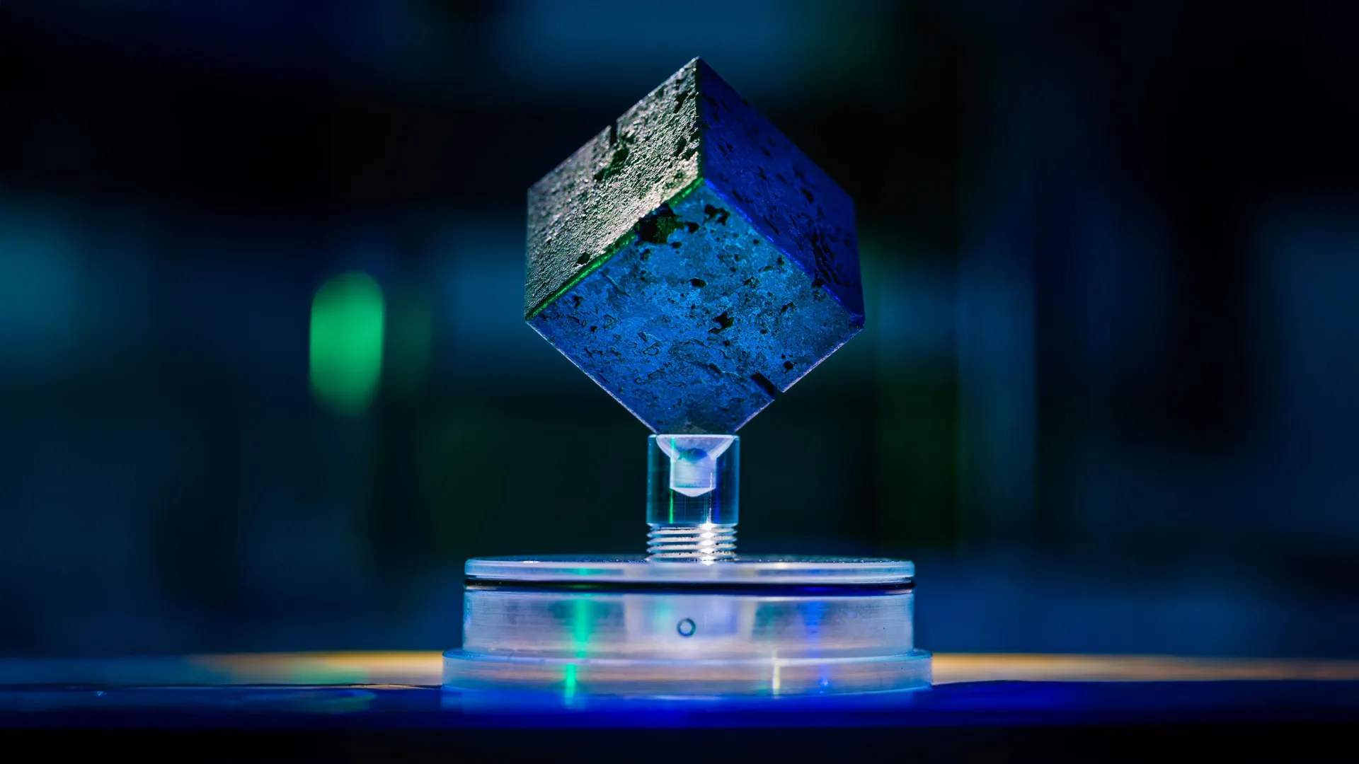
During World War II, the United States raced to develop the atomic bomb partly out of fear that Nazi Germany might build one first. But how close did Germany actually come to achieving that goal? A new scientific investigation suggests the Nazi nuclear program was much further from success than its leading physicist claimed.
By examining surviving uranium cubes and previously documented historical evidence, researchers have uncovered major limitations in Germany’s final experimental nuclear reactor. Their findings indicate that the project was not simply missing a few materials. The reactor would have required far more uranium and heavy water than Germany had available during the war.
The Race to Build an Atomic Bomb
The Manhattan Project, the secret American effort to develop nuclear weapons during World War II, was driven partly by the possibility that Nazi Germany might achieve a similar breakthrough first. Scientists knew that whoever succeeded could gain an unprecedented military advantage.
One possible route to an atomic bomb involved building a nuclear reactor capable of producing plutonium, a radioactive element that can be used in nuclear weapons. Inside a reactor, uranium atoms undergo nuclear fission, a process in which their nuclei split apart and release energy along with additional particles called neutrons. Under the right conditions, those neutrons trigger further fission reactions, creating a sustained chain reaction.
Although Nazi Germany pursued nuclear research through a program known as the Uranprojekt, historians have long debated how close its scientists came to developing a functioning reactor.
Germany’s final attempt was an experimental reactor known as the B8 pile, constructed in 1945 under the direction of physicist Werner Heisenberg. The device used cubes of natural uranium suspended in heavy water, a specialized substance that played an essential role in controlling the nuclear reaction.
Why Heavy Water Was Essential to Nazi Germany’s Reactor
Heavy water looks much like ordinary water, but its hydrogen atoms contain an additional neutron. This subtle difference makes it particularly useful in certain nuclear reactors.
In a reactor, heavy water serves as a neutron moderator, meaning it slows down the neutrons released during fission. Slower neutrons are more likely to trigger additional fission reactions in uranium, helping sustain the chain reaction needed for a working reactor.
For Germany, however, obtaining enough heavy water was a major obstacle.
During the war, the only European facility capable of supplying the heavy water needed by the German program was the Norsk Hydro plant in Vemork, Norway. Germany gained control of the facility after occupying Norway in April 1940.
Recognizing its importance to Germany’s nuclear ambitions, Allied forces targeted the plant through sabotage and bombing operations. A major American bombing raid struck Vemork on November 16, 1943, contributing to the eventual end of heavy water production there.
Scientists Reexamine Hitler’s Final Nuclear Reactor
When American forces discovered and dismantled the B8 reactor in 1945, it had never achieved criticality. In nuclear physics, criticality refers to the point at which a chain reaction becomes self-sustaining, with each generation of fission reactions producing enough neutrons to maintain the next.
Reaching this threshold is a fundamental requirement for operating a conventional nuclear reactor. Despite its name, criticality does not mean that a reactor is about to explode. It simply means the nuclear chain reaction can continue without relying on an external neutron source.
Heisenberg, who led the German reactor effort, maintained that the B8 experiment had come close to reaching this milestone. He argued that the device needed only a relatively modest increase in its uranium supply to become operational.
But a new study led by Timothy Koeth and his colleagues challenges that assessment.
The researchers examined two surviving cubes of natural uranium that had been used in the German nuclear project. They also studied historical records, technical reports, correspondence, and other archival documents to reconstruct the reactor’s capabilities and determine what would have been required for it to work.
Their findings paint a very different picture of Germany’s nuclear progress.
A Massive Uranium and Heavy Water Shortage
According to the researchers’ calculations, the B8 reactor would have needed approximately twice as much uranium and more than twice as much heavy water as it actually contained to achieve a sustained nuclear chain reaction.
The required quantities exceeded the materials available to Germany during the war. In particular, the heavy water needed for a functioning reactor was far beyond the amount the German program could obtain from its Norwegian supply.
The researchers also examined whether German scientists could have avoided the heavy water shortage by using graphite, a form of carbon that can also slow neutrons in nuclear reactors.
This approach had worked for American scientists, who achieved the world’s first controlled, sustained nuclear chain reaction using a graphite-moderated reactor in 1942.
However, the graphite available in German-controlled territory contained impurities that made it unsuitable for the same purpose. In particular, traces of boron could absorb neutrons that would otherwise help sustain the reaction, preventing the reactor from reaching criticality.
That meant Germany could not simply substitute graphite for heavy water to overcome its supply problems.
Why Nazi Germany’s Nuclear Program Was Doomed
The findings suggest that Heisenberg significantly underestimated how much additional material his reactor would have required.
Rather than standing on the verge of a functioning nuclear reactor, the German scientists faced shortages they could not overcome with the resources available to them.
The research also provides a clearer explanation for why Germany’s nuclear program failed to make the transition from experimental reactor designs to a working reactor capable of producing material for an atomic bomb.
Based on the designs and materials available during World War II, the researchers conclude that Nazi Germany lacked the resources necessary to make either its heavy water reactor or a graphite-based alternative operational.
More than eight decades after the war, the surviving uranium cubes have helped resolve a longstanding historical question, revealing just how far Nazi Germany remained from achieving a critical step toward building an atomic bomb.
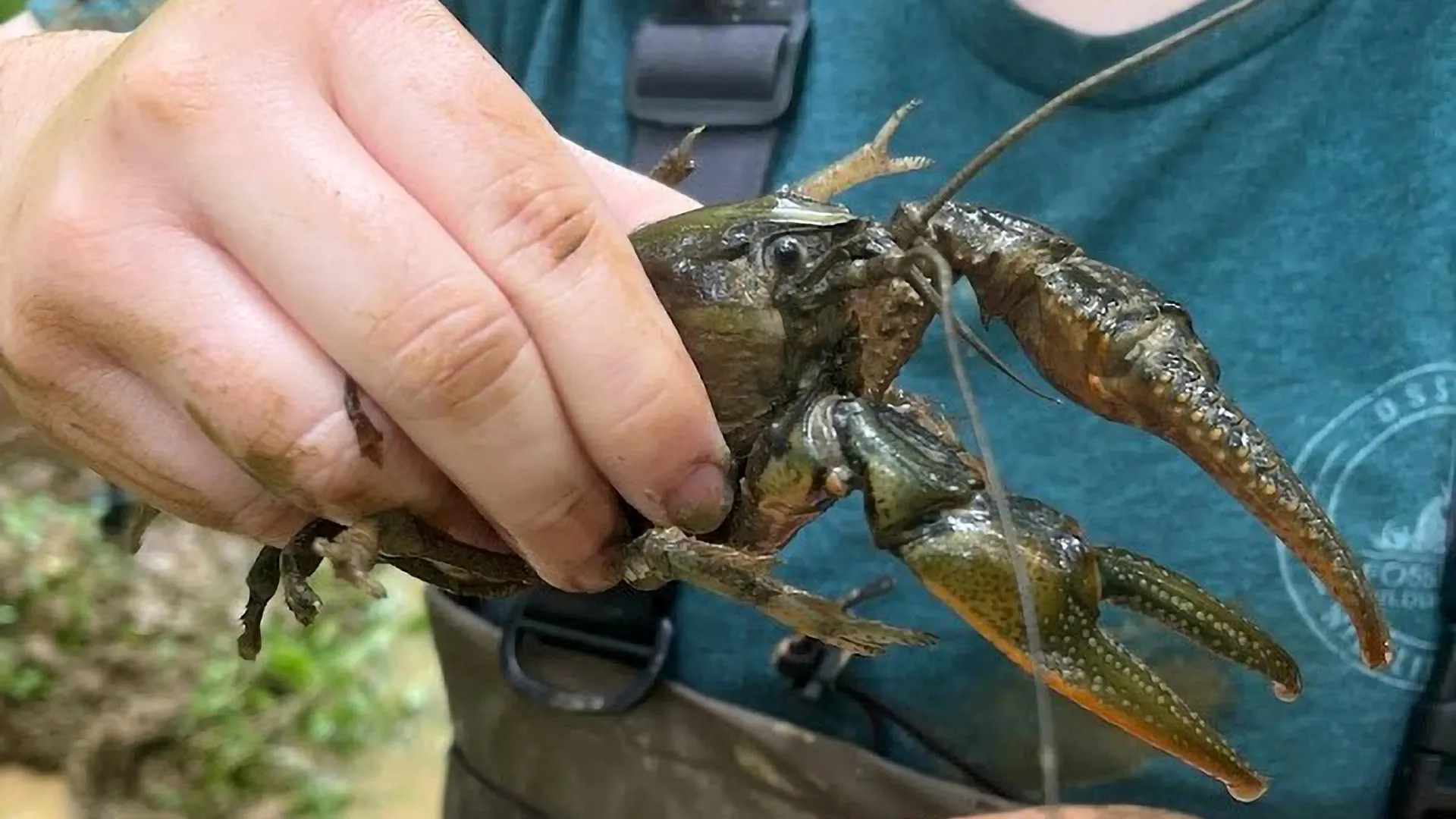
A giant crayfish nicknamed “Crawzilla” has sparked an unexpected scientific discovery, revealing a possible biological limit to how large North America’s crayfish can grow.
The investigation began when Caitlin Bloomer pulled an enormous Crawzilla crawdad from the mud during a biological survey in Kentucky. Crawzilla is the creature’s actual common name, and Bloomer was convinced she had captured the largest crayfish ever documented. But when she proudly showed her discovery to a colleague, the reaction was not quite what she expected.
“I thought, ‘There’s no doubt I have caught the biggest crayfish anyone’s ever seen!’ I brought it back to campus to show my colleague, naturally bragging about having caught the biggest-ever crayfish. But, as all biologists and sport fishermen do, he took one look and said, ‘No way, I’ve caught bigger,'” said Bloomer, a teaching assistant professor in the Department of Natural Resources and Environmental Sciences within the College of Agricultural, Consumer and Environmental Sciences at the University of Illinois Urbana-Champaign.
A Nationwide Hunt for North America’s Biggest Crayfish
What started as a little friendly rivalry soon turned into a nationwide search for record-breaking crayfish. Bloomer contacted biologists and museum curators across the country, asking them to examine their collections for unusually large specimens.
Using calipers, instruments designed to measure objects with great precision, the researchers compared the sizes of preserved crayfish. Their search produced a list of 20 exceptionally large individuals representing 18 different species. The results were published in the scientific journal Ecology.
Although Bloomer’s original Crawzilla did not claim the top spot, it still earned a place among the largest specimens identified.
“Mine was not the biggest, but it did make the top 20, so I’m super proud of it,” Bloomer said. “And it was also the only burrowing crayfish to make the list, so I’ll take the win.”
Scientists Identify the Largest Known North American Crayfish
The biggest specimen belonged to a Tennessee bottlebrush crayfish preserved in the collection at West Liberty University in West Virginia. It measured an impressive 86.6 millimeters, or approximately 3.4 inches, excluding its tail, claws, and antennae.
The record-holder was not a recent catch. It had been collected in 2011 by Dr. Zac Loughman and students from West Liberty University, making the discovery possible through the examination of existing museum specimens.
“I knew we had some massive crayfish in the West Liberty University Astacology Collection, but it was a surprise to learn we had the two largest specimens known from North America! Our record-holder, a Tennessee bottlebrush crayfish collected by Dr. Zac Loughman and West Liberty students back in 2011, shows just how amazing and diverse crayfish can be,” said Zackary Graham, an assistant professor of biology at West Liberty University. “Its carapace alone is about the length of a credit card, and with the tail and claws, it stretches nearly the width of a sheet of notebook paper. Both of these giants are now proudly displayed in my office, and I show them to just about everyone who walks in.”
Other institutions also contributed impressive specimens. The Illinois Natural History Survey, which belongs to the Prairie Research Institute at the University of Illinois, accounted for six of the 20 largest crayfish. The Smithsonian Institution’s National Museum of Natural History contributed another four.
Why Can’t North American Crayfish Grow Even Bigger?
Beyond settling the competition, the researchers uncovered a more intriguing scientific question. Their measurements provided some of the first estimates of the upper size limits for North American crayfish, and the results suggested that these animals may face a surprisingly consistent growth ceiling.
Despite differences among species and environments, the largest individuals tended to reach roughly 80 millimeters in the body measurement used by the researchers. That raised an obvious question: Why do North American crayfish appear to stop growing at around this size when some species elsewhere in the world, including the Tasmanian giant freshwater crayfish, can become dramatically larger?
“They all seemed to max out around that 80-millimeter mark, but we weren’t sure why, especially given that there are absolutely massive crayfish in other parts of the world,” Bloomer said. “We started looking at things like latitude, oxygen availability, and habitat type as possible correlates to explain why certain species and individuals were larger than others, but we weren’t seeing any patterns.”
The team examined whether geographic location, oxygen levels, or the type of environment where a crayfish lives could explain why some grow larger than others. However, none of these factors showed a clear relationship with maximum body size.
That unexpected result pointed the researchers toward a different possibility. Rather than being primarily controlled by environmental conditions, crayfish growth may be constrained by the animals’ own biology.
One potential explanation involves their exoskeletons, the hard outer coverings that protect their bodies. Differences in shell structure or muscle anatomy might impose physical limits on how large North American crayfish can become.
For now, that remains a hypothesis. Further research will be needed to determine whether these biological features actually restrict their growth.
“This was a case where we saw something unusual in the field and it sparked a future research direction,” Bloomer said. “We think that it could really be worthwhile to look at the physiology of crayfish to see if something there is influencing their body size.”
Giant Crayfish Reveal How Much Scientists Still Don’t Know
Crayfish are familiar inhabitants of streams, rivers, and other freshwater environments. Many people encounter them as children, making them among the most recognizable aquatic animals in North America.
Yet even with their widespread presence, scientists are still discovering basic facts about their biology, including how large they can grow and what determines their maximum size.
“Anyone that goes to a stream as a kid knows what crayfish are — they’re almost a universal onboarding for people to care about aquatic life. I still go out with people who are in their 50s or 60s, who recall catching crawfish in the creek when they were seven,” Bloomer said. “It’s just amazing how much we still have to learn about them.”
Bloomer also emphasized that the investigation would not have been possible without museum collections. By comparing specimens preserved over many years, researchers were able to answer a fundamental question that would have been difficult to investigate through fieldwork alone.
What began with one unusually large crayfish and a disagreement between colleagues has now opened a new line of scientific inquiry into the biological factors that shape the size of these familiar freshwater creatures.
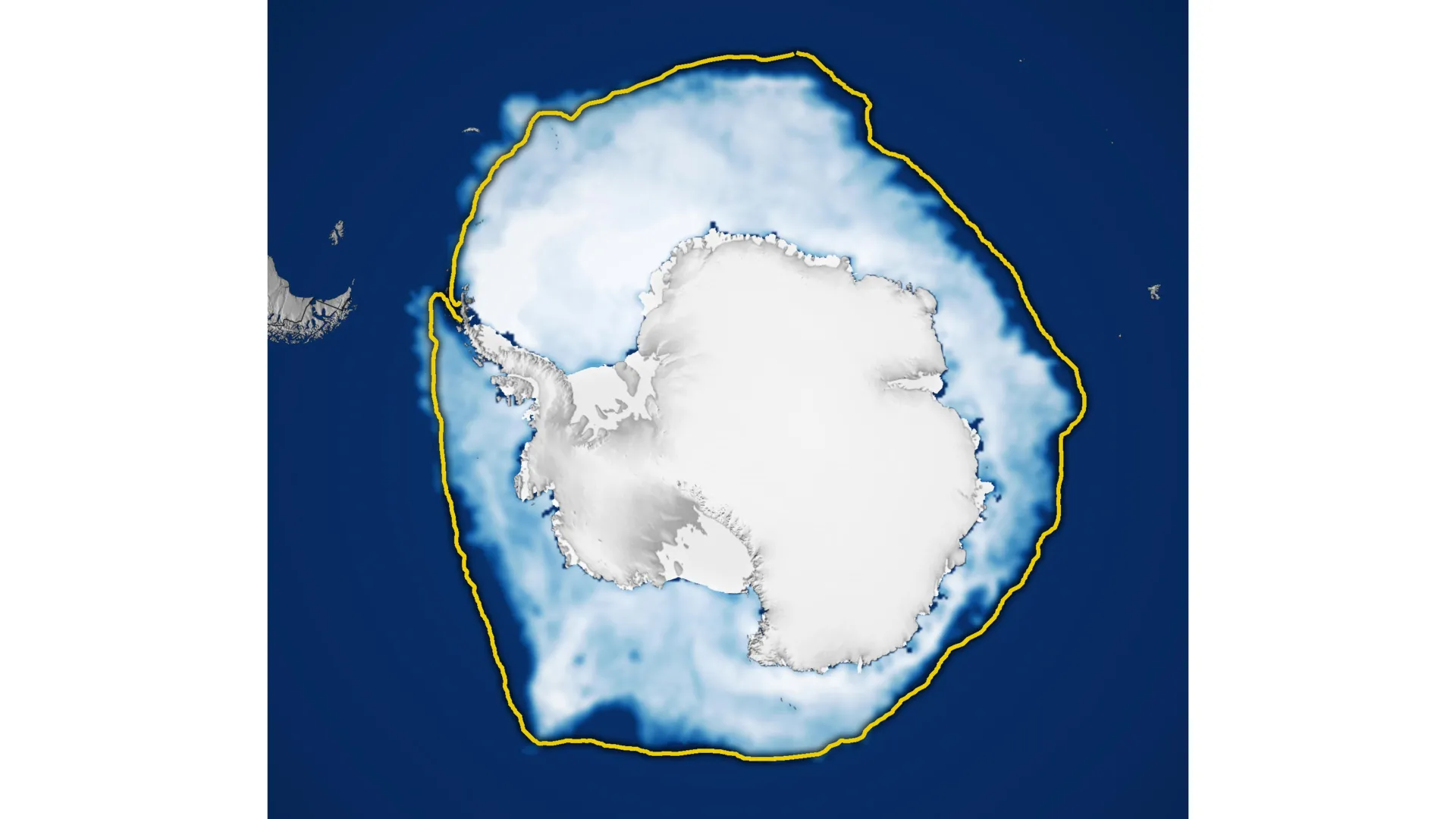
Antarctic sea ice reached its annual winter peak in September 2026, but the ice covered less ocean than in nearly any other year since satellite measurements began. Its maximum extent was the third lowest on record, adding to growing evidence of a dramatic shift in Antarctica’s frozen waters.
Antarctica is surrounded by a vast ocean, giving its sea ice plenty of room to spread outward during the Southern Hemisphere’s winter. This differs from the Arctic, where an ocean basin is enclosed by surrounding land. Although Antarctica’s geography allows winter sea ice to expand over enormous distances, it also pushes the ice into warmer latitudes, where melting becomes more likely during summer. As a result, Antarctic sea ice generally reaches its largest extent in September before shrinking to its annual minimum in February.
According to NASA and the National Snow and Ice Data Center (NSIDC), Antarctic sea ice likely reached its annual maximum extent on September 14 (above). At its peak, the ice stretched across approximately 17.59 million square kilometers (6.79 million square miles). That made 2026 the year with the third-lowest winter maximum in the satellite record.
The maps below illustrate how Antarctic sea ice changes between seasons, showing average ice concentrations in September (left) and the following February (right) from 1990 to 2026. Solid white regions represent areas with the greatest ice coverage, while dark blue indicates open ocean. Only locations where ice covers at least 15 percent of the water are included. Scientists use this threshold to determine which areas qualify as ice-covered when calculating the total “sea ice extent.”
Antarctic Sea Ice Shifts From Record Growth to Historic Lows
For decades, Antarctic sea ice appeared to be moving in the opposite direction from the rapidly shrinking ice in the Arctic. Between 1979 and 2014, sea ice extent around Antarctica increased by approximately 1 percent per decade. However, because Antarctic ice coverage naturally fluctuates considerably from year to year, researchers remain uncertain whether this gradual increase reflected a meaningful long-term change.
The expansion became particularly noticeable between 2012 and 2014, when Antarctic winter sea ice set new record highs for three consecutive years. These records stood in stark contrast to conditions at the opposite pole, where Arctic sea ice was undergoing a steep decline.
But Antarctica’s upward trend ended in 2015. Beginning in 2016, the region experienced substantial losses in sea ice coverage, marking a striking reversal from the preceding decades. Although some years have brought modest recoveries, Antarctic sea ice has generally remained below its historical average.
The most extreme conditions occurred in 2023, when Antarctica recorded both its lowest summer minimum and its lowest winter maximum since satellite observations began. The unusually low maximum in 2026 adds another significant measurement to this changing picture. Scientists are continuing to track the ice each year to determine whether these declines represent a lasting transformation in Antarctic sea ice conditions or fall within the range of statistical variability.
How Satellites Track Antarctic Sea Ice Through Clouds
Scientists measure Antarctic sea ice using satellites equipped with instruments called passive microwave sensors. Rather than relying on visible light, these instruments detect microwave energy naturally emitted by Earth’s surface.
Sea ice produces a stronger microwave signal than open ocean water, allowing researchers to distinguish frozen areas from ice-free seas. An important advantage of this approach is that microwaves can pass through clouds. This means scientists can repeatedly observe Antarctic sea ice even when thick cloud cover prevents conventional satellite cameras from seeing the surface.
The long-running satellite record began in late 1978 with observations from NASA’s Nimbus-7 spacecraft. Starting in 1987, satellites operated under the Defense Meteorological Satellite Program (DMSP) contributed additional measurements. NASA’s Aqua satellite also provided data between 2002 and 2011.
The monitoring effort continues to evolve as satellite technology changes. In 2025, researchers began incorporating observations from the Japan Aerospace Exploration Agency’s GCOM-W satellite, helping maintain the decades-long record used to understand Antarctica’s changing ice.
NASA Earth Observatory images by Michala Garrison, using data from the National Snow and Ice Data Center.
Metagenomics offers a new way of detecting infections in intensive care patients.
New guidance says trans patients should be cared for on single-sex hospital wards or bays that align with their biological sex.

Even when a person is fully unconscious under general anesthesia, their brain may still be doing something remarkable: processing language, recognizing patterns, and even anticipating which words will come next.
Researchers at Baylor College of Medicine have discovered that the brain retains surprisingly sophisticated abilities during anesthesia, despite the absence of conscious awareness. The findings, published in the journal Nature, challenge traditional assumptions about the relationship between consciousness and thinking. They could also provide new insights into how the brain processes memories and language, potentially leading to advances in technologies that help people communicate after neurological injuries.
“Our findings show that the brain is far more active and capable during unconsciousness than previously thought,” said Dr. Sameer Sheth, professor and Cullen Foundation Endowed chair of neurosurgery and a McNair Scholar at Baylor. “Even when patients are fully anesthetized, their brains continue to analyze the world around them.”
Recording Brain Activity During General Anesthesia
To investigate what happens inside the unconscious brain, Sheth, who is also a neurosurgeon at Baylor St. Luke’s Medical Center, and his colleagues studied patients undergoing epilepsy surgery while under general anesthesia.
These procedures offered an unusual opportunity to examine the hippocampus, a brain structure deeply involved in learning and memory. Located within the brain’s temporal lobe, the hippocampus helps organize information and plays an important role in forming new memories.
The researchers used advanced recording devices called Neuropixels probes to monitor electrical signals from hundreds of individual neurons in the hippocampus. These tiny probes can detect activity from many nerve cells simultaneously, providing a detailed picture of how groups of neurons respond to information.
According to the researchers, this technology had not previously been used to investigate this particular brain region in this way. It allowed the team to examine how the hippocampus responds to sounds and language while patients are unconscious and unable to report what they experience.
The first experiment involved playing a series of repeating tones, occasionally interrupted by a sound that differed from the others.
Although the patients were under anesthesia, neurons in their hippocampus responded differently to the unexpected sounds. Even more surprisingly, the brain cells became better at distinguishing these unusual tones as the experiment continued.
This improvement suggested that the hippocampus was showing signs of neural plasticity, the brain’s ability to change its responses through experience. In other words, some of the basic processes associated with learning appeared to remain active even without conscious awareness.
The Unconscious Brain Can Process Language and Predict Words
The researchers then tested whether the unconscious brain could handle something more complicated than simple sounds.
They played short stories to the anesthetized patients while continuing to record activity from neurons in the hippocampus.
The results revealed a surprising level of language processing. Distinct patterns of neural activity contained information about different categories of words, including nouns, verbs, and adjectives. This suggested that the hippocampus was responding to meaningful features of language rather than simply detecting that sounds were occurring.
But the most intriguing discovery went a step further.
The researchers found that neural activity also carried information that could be used to predict upcoming words in a sentence. Even though the patients were unconscious, their brains appeared to anticipate how the language unfolding around them might continue.
“The brain appears to anticipate what comes next in a story, even without conscious awareness,” said Sheth, who is also Director of The Gordon and Mary Cain Pediatric Neurology Research Foundation Laboratories within the Duncan Neurological Research Institute at Texas Children’s Hospital.
This ability is related to a process known as predictive coding. Rather than passively receiving information, the brain continually uses patterns and previous experience to anticipate what might happen next. When listening to someone speak, for example, the brain can use the words already heard to form expectations about what is likely to follow.
Scientists have typically studied this kind of sophisticated prediction in people who are awake and paying attention. Finding evidence of it during general anesthesia suggests that at least some of the underlying neural processes can operate independently of conscious awareness.
“This kind of predictive coding is something we associate with being awake and attentive, yet it’s happening here in an unconscious state,” said Dr. Benjamin Hayden, professor of neurosurgery at Baylor.
What Does This Discovery Mean for Consciousness?
The findings raise important questions about what consciousness actually contributes to the brain’s ability to process information.
Consciousness is generally associated with subjective awareness, including the experience of hearing, thinking, and understanding the world around us. Yet the study suggests that some neural processes involved in interpreting language and predicting future information can continue even when that awareness is absent.
This does not necessarily mean that the anesthetized patients consciously understood the stories being played to them. Instead, it indicates that the brain can carry out surprisingly complex aspects of language processing without a person being aware of doing so.
The distinction could help researchers better understand how consciousness emerges.
One possibility is that awareness depends not simply on whether individual brain regions remain active, but on how information is coordinated and shared across larger networks of brain regions.
Under this view, a structure such as the hippocampus might continue analyzing information even when the broader communication between brain regions needed for conscious experience is disrupted.
A Surprising Connection Between the Brain and Artificial Intelligence
The discovery also highlights an intriguing similarity between human brain activity and artificial intelligence (AI).
Large language models, the technology behind many modern AI chatbots, generate text by using patterns learned from enormous amounts of language data to predict what should come next.
The researchers observed a related predictive ability in the hippocampus, where neural signals reflected information about upcoming words even during unconsciousness.
Although biological brains and AI models operate in fundamentally different ways, both can use patterns in language to anticipate future information. Studying these similarities may help scientists better understand how prediction works in natural and artificial systems.
The findings could also have practical implications for brain-computer interfaces, which use signals recorded from the brain to communicate with external devices.
One potential application involves speech prosthetics, technologies designed to help people communicate when injuries or neurological conditions have impaired their ability to speak.
If scientists can reliably identify meaningful language signals in the brain, those signals might eventually be used to support communication technologies for patients who have lost speech abilities.
“Can we use these signals to deploy and run a speech prosthetic for some of the parts of the brain that are damaged by stroke or injury? These are questions that we can now consider in relation to this part of the brain,” said Dr. Vigi Katlowitz, first author and a neurosurgery resident with Baylor.
Scientists Are Rethinking the Limits of the Unconscious Brain
Despite the striking results, the researchers emphasize that several questions remain unanswered.
The experiments were conducted under one particular type of general anesthesia, so the findings cannot automatically be extended to other anesthetic methods or states of unconsciousness.
It is also unclear whether similar language processing occurs during sleep or coma, two conditions that differ significantly from medically induced unconsciousness.
Another important limitation is that the researchers focused on the hippocampus. Further studies will be needed to determine whether comparable abilities persist in other brain regions and how these areas interact when consciousness is absent.
Nevertheless, the results provide compelling evidence that unconsciousness does not necessarily mean the brain has stopped performing sophisticated information processing.
Instead, the brain may continue analyzing patterns, responding to unexpected events, and anticipating language even when a person has no conscious awareness of the surrounding world.
“This work pushes us to rethink what it means to be conscious,” said Sheth. “The brain is doing much more behind the scenes than we fully understand.”
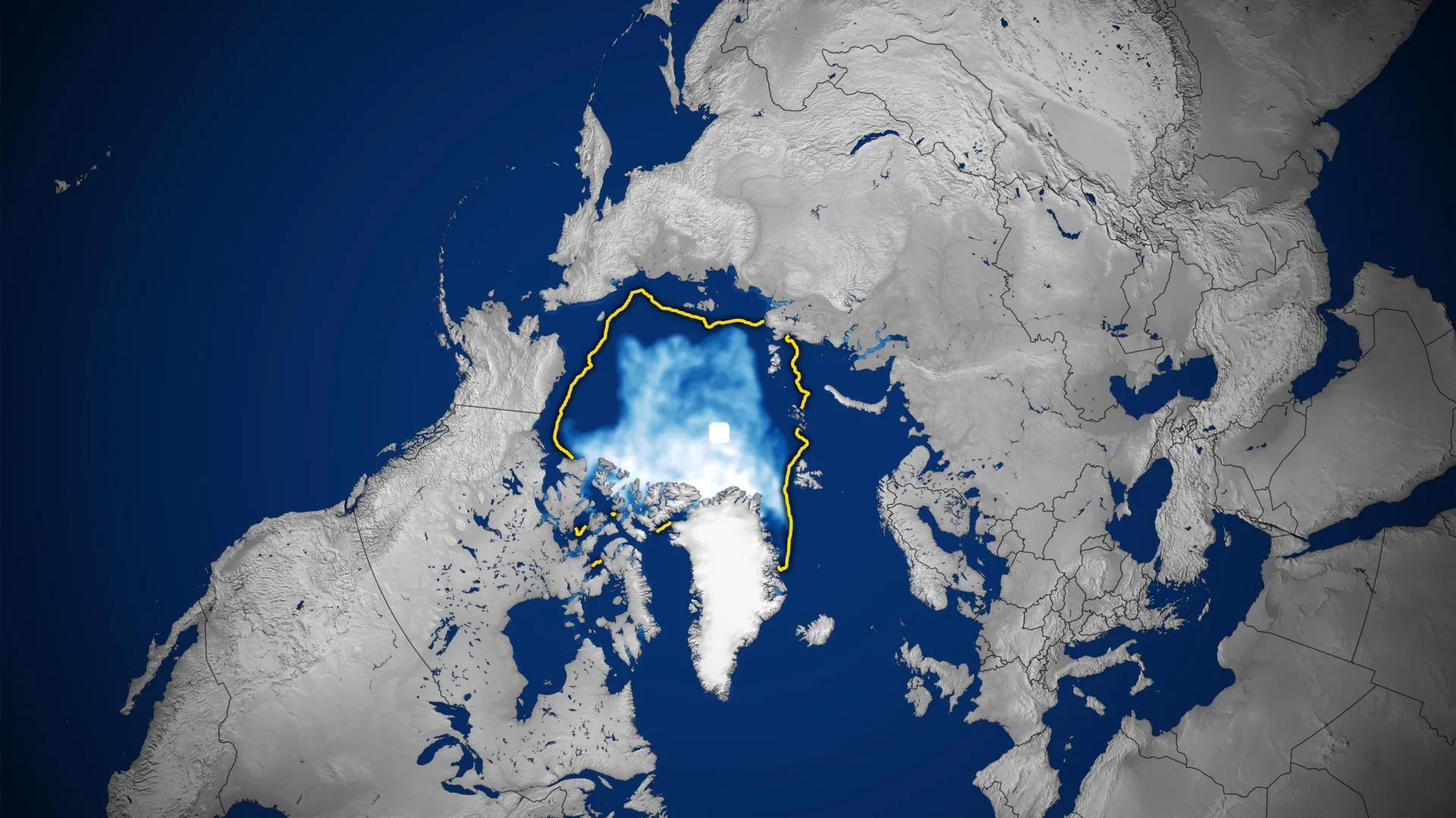
Nearly five decades of satellite observations reveal a troubling transformation in the Arctic, where sea ice is shrinking in both summer and winter, and much of the remaining ice is becoming younger and thinner.
The Arctic Ocean is covered by a vast expanse of frozen seawater that expands and contracts with the seasons. Ice typically builds up throughout the long, dark winter, reaching its greatest extent in March before retreating during the warmer months to a September minimum. This seasonal cycle has continued for thousands of years, but satellite measurements reveal that the amount of ice covering the ocean has declined substantially at both ends of the cycle.
In 2026, Arctic sea ice reached its annual minimum extent on September 12 (above), covering approximately 4.6 million square kilometers (1.78 million square miles). According to NASA and the National Snow and Ice Data Center (NSIDC), this tied the minimums recorded in 2008, 2010, and 2025 for the 10th-lowest level since satellite monitoring began.
The maps below illustrate how Arctic sea ice has changed over time, comparing September conditions since 1990 (left), when ice coverage is typically at its lowest, with the following March (right), when it approaches its annual peak. The sequence continues through March 2026.
Decades of Arctic Sea Ice Loss
Satellites have continuously tracked the seasonal expansion and retreat of Arctic sea ice since late 1978, providing scientists with nearly five decades of observations. Those measurements reveal a long-term decline that accelerated during the 2000s, leading to a succession of exceptionally low ice levels.
One particularly striking pattern is that every summer from 2007 through 2026 ranks among the 20 lowest September sea ice extents in the satellite record. The smallest extent ever observed occurred in September 2012.
However, the rate of summer ice loss has not been consistent. During the past decade, September sea ice coverage has remained comparatively stable rather than continuing its earlier rapid decline. Scientists emphasize that short-term weather conditions can strongly influence how much ice disappears in any given summer.
For instance, increased cloud cover over the Arctic during recent years has reduced the amount of sunlight reaching the ice, helping to limit additional melting. NASA researchers have identified this as one factor contributing to the recent slowdown. Even with that temporary stabilization, however, September ice coverage remains far below its historical average.
The Arctic Is Losing Its Oldest, Thickest Ice
The changes extend beyond the shrinking area covered by sea ice. Scientists have also documented a major shift in the ice itself, which is becoming increasingly young and thin.
Historically, much of the Arctic contained multiyear ice, which survives at least one summer melting season. Because this ice can persist and accumulate over multiple years, it generally becomes thicker and more resilient than ice that forms during a single winter.
Satellite observations now show that multiyear ice has declined significantly. As a result, the Arctic is increasingly covered by thinner first-year ice, which forms during the cold months but is more susceptible to melting when summer returns.
The decline is also evident during winter, when Arctic sea ice normally reaches its greatest coverage. In March 2026, the annual maximum statistically tied the March 2025 maximum for the lowest winter peak in the satellite record.
Could the Arctic Become Nearly Ice-Free?
Natural climate patterns influence Arctic sea ice from year to year. One example is the Arctic Oscillation, a pattern of atmospheric circulation that affects temperatures, winds, and ice conditions across the region.
However, natural fluctuations cannot fully account for the persistent decline recorded by satellites since 1978. Rising global temperatures, together with natural climate variability, have contributed to the growing loss of Arctic sea ice.
Looking ahead, scientists project that the Arctic Ocean could become essentially free of sea ice during the summer at least once before the middle of the 21st century. Such an event would mark a profound change in a region that has maintained extensive seasonal ice cover for millennia.
How Satellites Track Arctic Ice Through Clouds
Scientists measure Arctic sea ice coverage using satellites equipped with passive microwave sensors. These instruments detect microwave energy naturally emitted by Earth’s surface, allowing researchers to distinguish frozen seawater from the surrounding open ocean.
Sea ice emits more microwave energy than liquid water, producing a recognizable contrast in satellite observations. Unlike visible-light imaging, microwave measurements can penetrate cloud cover, making it possible to monitor the Arctic consistently even when storms or persistent clouds obscure the surface.
The continuous satellite record began in 1978 with NASA’s Nimbus-7 spacecraft. Additional observations came from the Defense Meteorological Satellite Program (DMSP), beginning in 1987, and NASA’s Aqua satellite, which provided measurements between 2002 and 2011.
In 2025, researchers began incorporating observations from the Japan Aerospace Exploration Agency’s GCOM-W satellite, continuing the long-running effort to measure changes in the planet’s polar ice.
The satellite maps also contain a white circular region at the North Pole known as the “pole hole.” This represents an area beyond the historical coverage of the satellite sensors, where direct measurements were unavailable. When calculating total sea ice extent, scientists assume this region is covered by ice.
NASA Earth Observatory images by Michala Garrison, using data from the National Snow and Ice Data Center.
Fewer than 800 Scots have received jabs for weight loss on the NHS despite them being approved for use three years ago, BBC News has found.
