How the body really ages: 7 million cells mapped across 21 organs

Growing older brings a higher risk of serious illnesses such as cancer, heart disease, and dementia. For years, researchers have tackled these conditions individually. Now, many scientists are stepping back to ask a broader question. Instead of treating diseases one by one, could slowing the aging process reduce the risk of several at once? To answer that, they first need to understand what sparks the biological changes that come with age.

A new study published in Science offers an unprecedented look at that process. Researchers at The Rockefeller University built the most detailed atlas so far of how aging affects thousands of cell subtypes across 21 mammalian tissues. By examining nearly 7 million individual cells from mice at three different ages, the team identified which cells are most vulnerable over time and what factors may be driving their decline.

“Our goal was to understand not just what changes with aging, but why,” says Junyue Cao, who heads the Laboratory of Single Cell Genomics and Population Dynamics. “By mapping both cellular and molecular changes, we can identify what drives aging. That opens the door to interventions that target the aging process itself.”

One of the most striking findings was that many age-related shifts happen in sync across multiple organs. The researchers also found that nearly half of these changes differ between males and females.

A Massive Cellular Census Across 21 Organs

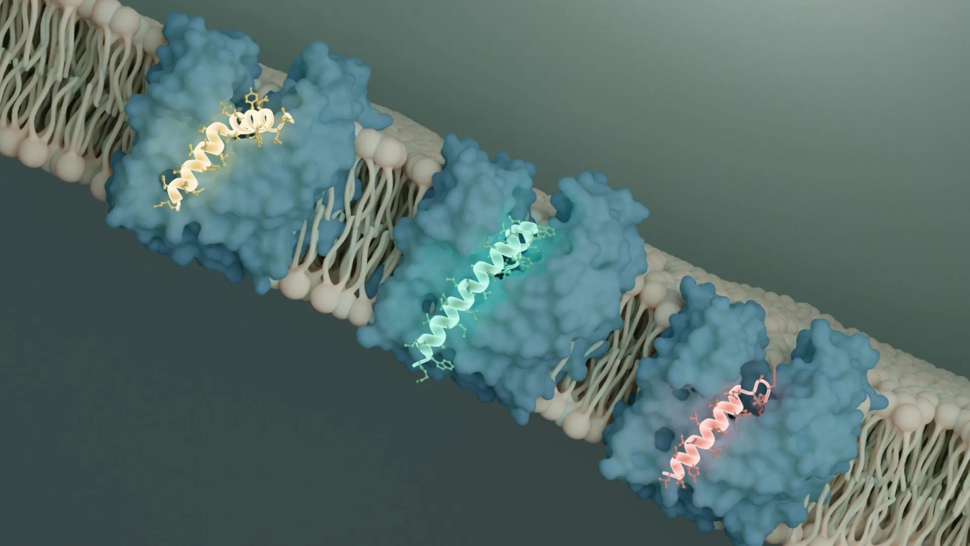
To map aging at this scale, Cao’s team, led by graduate student Ziyu Lu, refined a method known as single-cell ATAC-seq. This approach looks at how DNA is packaged inside each cell, revealing which regions of the genome are accessible and active, a key indicator of a cell’s state and function.

The researchers applied this technique to millions of individual cells taken from 21 organs in 32 mice at three ages: one month (young adult), five months (middle-aged), and 21 months (elderly).

“What’s remarkable is that this entire atlas was generated by a single graduate student,” Cao says. “Most large atlases like this require large consortia with dozens of laboratories but our method is far more efficient than other approaches.”

In total, the lab identified more than 1,800 distinct cell subtypes, including many rare groups that had never been fully described. The team then tracked how the numbers of these cells changed as the mice moved from young adulthood to middle age and then to old age.

Early and Coordinated Cellular Shifts

For decades, scientists believed aging mainly altered how cells function, not how many of each type exist. This new analysis challenges that view. About one quarter of all cell types showed significant changes in abundance over time. Certain muscle and kidney cell populations declined sharply, while immune cells expanded considerably.

“The system is far more dynamic than we realized,” says Cao. “And some of these changes begin surprisingly early. By five months of age, some cell populations had already begun to decline. This tells us that aging isn’t just something that happens late in life; it’s a continuation of ongoing developmental processes.”

Equally surprising was how synchronized these changes were. Similar cellular states rose and fell together across different organs. This pattern suggests that shared signals, possibly factors circulating in the bloodstream, help coordinate aging throughout the body.

The study also revealed pronounced differences between males and females. Roughly 40 percent of aging-associated changes varied significantly by sex. For example, females showed much broader immune activation as they aged.

“It’s possible this could explain the higher prevalence of autoimmune diseases in women,” Cao speculates.

Genetic Hotspots and Future Anti-Aging Therapies

Beyond counting how cell populations shifted, the researchers examined how accessible regions of DNA changed within those cells over time. Out of 1.3 million genomic regions analyzed, about 300,000 displayed significant aging-related alterations. Around 1,000 of those changes appeared across many different cell types, reinforcing the idea that common biological programs drive aging across the body. Many of these shared regions were linked to immune function, inflammation, or stem cell maintenance.

“This challenges the idea that aging is just random genomic decay,” Cao says. “Instead, we see specific regulatory hotspots that are particularly vulnerable, and these are precisely the regions we should be studying if we want to understand what drives the aging process.”

When the team compared their findings with earlier research, they discovered that immune signaling molecules called cytokines can trigger many of the same cellular changes observed during aging. Cao suggests that drugs designed to adjust these cytokines could potentially slow coordinated aging processes across multiple organs.

“This is really a starting point,” Cao says. “We’ve identified the vulnerable cell types and molecular hotspots. Now the question is whether we can develop interventions that target these specific aging processes. Our lab is already working on that next step.”

The full aging atlas is available to the public at epiage.net.

Share Button

A major climate hope in Antarctica just melted away

For years, researchers studying the Southern Ocean have pointed to one possible upside in an otherwise troubling climate outlook. A widely discussed idea known as iron fertilization proposed that as Antarctica warms and glaciers melt, iron trapped in the ice would be released into nearby waters. That iron would fuel blooms of microscopic algae, which absorb heat trapping carbon dioxide as they grow.

But new evidence suggests that expectation may not be accurate.

In what the team calls the most precise measurement so far of iron flowing from an Antarctic glacier, scientists from Rutgers University-New Brunswick found that meltwater from an ice shelf contributes far less iron to surrounding ocean waters than previously believed.

The study, published in Communications Earth and Environment, raises new questions about where iron in the Southern Ocean actually originates. According to the researchers, the results could influence how climate change forecasts and models are developed.

“It has been widely assumed that glacial melting underneath ice shelves contributes considerable bioavailable iron to these shelf waters, in a process of natural glacier-driven iron fertilization,” said Rob Sherrell, a professor in the Department of Marine and Coastal Sciences at the Rutgers School of Environmental and Biological Sciences and the study’s principal investigator.

Sherrell said the findings revise those assumptions. The amount of iron carried by meltwater is several times lower than earlier estimates. In addition, much of that iron appears to come from a different form of meltwater than the kind produced directly by melting ice shelves.

Why Iron in the Southern Ocean Matters

Even though Antarctic waters are dark for months at a time, the Southern Ocean supports abundant phytoplankton growth. These microscopic plants form the foundation of the food web, feeding krill that sustain penguins, seals, and whales. As phytoplankton grow, they remove large quantities of carbon dioxide from the atmosphere through photosynthesis, making this region the world’s largest oceanic sink for the climate warming gas.

Until now, much of what scientists understood about iron sources in these waters came from simulations and computer models. Sherrell and colleagues from Rutgers and partner institutions in the United States and the United Kingdom chose to gather direct field measurements instead.

In 2022, the researchers traveled aboard the now-decommissioned U.S. icebreaker, the Nathaniel B. Palmer, to the Dotson Ice Shelf in the Amundsen Sea of West Antarctica. The Amundsen Sea accounts for most of the sea level rise driven by Antarctic melting. Their goal was to collect glacial meltwater at its source.

Sampling Beneath the Ice Shelf

In the Amundsen Sea, meltwater forms under floating ice shelves, which extend from glaciers on land into the ocean. The melting is driven mainly by relatively warm water from the deep ocean that flows into cavities beneath the ice.

At the Dotson Ice Shelf, the team located where seawater flows into one of these cavities and where it exits after mixing with meltwater. Water samples were taken at both entry and exit points.

Back in New Jersey, Venkatesh Chinni, a postdoctoral scholar and lead author of the study, measured iron concentrations in the samples, analyzing both dissolved iron and iron attached to suspended particles. Collaborators Jessica Fitzsimmons and Janelle Steffen at Texas A&M University examined isotopic ratios to “fingerprint” the iron and trace its origin. Steffen performed the initial isotopic analyses in the laboratory of Tim Conway at the University of South Florida.

Using these measurements, Chinni and the team calculated how much additional iron was present in water leaving the cavity compared with water entering it. The isotopic signatures also helped identify which melting processes were responsible.

Deep Water and Sediments Supply Most Iron

The results were unexpected, Sherrell said. Meltwater accounted for only about 10% of the dissolved iron flowing out of the cavity. Most of the iron came from deep ocean water (62%), while another 28% originated from sediments on the continental shelf.

“Roughly 90% of the dissolved iron coming out of the ice shelf cavity comes from deep waters and sediments outside the cavity, not from meltwater,” Chinni said.

The isotope data also point to processes occurring beneath the glacier itself. The samples suggest the presence of a liquid meltwater layer that lacks dissolved oxygen. Under such conditions, solid iron oxides in bedrock can dissolve more readily, releasing iron into the water. According to Chinni, this mechanism may contribute more iron than melting ice shelves do.

Rethinking Antarctic Iron and Climate Models

Together, these findings challenge long standing assumptions about iron sources in the Southern Ocean as the planet warms. The researchers emphasize that more work is needed to fully understand how subglacial processes influence iron release.

“Our claim in this paper is that the meltwater itself carries very little iron, and that most of the iron that it does carry comes from the grinding up and dissolving of bedrock into the liquid layer between the bedrock and the ice sheet, not from the ice that is driving sea level rise,” Sherrell said.

He added that many scientists may find this conclusion surprising.

Share Button

Scientists discover a bacterial kill switch and it could change the fight against superbugs

Scientists have revealed how viruses that infect bacteria shut down MurJ, a protein essential for building the bacterial cell wall. Remarkably, different viruses evolved separate proteins that all block MurJ in the same way, highlighting it as a promising new antibiotic target.

The findings appear in the February 26 issue of Nature. The research was led by Yancheng Evelyn Li, a graduate student in the lab of Bil Clemons at Caltech. Clemons, the Arthur and Marian Hanisch Memorial Professor of Biochemistry, is the corresponding author.

The Urgent Need for New Antibiotics

Bacteria evolve quickly, and that adaptability is fueling a growing public health crisis. As Clemons explains, “Evolution is powerful, and in bacteria, resistance to antibiotics develops quickly. This means that we now deal with bacteria that are resistant to all the medicines that we have.” He adds, “In the USA alone, tens of thousands of people die every year from antibiotic-resistant bacterial infections, and that number is rising rapidly. We need new antibiotics to combat this.”

With existing drugs losing effectiveness, researchers are searching for entirely new bacterial weak points.

Targeting the Bacterial Cell Wall

One long standing focus has been the pathway bacteria use to construct peptidoglycan, the rigid material that forms their cell wall. This process, called the peptidoglycan biosynthesis pathway, is especially attractive because peptidoglycan is found in bacteria but not in human cells. As Clemons notes, “Peptidoglycan is a unique feature of bacteria, and that makes it an attractive antibiotic target.”

Several antibiotics already disrupt this pathway. Penicillin, discovered by Alexander Fleming in the mid 20th century, blocks a late stage of peptidoglycan production. Related drugs such as amoxicillin work in a similar way.

Key Proteins MraY, MurG, and MurJ

Three essential proteins drive the movement of peptidoglycan building blocks across the bacterial inner membrane: MraY, MurG, and MurJ. These proteins help transport the components needed to assemble the cell wall outside the inner membrane barrier. If any one of them fails, peptidoglycan cannot be produced and the bacterium dies, making them promising drug targets.

Although researchers understand much about how these proteins function, Clemons points out that important mechanistic details remain unclear.

At present, no approved drugs directly inhibit these three proteins. Still, Clemons says there is potential. “We do know that we can find small molecules, either derived from nature or synthesized in chemical libraries, that will inhibit these proteins. Excitingly, recent discoveries have shown that bacteriophages have figured out how to target this pathway.”

How Bacteriophages Break Through Bacterial Defenses

Bacteriophages, or phages, are viruses that infect bacteria. To survive, they must enter a bacterial cell, replicate, and then escape to infect others. Breaking out requires getting through the peptidoglycan layer. Clemons explains, “Getting back out means that they have to get past the peptidoglycan layer. Because it acts like chainmail, the phages get stuck if they can’t break through it.”

The Clemons lab studies small phages that contain single stranded DNA or RNA. These viruses have compact genomes and rely on simple strategies to kill bacteria. In 2023, the team reported in Science on φX174, a phage with a long research history at Caltech.

Viral Proteins That Disable MurJ

Small phages rely on specialized protein antibiotics called single-gene lysis proteins, or Sgls (pronounced like “sigils”), to kill bacteria. Li and Clemons have focused on Sgls that target MurJ, one of the key cell wall proteins.

MurJ acts as a flippase. It transports peptidoglycan precursors from the inside of the cell across the membrane so they can be incorporated into the growing cell wall. Earlier work from collaborators showed that two unrelated Sgls, SglM and SglPP7, both kill bacteria by blocking MurJ.

To understand how this happens, Li used cryo electron microscopy at Caltech’s Beckman Institute Biological and Cryogenic Transmission Electron Microscopy (Cryo-EM) Resource Center. Flippases such as MurJ move molecules by alternately exposing them to each side of the membrane without forming a permanent opening. When MurJ binds its cargo inside the cell, it changes shape to release the molecule on the outside.

Li found that both SglM and SglPP7 attach to a groove in MurJ, preventing the structural shift required for transport.

“It is clear that both of these Sgls bind to MurJ in an outward-facing conformation, locking it into this position,” Li says. Researchers are encouraged by this because the outward-facing form of MurJ is exposed to the surrounding environment, which could make it more accessible to future drugs than a conformation that faces inward.

Convergent Evolution Highlights a Promising Drug Target

Clemons says the result was surprising for another reason. “These peptides, which have no evolutionary links to each other, have both figured out how to target MurJ in a very similar way. These are two examples of convergent evolution, in which different evolutionary paths arrive at the same solution. We were surprised!”

Because viruses evolve rapidly, the team believes many more phages likely carry similar Sgls. Phages are relatively easy to isolate, and studying their genomes could reveal additional biological insights and new antibiotic targets.

In the Nature study, the researchers analyzed another phage genome with the help of a collaborator. They identified a new Sgl called SglCJ3 (from a genome sequence that is predicted to be a phage and is called Changjiang3) and examined it using cryo electron microscopy. Li determined the structure of SglCJ3 bound to MurJ and found that it also locks the protein in the same outward-facing conformation.

“This is a third genome that evolved a distinct peptide to inhibit the same target in a similar way,” Clemons says. “It is the first strong evidence that evolution identifies MurJ as a great target for killing bacteria, which means we should follow evolution’s lead and develop therapeutics that target MurJ. This demonstrates the power of basic biology to help us solve problems in medicine. Our path is set on leveraging Sgl discovery, and we hope to continue to be supported to turn these concepts into realities.”

Authors and Funding

The paper is titled “Convergent MurJ flippase inhibition by phage lysis proteins.” In addition to Clemons and Li, the authors include Caltech graduate student Grace F. Baron and Francesca S. Antillon, Karthik Chamakura, and Ry Young of Texas A&M University. The research was supported by the Chan Zuckerberg Initiative, the National Institutes of Health, the G. Harold and Leila Y. Mathers Foundation, and the Center for Phage Technology at Texas A&M, jointly sponsored by Texas A&M AgriLife.

Share Button

I need 120 pills a week to deal with the agony caused by vaginal mesh

Kerry Watson is one of 25 women to get compensation after operations done by a single surgeon.

Share Button

I take 120 tablets a week after mesh operation

Twenty-five women have received compensation following gynaecological surgery.

Share Button

James Webb reveals a barred spiral galaxy shockingly early in the Universe

A research team led by Daniel Ivanov, a physics and astronomy graduate student in the Kenneth P. Dietrich School of Arts and Sciences at Pitt, has identified a strong candidate for one of the earliest known spiral galaxies with a stellar bar. These bright, elongated structures can strongly influence how galaxies grow and change over time. The Milky Way itself contains a stellar bar at its center.

The newly studied galaxy, named COSMOS-74706, appears to have existed about 11.5 billion years ago. By analyzing its light, researchers were able to determine its place in cosmic history and narrow down when barred structures may have first formed in the universe.

“This galaxy was developing bars 2 billion years after the birth of the universe,” Ivanov said. “Two billion years after the Big Bang.”

The results were presented at the 247th meeting of the American Astronomical Society.

What Is a Stellar Bar?

As the name suggests, a stellar bar is a straight, elongated feature that stretches across the central region of a spiral galaxy. “A stellar bar is a linear feature at the center of the galaxy,” Ivanov said. Rather than being a single object, the bar is made up of tightly packed stars and gas. When viewed from above or below the galaxy’s disk, this alignment creates the appearance of a bright line cutting through the middle.

These bars are more than just visually striking. They can shape a galaxy’s long term development by channeling gas from the outer regions inward. This inward flow can fuel the supermassive black hole at the galaxy’s core and reduce star formation across the surrounding disk.

Why This Discovery Stands Out

Other teams have previously reported possible barred spiral galaxies from even earlier periods. However, those findings relied on less precise measurements of redshift. In contrast, COSMOS-74706 was confirmed using spectroscopy, which provides more reliable distance data. In some earlier cases, the galaxy’s light was also distorted by passing near a massive object, an effect known as gravitational lensing.

In essence, Ivanov said, “It’s the highest redshift, spectroscopically confirmed, unlensed barred spiral galaxy.”

Although the galaxy dates back to a very early era, Ivanov was not entirely surprised. Computer simulations have suggested that stellar bars could begin forming at redshift 5, or roughly 12.5 billion years ago. Still, he noted that such objects are not expected to be common at that stage of cosmic history.

“In principle, I think that this is not an epoch in which you expect to find many of these objects. It helps to constrain the timescales of bar formation. And it’s just really interesting.”

Powered by the James Webb Space Telescope

The research relied in part on observations from the NASA/ESA/CSA James Webb Space Telescope. Data were obtained through the Space Telescope Science Institute, which is operated by the Association of Universities for Research in Astronomy, Inc., under NASA contract NAS 5-03127, which is supported by NASA. The project also received support from the Brinson Foundation.

Share Button

MIT study finds Earth’s first animals were likely ancient sea sponges

Researchers at MIT have uncovered new chemical clues in extremely old rocks that suggest some of the earliest animals on Earth were likely ancestors of modern sea sponges.

Reporting in the Proceedings of the National Academy of Sciences, the team describes the discovery of “chemical fossils” preserved in rocks more than 541 million years old. These chemical fossils are traces of biological molecules once produced by living organisms that were later buried, altered, and locked into sediment for hundreds of millions of years.

The newly identified molecules belong to a group called steranes, which are stable remnants of sterols such as cholesterol that form part of the cell membranes of complex life. By analyzing their structure, the scientists linked these steranes to demosponges, a major group of sea sponges. Today, demosponges appear in many shapes, sizes, and colors and live throughout the world’s oceans as soft filter feeders. Their ancient relatives were likely similar in being soft bodied marine organisms.

“We don’t know exactly what these organisms would have looked like back then, but they absolutely would have lived in the ocean, they would have been soft-bodied, and we presume they didn’t have a silica skeleton,” says Roger Summons, the Schlumberger Professor of Geobiology Emeritus in MIT’s Department of Earth, Atmospheric and Planetary Sciences (EAPS).

The presence of these sponge specific chemical signatures strengthens the case that ancestors of demosponges were among the first animals to evolve, emerging well before most other major animal groups.

The research team includes lead author Lubna Shawar, a former MIT EAPS Crosby Postdoctoral Fellow who is now a research scientist at Caltech, along with Summons and colleagues Gordon Love of the University of California at Riverside, Benjamin Uveges of Cornell University, Alex Zumberge of GeoMark Research in Houston, Paco Cárdenas of Uppsala University in Sweden, and José Luis Giner of the State University of New York College of Environmental Science and Forestry.

Revisiting a 2009 Discovery in Precambrian Rocks

This work builds on a study the group first published in 2009. At that time, they analyzed rocks from an outcrop in Oman and detected an unusually high concentration of steranes derived from 30-carbon (C30) sterols. These rare steroid molecules appeared to originate from ancient sea sponges.

The rocks dated to the Ediacaran Period, which lasted from about 635 million to 541 million years ago, just before the Cambrian Period when complex multicellular life rapidly diversified. The earlier findings suggested that sponges existed long before the Cambrian explosion and may have been among the planet’s earliest animals.

Not everyone agreed. Some researchers proposed that the C30 steranes might have been produced by other organisms or even formed through nonbiological geological processes.

The new study adds weight to the sponge hypothesis. The team identified another distinctive chemical fossil in the same Precambrian rocks that is highly likely to have come from living organisms rather than from chemistry alone.

Rare Sterols and the Search for Early Animal Life

As in their previous investigations, the researchers examined Ediacaran age rocks collected from drill cores and outcrops in Oman, western India, and Siberia. They searched for steranes, which are stable versions of sterols found in all eukaryotes (plants, animals, and any organism with a nucleus and membrane-bound organelles).

“You’re not a eukaryote if you don’t have sterols or comparable membrane lipids,” Summons says.

Sterols share a core structure made of four connected carbon rings. Different organisms modify that structure by adding carbon side chains and other chemical groups, depending on the genes they carry. In humans, cholesterol contains 27 carbon atoms, while plant sterols typically contain 29.

“It’s very unusual to find a sterol with 30 carbons,” Shawar says.

The earlier research had identified a 30-carbon sterol tied to a specific enzyme encoded by a gene common in demosponges. In the new analysis, the team realized that the same gene could also produce an even rarer 31-carbon sterol (C31). When they reexamined their rock samples, they detected abundant C31 steranes alongside the previously identified C30 forms.

“These special steranes were there all along,” Shawar says. “It took asking the right questions to seek them out and to really understand their meaning and from where they come.”

Laboratory Tests Confirm Biological Origin

To confirm the source, the scientists studied living demosponges and found that some species produce C31 sterols, the biological precursors of the C31 steranes preserved in rock. They then synthesized eight different C31 sterols in the laboratory to serve as reference compounds. After subjecting these molecules to conditions that mimic burial and geological transformation over millions of years, they compared the results with the ancient samples.

Only two of the eight synthesized sterols transformed into compounds that matched the C31 steranes found in the rocks. The absence of the other six products indicates that the molecules were not created by random chemical reactions in the environment.

Together, evidence from rock chemistry, modern sponges, and laboratory experiments supports the conclusion that the steranes originated from living organisms. Those organisms were most likely early ancestors of demosponges, which still retain the ability to produce similar compounds today.

“It’s a combination of what’s in the rock, what’s in the sponge, and what you can make in a chemistry laboratory,” Summons says. “You’ve got three supportive, mutually agreeing lines of evidence, pointing to these sponges being among the earliest animals on Earth.”

“In this study we show how to authenticate a biomarker, verifying that a signal truly comes from life rather than contamination or non-biological chemistry,” Shawar adds.

Expanding the Search for the First Animals

Now that C30 and C31 sterols appear to be reliable indicators of ancient sponges, the researchers plan to examine rocks from other parts of the world. So far, the samples indicate that these sponges lived during the Ediacaran Period. With additional material, the team hopes to pinpoint more precisely when some of the earliest animals first emerged.

This research was supported, in part, by the MIT Crosby Fund, the Distinguished Postdoctoral Fellowship program, the Simons Foundation Collaboration on the Origins of Life, and the NASA Exobiology Program.

Share Button

A lost moon may have created Titan and Saturn’s rings

New research suggests that Saturn’s brilliant rings and its largest moon, Titan, may share a violent past shaped by collisions between moons. Although NASA’s Cassini spacecraft transformed our understanding of Saturn during its 13 year mission, it also uncovered new puzzles, including the surprisingly young age of Saturn’s rings and Titan’s shifting orbit. A new study led by SETI Institute scientist Matija Ćuk proposes that these mysteries are connected and that Titan itself may have formed when two earlier moons merged.

Toward the end of its mission, Cassini measured how mass is distributed inside Saturn. That internal structure controls the planet’s slow wobble in space, known as precession. For many years, researchers believed Saturn’s precession matched Neptune’s, allowing their gravitational interactions to gradually tilt Saturn and make its rings more visible from Earth.

However, Cassini’s final measurements revealed that Saturn’s mass is more concentrated toward its center than scientists had expected. This subtle difference changes Saturn’s precession rate so that it no longer aligns with Neptune’s. To account for the mismatch, researchers at MIT and UC Berkeley proposed that Saturn once had an additional moon. According to their idea, that moon was flung away after a close encounter with Titan and later broke apart, creating the rings.

Hyperion’s Orbit Offers a Clue

The SETI Institute team tested whether such an extra moon could have moved close enough to Saturn to form the rings. Computer simulations showed that the most likely outcome was not ring formation directly, but a collision between the extra moon and Titan.

An important clue comes from Hyperion, Saturn’s small, irregularly shaped moon that tumbles chaotically in space. Hyperion’s orbit is locked with Titan’s.

“Hyperion, the smallest among Saturn’s major moons provided us the most important clue about the history of the system,” said Ćuk. “In simulations where the extra moon became unstable, Hyperion was often lost and survived only in rare cases. We recognized that the Titan-Hyperion lock is relatively young, only a few hundred million years old. This dates to about the same period when the extra moon disappeared. Perhaps Hyperion did not survive this upheaval but resulted from it. If the extra moon merged with Titan, it would likely produce fragments near Titan’s orbit. That is exactly where Hyperion would have formed.”

In other words, Hyperion may not have simply survived past chaos. It may have formed from debris created when Titan merged with another moon.

A Collision Between Proto Moons

The new model proposes that Titan formed when two earlier moons combined. One was a large body called “Proto-Titan,” nearly as massive as Titan today. The other was a smaller companion referred to as “Proto-Hyperion.”

Such a merger could explain why Titan has relatively few impact craters. A massive collision would have resurfaced the moon, erasing much of its earlier crater record. Titan’s current orbit, which is slightly elongated but gradually becoming more circular, also hints at a relatively recent disturbance consistent with a past merger.

Before the collision, Proto-Titan may have resembled Jupiter’s moon Callisto, heavily cratered and lacking an atmosphere. The team also found that before it disappeared, Proto-Hyperion could have tilted the orbit of Saturn’s distant moon Iapetus, potentially solving another longstanding mystery about the Saturn system.

How Titan’s Merger May Have Created Saturn’s Rings

If Titan formed from a moon merger, the question remains: where did Saturn’s rings come from?

More than a decade ago, members of the SETI Institute team suggested that the rings formed from debris created when medium sized moons closer to Saturn collided. Later simulations by researchers at the University of Edinburgh and NASA Ames Research Center supported this idea. Those studies showed that most of the debris from such impacts would eventually clump back together into moons, but some material would be scattered inward and remain as rings.

Previously, scientists believed the Sun may have triggered the instability that caused those inner moon collisions. The new research suggests a different chain of events. Titan’s merger may have set off the process.

Titan’s slightly elongated orbit can disturb inner moons when their orbital periods become simple fractions of Titan’s. This configuration, known as orbital resonance, strengthens gravitational interactions. Although such alignments are unlikely at any given moment, Titan’s outward migration sometimes creates these resonances.

When that happens, smaller moons can be pushed into more stretched out orbits, increasing the chances that they collide with neighboring moons. The timing of this second round of destruction is uncertain, but it must have occurred after Titan’s merger. That sequence fits with estimates that Saturn’s rings are about 100 million years old.

Dragonfly Mission Could Test the Theory

NASA’s Dragonfly mission, scheduled to arrive at Titan in 2034, could provide crucial evidence. The nuclear powered octocopter will study Titan’s surface geology and chemistry in detail. If Dragonfly finds signs of large scale resurfacing or other clues tied to a massive collision about half a billion years ago, it would support the idea that Titan was shaped by a dramatic moon merger.

The study has been accepted for publication in the Planetary Science Journal, and the preprint is available on arXiv.

Share Button

Parents of gravely ill child refused respite care

Birmingham Children’s Trust refused respite help and suggested training grandparents to help.

Share Button

Taking collagen keeps skin more elastic but won’t stop wrinkles, say scientists

The new review brings together the strongest evidence to date on collagen supplementation, say experts.

Share Button